IgA nephropathy is a common form of glomerulonephritis, classically manifested by asymptomatic hematuria. Although the exact pathophysiologic mechanism is ...
Case Report Bronchiolitis Obliterans and IgA Nephropathy A New Cause of Pulmonary–Renal Syndrome JOSÉ L. HERNÁNDEZ, JAVIER GÓMEZ-ROMÁN, EMILIO RODRIGO, JOSÉ M. OLMOS, CARMEN GONZÁLEZ-VELA, JUAN C. RUIZ, JOSÉ F. VAL, and JOSÉ A. RIANCHO Department of Internal Medicine, Department of Pathology, and Division of Nephrology, Hospital Marqués de Valdecilla, University of Cantabria, Santander, Spain
IgA nephropathy is a common form of glomerulonephritis, classically manifested by asymptomatic hematuria. Although the exact pathophysiologic mechanism is still unknown, renal damage has been related to mesangial deposition of IgA-containing immune complexes. In recent years, some lung diseases have been associated with IgA nephropathy, including pulmonary hemorrhage and sarcoidosis. We report a patient with idiopathic bronchiolitis obliterans who developed a rapidly progressive glomerulonephritis due to IgA deposits. Extensive deposits of IgA were also found in the lungs, thus suggesting a pathogenetic role for IgA in tissue injury at both organ levels. To our knowledge this association has not been previously described in the literature. Hernández JL, Gómez-Román J, Rodrigo E, Olmos JM, González-Vela C, Ruiz JC, Val JF, Riancho JA. Bronchiolitis obliterans and IgA nephropathy: a new cause of pulmonary–renal syndrome. AM J RESPIR CRIT CARE MED 1997;156:665–668.
IgA nephropathy (IgAN) is a glomerular disease, usually manifested by asymptomatic hematuria, that may occasionally present as a rapidly progressive glomerulonephritis (1). Although the exact pathophysiologic mechanism is still unknown, renal damage has been related to mesangial deposition of IgA-containing immune complexes (2). In recent years some lung diseases, such as pulmonary hemorrhage (3) and sarcoidosis (4–6), have been associated with IgAN. On the other hand, a number of factors have been implicated in the pathogenesis of bronchiolitis obliterans (BO), a disease characterized by inflammation and obstruction of the bronchioles. They include drugs, allergic reactions, infections, connective tissue disorders, dusts and toxic fumes exposure, and organ transplantation (7). We report a patient with pathologically proven BO who developed a rapidly progressive glomerulonephritis due to mesangial IgA deposits. To our knowledge, this association has not been previously reported.
CASE REPORT A 78-yr-old man was admitted to the hospital because of malaise and increasing dyspnea. The patient had smoked half a pack of cigarettes daily for 40 yr, but now he smoked two cigars daily. There was a his-
(Received in original form October 16, 1996 and in revised form March 3, 1997) Correspondence and requests for reprints should be addressed to José A. Riancho, M.D., Department of Internal Medicine, Hospital Marqués de Valdecilla, University of Cantabria, 39008 Santander, Spain. Am J Respir Crit Care Med
Vol. 156. pp. 665–668, 1997
tory of non–insulin-dependent diabetes mellitus, and he had suffered an acute myocardial infarction 20 yr earlier. There was no history of allergy or exposure to industrial or environmental agents with known pulmonary toxicity. His medications included diltiazem, digoxin, and glibenclamide. The patient had been well until 1 mo earlier, when he developed symptoms of an upper respiratory tract infection with cough, exertional dyspnea, and weakness. Renal function tests, urinary sediment, and radiograph of the chest obtained at that moment were all normal. Fifteen days before admission, he noticed increasing dyspnea followed by orthopnea, oliguria, and chest pain. On admission, his temperature was 378 C, pulse 130, respirations 30, and blood pressure 140 ⁄ 70. The heart sounds were arrhythmic without murmurs, and inspiratory crackles were heard over the lower two-thirds of both lungs. The remainder of the examination was unremarkable. The hematocrit was 35%, and the hemoglobin was 11.5 g/dl. The white cell count was 6.1 K/mm3, with 50% neutrophils, 26% lymphocytes, and 14% eosinophils. The erythrocyte sedimentation rate was 55 mm/h. Serum urea was 80 mg/dl (13.3 mmol/L), serum creatinine 1.3 mg/dl (114.9 mmol/L), and lactate dehydrogenase 674 UI/L. Other biochemical results, including thyroxin levels, were within normal limits. The urine sediment contained 8 to 10 red cells per high-power field. A specimen of arterial blood, drawn while breathing room air, showed that the PO2 was 44 mm Hg, PCO2 37 mm Hg, and pH 7.48. Chest X-ray showed bilateral alveolar and interstitial infiltrates (Figure 1). Intravenous furosemide and amoxicillin-clavulanate were given, but the patient’s condition was unchanged and pulmonary infiltrates did not clear. A tuberculin skin test was negative. Repeated sputum cultures yielded normal respiratory flora, and no acid-fast bacilli were observed. Parasites and ova were not detected in stools. Serological tests for common viruses (adenoviruses, respiratory syncytial virus, influenza viruses types A and B, parainfluenza viruses types 1–3, rubella,
666
AMERICAN JOURNAL OF RESPIRATORY AND CRITICAL CARE MEDICINE
VOL. 156
1997
Figure 2. CT scan showing thickened septa, patchy airspace opacities, and emphysematous changes.
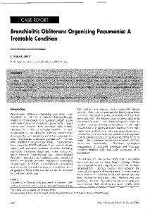
Figure 1. Chest X-ray on admission, showing bilateral alveolointerstitial infiltrates, predominantly in the lower lobes.
measles, mumps, enteroviruses, herpes simplex virus, varicella-zoster virus, and cytomegalovirus), Mycoplasma pneumoniae, Legionella pneumophila, Coxiella burnetti, Chlamydia psittaci, Chlamydia pneumoniae, Fasciola hepatica, Echinococcus granulossus, Aspergillus spp, and human immunodeficiency virus types 1 and 2 were all negative. No malignant cells were found in sputum. Rheumatoid factor, antinuclear antibodies, anti-double-stranded DNA, anti-Sm, anti-RNP, anti-SS-A, anti-SS-B, and anti-neutrophil cytoplasmic autoantibodies were negative. Serum C3 level was 36 mg/dl (normal 43–120 mg/dl). C4, CH50, and serum immunoglobulin levels were all normal. Antiglomerular basement membrane antibodies were negative. A coagulation profile was normal, and there was no evidence of lupus anticoagulant. A standard prick-test was negative. The patient did not cooperate for pulmonary function studies or bronchoscopy. An echocardiogram showed an inferior akinesia and a normal ejection fraction. A gallium-67 scan showed no pathological uptake. A CT scan of the chest showed small areas of centrilobular emphysema in the upper lobes and a mixed alveolar and interstitial pattern, predominantly peripheral, in the lower lobes, with thickened interlobular septa. No lymphadenopathy or pleural effusion were found (Figure 2). Based on the findings of pulmonary infiltrates with eosinophilia, oral prednisone (1 mg/kg/d) was given. During the next few days, the patient’s status improved and the arterial PO2 increased to 86 mm Hg (breathing room air). However, bilateral infiltrates, mostly interstitial type, persisted in an X-ray done 2 wk later. A few days later, the patient developed a self-limited episode of macroscopic hematuria. Afterwards, the urine gave 111 test for protein, and the sediment contained 80 to 100 red cells per high-power field (80% dysmorphics). The urinary protein excretion was 5.5 g/d. Over the next few days, serum urea and creatinine increased to 200 mg/dl (33.2 mmol/L) and 4.9 mg/dl (433.2 mmol/L), respectively (Figure 3). A diagnosis of rapidly progressive glomerulonephritis was made. A percutaneous renal biopsy was performed, and the patient received three boluses of methylprednisolone (0.5 g daily) followed by oral prednisone at the previous dose. In the renal biopsy glomeruli showed mesangial widening with PAS-positive deposits and mild cell proliferation. About one-third
had fibroepithelial crescents. There were some foci of interstitial fibrosis as well as tubular atrophy with scattered groups of inflammatory cells. Immunofluorescence revealed mesangial deposits of IgA (111) and C3 (11) (Figure 4). Those findings were characteristic of mesangial proliferative IgA nephropathy. A video-assisted thoracoscopic biopsy of the right lung was performed. The most striking finding was the filling of terminal and respiratory bronchioles by plugs of mucus, inflammatory cells, and proliferating fibroblasts, with some foamy macrophages. The alveolar septa located near bronchovascular bundles were expanded by an infiltrate of lymphocytes and occasional plasma cells. There were scattered honeycomb areas. No microorganisms were identified (Figure 5). Renal function deteriorated in spite of corticosteroid therapy, and a bolus of cyclophosphamide was given. Hemodialysis was required. An empyema complicated the postoperative course, and the patient died. At autopsy, some bronchioles showed regrowth of metaplastic epithelium over the granulation plugs. There was an extensive fibrotic reaction with atresia of the airways and honeycomb changes. By immunofluorescence, deposits of IgA (111) were demonstrated in alveolar and capillary walls (Figure 5). The kidneys showed a diffuse crescentic glomerulonephritis with large occluding fibrocellular crescents, endocapillary proliferation, and mesangial widening.
DISCUSSION IgAN is a type of glomerulonephritis characterized by diffuse granular deposits of IgA and C3 in the mesangium, commonly manifested by hematuria and proteinuria. The prognosis is usually good, with a rate of progression to end-stage renal failure between 13% and 24% after 15 yr of follow-up (8). However, some cases may present as a rapidly progressive glomerulonephritis or acute renal failure. Systemic complications of IgAN are uncommon. Nevertheless, a few cases of pulmonary hemosiderosis, acute pulmonary hemorrhage, and interstitial pneumonitis, sometimes associated with IgA deposits in the lung, have been reported (3, 9–11). The etiology of BO is diverse, reflecting the fact that it likely represents a nonspecific final tissue reaction to a variety of noxious factors, including infectious agents, drugs, toxic fumes, and autoimmune reactions (7, 12). Idiopathic cases are not uncommon. A respiratory tract infection could be the triggering factor in the present case, but all the microbiological investigations were unrewarding. It has been suggested that the overexpression of MHC class II molecules, leading to autoan-
667
Case Report
Figure 3. Time course of serum creatinine and partial pressure of oxygen. * 5 Start of prednisone; † 5 bolus of methylprednisolone; ‡ 5 bolus of cyclophosphamide; § 5 start of hemodialysis.
tigen presentation and lymphocyte activation, might play a critical role in the development of the inflammatory reaction finally resulting in bronchiolitis (2). IgA is known to exist in respiratory secretions, where it participates in the defense mechanisms of the lung. Whereas IgA in the alveoli seems to be of circulating origin, secretory IgA (dimeric), synthesized by submucosal plasma cells, is the main form present in the airways (13). The type of IgA deposited in the tissues of this patient was not established. Nevertheless, whatever it was, IgA immune complexes, through complement activation and other mechanisms, could be implicated in tissue injury, as previously suggested (11). Mesangial deposit of IgA is the hallmark of idiopathic IgAN. The type and sources of IgA molecules are controversial, although the fact that episodes of macroscopic hematuria are frequently precipitated by upper respiratory tract and gastrointestinal infections suggests the participation of the immune mucosal system (14). Immunoglobulin deposition in the lung may sometimes reflect nonspecific damage to alveolar walls, allowing circulating immune complexes to be deposited secondarily (15). In the present case, IgAN became clinically
Figure 4. Renal biopsy. (A) Glomerulus with mesangial widening and a fibroepithelial crescent (arrow) (HE). (B) Mesangial IgA deposits revealed by immunofluorescence.
evident several weeks after the development of BO, and IgA deposits were found not only in the kidneys but also in the alveolar walls and lung capillaries. It is tempting to speculate that IgA deposition was implicated in the injury mechanisms at both the lung and glomerular levels, according to the following sequence of events. An unidentified noxious agent reached the respiratory tract, triggered an inflammatory reaction and activated the submucosal lymphoid elements. It resulted in bronchiolar wall injury and increased IgA synthesis. IgA deposition, associated with other mechanisms, contributed to bronchiolar wall inflammation. On the other hand, circulating IgA or IgA antigen–antibody complexes, would deposit in the mesangium, thus triggering glomerular injury. This sequence, albeit speculative, sounds plausible in view of the clinical course of the patient reported here. IgA may activate the complement cascade through the alternative pathway. The decrease in circulating C3 levels and the finding of C3 deposits in the mesangium are consistent with such a mechanism.
Figure 5. (A) Lung biopsy showing obliterative bronchiolitis (asterisk) with regrowth of metaplastic epithelium (thick arrow) (HE). (B) Immunofluorescence of lung tissue obtained at autopsy showing IgA deposition in the capillary (thin arrow) and alveolar walls (thick arrow).
668
AMERICAN JOURNAL OF RESPIRATORY AND CRITICAL CARE MEDICINE
To our knowledge, this is the first reported case of IgAN associated with BO. However, there are other examples of IgAN associated with lung diseases. In fact, Endo and Hara (16) found that Japanese patients with diffuse panbronchiolitis frequently had glomerular IgA deposits at autopsy. Moreover, IgAN has also been reported as associated with a variety of acute respiratory infections and chronic lung diseases, including tuberculosis, sarcoidosis, silicosis, pneumonitis, and carcinoma (16, 17). Some clinical and radiological features of the present case would suggest the diagnosis of bronchiolitis obliterans organizing pneumonia (BOOP). However, lung samples taken at biopsy or autopsy showed no organizing pneumonia but changes consistent with evolving obliterative or constrictive bronchiolitis (18). Nevertheless, it is unclear to what extent previous therapy with corticosteroids (which actually resulted in a marked improvement in gas exchange) may have partially cleared alveolar inflammation. Furthermore, the exact relationship between BOOP and constrictive bronchiolitis is uncertain (19), and in some cases they might represent different temporal phases of lung injury. Recent studies have demonstrated an increase of eosinophils in bronchoalveolar lavage (BAL) fluid of patients with idiopathic BOOP (20, 21). Thus, it has been suggested that the eosinophil may be responsible, at least in part, for the lung tissue damage in these patients. In this sense, Olopade and coworkers (22) found eosinophils and eosinophil degranulation in open-lung biopsy specimens of patients with idiopathic BOOP. In our case, peripheral eosinophilia was present, but BAL was not performed, and no eosinophils were seen in either the lung or kidney. Nevertheless, corticosteroid treatment could have changed the appearance of the tissue samples. Thus, the exact role of eosinophils in the case reported here is uncertain. The pulmonary–renal syndrome includes a variety of processes affecting both the lungs and the kidneys, including vasculitis, connective-tissue diseases, and Goodpasture’s syndrome. Although this is a rather heterogeneous group of disorders, the concept is clinically useful and helpful in the diagnostic approach to patients with lesions of both organs. The association of IgAN and BO reported here further expands the clinical spectrum of this syndrome and illustrates the pathogenetic role of abnormal IgA deposition at various organ levels. In view of this case and the data reported by Endo and Hara (16), it would be desirable to study renal function systematically in patients with BO in order to establish the actual frequency of clinically significant IgAN in this setting. References 1. Julian, B. A., B. Waldo, A. Rifai, and J. Mestecky. 1988. IgA nephropathy, the most common glomerulonephritis worldwide: a neglected disease in the United States? Am. J. Med. 84:129–132. 2. Endo, Y., and H. Kanbayashi. 1994. Etiology of IgA nephropathy syn-
VOL. 156
1997
drome. Pathol. Int. 44:1–13. 3. Lai, F. M., E. K. Li, M. W. Suen, S. F. Lui, P. K. Li, and K. N. Lai. 1994. Pulmonary hemorrhage: a fatal manifestation in IgA nephropathy. Arch. Pathol. Lab. Med. 118:542–546. 4. Chung-Park, M., M. Lam, and A. M. Yazdy. 1990. IgA nephropathy associated with sarcoidosis. Am. J. Kidney Dis. 15:601–602. 5. Anwar, N., and R. Gokal. 1993. Simultaneous occurrence of IgA nephropathy and sarcoidosis in the context of pre-existent minimal change nephrotic syndrome. Nephron 65:310–312. 6. Tateno, S., Y. Kobayashi, and F. Kobayashi. 1994. A case of sarcoidosis revealed in the course of IgA nephropathy. Pathol. Int. 44:387–390. 7. King, T. E., Jr. 1993. Overview of bronchiolitis. Clin. Chest Med. 14:607– 610. 8. Ibels, L. S., and A. Z. Györy. 1994. IgA nephropathy: analysis of the natural history, important factors in the progression of renal disease, and a review of the literature. Medicine (Baltimore) 73:79–102. 9. Yum, M. N., L. M. Lampton, P. M. Bloom, and J. S. Edwards. 1978. Asymptomatic IgA nephropathy associated with pulmonary hemosiderosis. Am. J. Med. 64:1056–1060. 10. Travis, W. D., T. V. Colby, C. Lombard, and H. A. Carpenter. 1990. A clinicopathologic study of 34 cases of diffuse pulmonary hemorrhage with lung biopsy confirmation. Am. J. Surg. Pathol. 14:1112–1125. 11. Harland, R. W., C. G. Becker, J. C. Brandes, C. Fritsche, and D. Y. Rosenzweig. 1992. Immunoglobulin A (IgA) immune complex pneumonitis in a patient with IgA nephropathy. Ann. Intern. Med. 116:220– 222. 12. Ezri, T., S. Kunichezky, A. Eliraz, D. Soroker, D. Halperiz, and A. Schattner. 1994. Bronchiolitis obliterans: current concepts. Q. J. Med. 87:1–10. 13. Montinaro, V., L. Gesualdo, and F. P. Schena. 1992. Primary IgA nephropathy: the relevance of experimental models in the understanding of human disease. Nephron 62:373–381. 14. Harper, S. J., and J. Feehally. 1993. The pathogenetic role of immunoglobulin A polymers in immunoglobulin A nephropathy. Nephron 65: 337–345. 15. Leatherman, J. W., S. F. Davies, and J. R. Hoidal. 1984. Alveolar hemorrhage syndromes: diffuse microvascular lung hemorrhage in immune and idiopathic disorders. Medicine (Baltimore) 63:343–361. 16. Endo, Y., and M. Hara. 1986. Glomerular IgA deposition in pulmonary diseases. Kidney Int. 29:557–562. 17. D’Amico, G., E. Imbasciati, G. B. Di Belgioioso, S. Bertoli, G. Fogazzi, F. Ferrario, G. Fellin, A. Ragni, G. Colasanti, L. Minetti, and C. Ponticelli. 1985. Idiopathic IgA mesangial nephropathy: clinical and histological study of 374 patients. Medicine (Baltimore) 64:49–60. 18. Yousem, S. A. 1991. Small airways disease. Pathol. Annu. 26:109–143. 19. Myers, J. L., and T. V. Colby. 1993. Pathological manifestations of bronchiolitis, constrictive bronchiolitis, cryptogenic organizing pneumonia, and diffuse panbronchiolitis. Clin. Chest Med. 14:611–622. 20. Costabel, U., H. Teschler, and J. Guzman. 1992. Bronchiolitis obliterans organizing pneumonia: the cytological and immunocytological profile of bronchoalveolar lavage. Eur. Respir. J. 5:791–797. 21. Mukae, H., J. Kadota, S. Kohno, S. Matsukura, and K. Hara. 1995. Increase of activated T-cells in BAL fluid of Japanese patients with bronchiolitis obliterans organizing pneumonia and chronic eosinophilic pneumonia. Chest 108:123–128. 22. Olopade, C. O., T. B. Crotty, W. W. Douglas, T. V. Colby, and S. Sur. 1995. Chronic eosinophilic pneumonia and idiopathic bronchiolitis obliterans organizing pneumonia: comparison of eosinophil number and degranulation by immunofluorescence staining for eosinophilderived major basic protein. Mayo Clin. Proc. 70:137–142.