169
BioScience Trends. 2015; 9(3):169-181.
Original Article
DOI: 10.5582/bst.2015.01011
Bu-Shen-Ning-Xin decoction suppresses osteoclastogenesis via increasing dehydroepiandrosterone to prevent postmenopausal osteoporosis Yuyan Gui1,2,*, Xuemin Qiu1,2,*, Yingping Xu1,2, Dajin Li1,2,**, Ling Wang1,2,3,** 1
Obstetrics and Gynecology Hospital, Fudan University, Shanghai, China; Laboratory for Reproductive Immunology, Hospital & Institute of Obstetrics and Gynecology, IBS, Fudan University Shanghai Medical College, Shanghai, China; 3 Shanghai Key Laboratory of Female Reproductive Endocrine Related Diseases, Shanghai, China. 2
Summary
Bu-Shen-Ning-Xin decoction (BSNXD), a traditional Chinese medicine, has been used to prevent and treat age-related diseases such as postmenopausal osteoporosis (PMO) for decades. This study sought to investigate the underlying mechanisms of BSNXD in terms of receptor activation of nuclear factor κB ligand (RANKL)-induced osteoclastogenesis in vitro because of the critical roles of bone resorption in the development and progression of osteoporosis. In mice, serum levels of dehydroepiandrosterone (DHEA), dehydroepiandrosterone sulfate (DHEAS), and 17-β-estradiol (E2) were evaluated with an enzyme immunoassay kit after ovariectomy. Levels of DHEA and DHEAS increased significantly following administration of BSNXD while the level of E2 did not. In addition, tartrate-resistance acid phosphatase staining showed that DHEA profoundly inhibited RANKL-induced osteoclastogenesis in vitro in a dose-dependent manner via estrogen receptor α (ERα) but not via estrogen receptor β or androgen receptors. Cytotoxicity was not detected in the 3-(4,5-dimethylthiazol-2-yl)-2,5-diphenyltetrazolium bromide (MTT) assay. These data suggest that BSNXD prevents PMO by increasing DHEA via the ERα pathway to suppress osteoclastogenesis. Keywords: Bu-Shen-Ning-Xin decoction, DHEA, osteoclastogenesis, estrogen receptor α
1. Introduction Osteoporosis, characterized by a decrease in bone mass and micro-architectural alterations, is a progressive bone disease resulting in bone fragility and an increased risk of fractures. The disease may be classified as primary or secondary. Advanced age and being female are the major risk factors for primary osteoporosis (1). Accordingly, the form of osteoporosis most common in women after menopause is referred to as primary type 1 or postmenopausal osteoporosis (PMO), and this
*These authors contributed equally to this works. **Address correspondence to: Dr. Ling Wang and Dr. Dajin Li, Obstetrics & Gynecology Hospital of Fudan University, 413 Zhaozhou Road, Shanghai 200011, China. E-mail:
[email protected] (Wang L.); djli@shmu. edu.cn (Li DJ)
condition is due to a dearth of estrogen. The underlying mechanism in osteoporosis is an imbalance between bone resorption and formation regulated by osteoclasts and osteoblasts, respectively. A dearth of estrogen after menopause increases bone resorption, and this is associated with increased production of pro-inflammatory cytokines such as the receptor activation of nuclear factor κB ligand (RANKL) (2). RANKL plays a pivotal role in osteoclastogenesis. Mature osteoclasts are multinucleated giant cells that differentiate from hematopoietic stem cells following stimulation by two key factors, macrophage-colonystimulating factor (M-CSF) and RANKL (3). Hormone replacement therapy (HRT) in postmenopausal women increases estrogen levels and prevents postmenopausal bone loss. However, there is debate about its safety. According to the Women's Health Initiative Study, longer term estrogen replacement was found to cause an unacceptable increase in the risk of heart attack, stroke,
www.biosciencetrends.com
BioScience Trends. 2015; 9(3):169-181. and breast and uterine cancer (4). Consequently, other potential therapeutic interventions have been examined, including traditional Chinese medicines (TCMs). TCMs have been used in Asian countries such as China, Japan, and Korea for more than 2,500 years to prevent and treat various diseases (5). In these areas, TCMs have developed into an integral part of complementary and alternative medicine. Traditional Chinese herbal medicines often consist of a combination of individual herbs specifically formulated to increase therapeutic efficacy and reduce adverse effects (5). Bu-Shen-Ning-Xin decoction (BSNXD), a traditional Chinese medicine, contains dried Rehmannia Root, common Anemarrhena rhizome, bark of the Chinese Corktree, Barbary Wolfberry fruit, Chinese Dodder seed, Shorthorned Epimedium, Spina Date seed, and Oriental Waterplantain rhizome (Table 1). BSNXD has been used for hundreds of years to treat and prevent menopauserelated disorders and age-related diseases (6), including osteoporosis. BSNXD has a beneficial effect on bone metabolism (7), but the mechanisms of that effect are unclear. In order to ascertain the effects of BSNXD on PMO, an animal model of ovariectomized (OVX) female mice was used to deplete ovarian hormones. The current authors previously found that pharmacological serum from OVX mice treated with BSNXD promoted the proliferation and inhibited the apoptosis of mouse osteoblasts (7). The aim of the present study was to investigate how BSNXD improved PMO in terms of osteoclasts in OVX mice. This study measured the serum levels of dehydroepiandrosterone (DHEA), dehydroepiandrosterone sulfate (DHEAS), and 17-β-estradiol (E2) in OVX mice administered a low, moderate, or high dose of BSNXD. The effects of DHEA on murine osteoclastogenesis were then tested in vitro. Results indicated that DHEA played an important role in the inhibitory effect of BSNXD against osteoclastogenesis. 2. Materials and Methods 2.1. Chemicals and reagents Minimum essential medium (MEM) without phenol red and fetal bovine serum (FBS) were obtained from Gibco-BRL (Gaithersburg, MD, USA). Penicillin, streptomycin, and a kit for the 3-(4,5-dimethylthiazol-2yl)-2,5-diphenyltetrazolium bromide (MTT) assay were purchased from the Beyotime Institute of Biotechnology (Shanghai, China). M-CSF was supplied by R&D Systems (Minneapolis, MN, USA). RANKL was obtained from Peprotech (Rocky Hill, NJ, USA). DHEA, E2, flutamide, and a Leukocyte Acid Phosphatase Kit were purchased from Sigma-Aldrich Co (Saint Louis, MO, USA). MPP and R,RTHC were obtained from Tocris Cookson Inc. (Ellisville, MO, USA). An active
170
DHEA enzyme immunoassay (EIA) kit was purchased from Diagnostic System Laboratories, Inc. (DSL, Webster, Texas, USA). A DHEAS EIA kit was supplied by IBL (Fujioka, Japan). An E2 EIA kit was supplied by BioCheck Inc. (Burlingame, CA, USA). 2.2. Preparation of BSNXD extract BSNXD was obtained from the pharmacy of the Hospital of Obstetrics and Gynecology, Fudan University, Shanghai, China. BSNXD includes 8 crude herbs as listed in Table 1, and BSNXD was formulated in accordance with traditional Chinese medicine theory and the clinical experience of the authors. The ingredients were decocted in a 10-fold volume of water at 90°C for 60 min. An aqueous extract was prepared by boiling the herbs three times to yield a decoction (1 g/mL, w/v). Products were prepared in accordance with good manufacturing practices (GMPs) at the Institute of Obstetrics and Gynecology, Fudan University. 2.3. Mice The mice used were 95 8-week-old female BALB/c mice with a body mass between 20 and 30 g that were purchased from the Laboratory Animal Facility of the Chinese Academy of Sciences (Shanghai, China). The experimental animals were housed and handled in accordance with the guidelines of the Chinese Council for Animal Care. All mice were habituated to housing conditions for 3 days and then housed four per cage on a reversed 12 h light and 12 h darkness cycle. Food and water were available ad libitum at room temperature. 2.4. Experimental and drug administration In accordance with the Principles of Laboratory Animal Care (National Institutes of Health publication number 85-23, revised 1985), 80 mice underwent bilateral oophorectomy, while a sham-operated group (15 mice) underwent surgery but no ovariectomy. The OVX mice were randomly divided into 5 groups (OVX, OVX + low-dose BSNXD, OVX + mod-dose BSNXD, OVX + high-dose BSNXD, and OVX + E2 group; n = 16 each group). Unfortunately, 5 mice died during anesthesia. These dead mice were excluded from analysis. OVX mice treated with saline (n = 15) served as controls. OVX + high-dose BSNXD group, OVX + mod-dose BSNXD group, and OVX + low-dose BSNXD group mice were orally administered 0.5 mL of evaporated BSNXD extract twice daily (with 2, 1, or 0.5 g/mL of raw herbs respectively, w/v, n = 15). These doses correspond to 18-, 9-, and 4.5-fold of the human adult dose based on an established formula for human-mouse drug conversion. As previously described, the OVX + E2 group was treated with E2 (100 μg/kg per day orally, n = 15) (8,9).
www.biosciencetrends.com
171
BioScience Trends. 2015; 9(3):169-181.
Table 1. The composition and preparation of the herbal preparation BSNXD Crude herb Dried Rehmannia root Common Anemarrhena rhizome Bark of Chinese Corktree Barbary Wolfberry fruit Chinese Dodder seed Shorthorned Epimedium Spina Date seed Oriental Waterplantain rhizome
Latin name
Content
Radix Rehmanniae Exsiccata Anemarrhena asphodeloides Bunge Phellodendron amurense Rupr. Fructus Lycii barbari Cuscuta chinensis Epimedium brevicornum Maxim Ziziphus jujuba Mill. var. spinosa Alisma plantago-aquatica Linn.
15 g 15 g 9g 15 g 12 g 12 g 9g 12 g
Notes: Prepared according to the traditional method. The crude herbs above (×15) were mixed, immersed in deionized water (10 times the total weight of herbs), and then boiled at 90°C for 60 min for the first decoction. An aqueous extract was prepared by boiling the herbs three times to make a decoction. The three extracts were combined and concentrated with a rotary evaporator (Model N1000, Eyela, Japan). The yield of the BSNXD extract was 742.5 mL with 2 g/mL (w/v) of raw herbs.
All mice were weighed and then sacrificed after the last treatment in order to collect blood samples and tissues for further investigation at 20 weeks of age. The ovariectomy was successful in all OVX animals based on a lack of ovarian tissue and atrophied uterine horns.
means of the Leukocyte Acid Phosphatase Kit. TRAPpositive multinucleated cells (TRAP+ MNCs; more than five nuclei) were counted under a microscope. Results from at least six independent experiments are shown. 2.7. Cell viability assay
2.5. Determination of DHEA, DHEAS, and E2 in drugderived serum samples At the end of experiment, all the mice were sacrificed by asphyxiation with CO2. Blood was collected via a cardiac puncture, and volumes up to 1 mL were often obtained from each mouse. Serum samples were prepared using centrifugation and then stored at -20°C for determination of DHEA, DHEAS, and E2 concentrations. A DHEA, DHEAS, or E2 EIA kit was used to measure these steroid concentrations in accordance with the manufacturer's protocol. 2.6. Primary osteoclast culture and osteoclastogenesis in vitro A primary osteoclast culture and osteoclastogenesis in vitro was accomplished as previously described (10). Briefly, bone marrow-derived monocyte/macrophage precursor cells (BMMs) from the femurs and tibias of 20-week-old mice in the OVX group were cultured in MEM without phenol red supplemented with FBS in the presence of 10 ng/mL M-CSF for 2 days. These cells were then allowed to differentiate into osteoclasts using 50 ng/mL RANKL and 10 ng/ml M-CSF for 3 days. To estimate the effect of DHEA on RANKL-induced osteoclastogenesis in vitro, the cells were exposed to a series of concentrations of DHEA (10-6-10-9 M DHEA) or a solvent control along with RANKL stimulation for 72 h. One hour before exposure to 10-7 M DHEA, other osteoclasts were also treated with 10-6 M MPP (estrogen receptor α antagonist), 10 nM R,RTHC (estrogen receptor β antagonist), or 10 μM flutamide (androgen receptor antagonist) in MEM without phenol red (11-13). Afterwards, osteoclastogenesis was evaluated with tartrate-resistance acid phosphatase (TRAP) staining by
Cell viability was determined using the MTT assay in accordance with the protocol provided by the manufacturer. Briefly, BMMs (5 × 10 4 cells/well) were cultured in M-CSF-containing MEM with serial concentrations of DHEA for 12, 24, and 48 h. Afterwards, 0.5 mg/mL of MTT reagent was added at 37°C for 4 h before the end of culturing. Following removal of the supernatant, insoluble formazan dye was dissolved in 200 μL of dimethyl sulfoxide (DMSO), and the absorbance at 550 nm was measured by using a microplate reader. The cell viability was quantified as the relative decrease in the absorbance at 550 nm in comparison to untreated control cells. Values are expressed as the percent viability in the sample vs. the control culture, which was set as 100%. 2.8. Statistical analysis All values were expressed as the mean ± SEM. The difference between experimental groups was analyzed using ANOVA and a Kruskal-Wallis test, with p < 0.05 being considered significant. Data were analyzed using SPSS software. 3. Results 3.1. Administration of BSNXD increased serum DHEA but not E2 A previous study showed that BSNXD up-regulated serum levels of the estrogen receptor (ER) without affecting E2 in OVX rabbits (6). Hence, a question was whether BSNXD prevented osteoporosis by affecting the production/metabolism of ER ligands, such as DHEA, DHEAS, and E2, in mice. The serum levels of these
www.biosciencetrends.com
BioScience Trends. 2015; 9(3):169-181.
172
Figure 1. Administration of BSNXD increased serum levels of DHEA and DHEAS but not E2. Mice were sacrificed after 3 months of treatment, and sera were collected for determination of DHEA (A), DHEAS (B), and E2 (C) concentrations. Steroid concentrations in sera were measured using a DHEA, DHEAS, or E2 enzyme immunoassay kit. Data are expressed as the mean ± SEM (n = 10). * p < 0.05, ** p < 0.01.
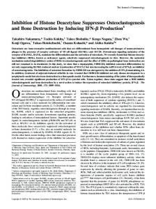
Figure 2. DHEA profoundly inhibited the formation of TRAP-positive cells in a dose-dependent manner. BMMs from OXV mice were exposed to a series of concentrations (10-6-10-9 M) of DHEA or a solvent control at the same time as RANKL stimulation for 72 h to estimate the effect of DHEA on osteoclastogenesis. Osteoclastogenesis was evaluated with TRAP staining (A). Data are expressed as the mean ± SEM (n = 10). * p < 0.05, ** p < 0.01 (B).
steroid hormones decreased significantly in OVX mice compared to the sham-operated mice. A moderate and high dose of BSNXD significantly increased serum levels of DHEA and DHEAS (p < 0.01) but had no effect on E2 (p > 0.05, Figures 1A and 1B). Administration of E2 significantly increased the serum concentration of E2 (p < 0.01), but levels of DHEA and DHEAS were relatively unaltered (p > 0.05, Figure 1C).
3.3. DHEA had no effect on osteoclast viability To determine the mechanism by which DHEA inhibited osteoclastogenesis in vitro, the cytotoxicity of DHEA was examined using the MTT assay. BMMs were viable at each time-point, as shown in Figure 3. As indicated by the MTT assay, treatment with 10-5-10-9 M DHEA did not significantly affect the cell viability of BMMs in comparison to the controls (p > 0.05, Figure 3).
3.2. DHEA inhibited osteoclastogenesis To investigate the effect of DHEA on RANKLinduced osteoclastogenesis in BMMs, BMMs from OVX mice were exposed to a series of concentrations of DHEA (10 -6-10 -9 M DHEA) or a solvent control along with RANKL stimulation for 72 h. Results i n d i c a t e d t h a t 1 0 -7- 1 0 -8 M D H E A p r o f o u n d l y inhibited the formation of TRAP-positive cells (p < 0.01, Figures 2A and 2B).
3.4. DHEA inhibited osteoclastogenesis via an estrogen receptor α-dependent pathway Since DHEA did not affect osteoclast viability, DHEA might possibly inhibit osteoclast differentiation via some other pathway. Although no specific intra-nuclear receptors of DHEA have been identified to date (14), studies have found that both ERs (15) and androgen receptors (ARs) (15) are involved in mediating the
www.biosciencetrends.com
173
BioScience Trends. 2015; 9(3):169-181.
effects of DHEA in an organ-dependent manner. Accordingly, a concentration of 10-7 M of DHEA was used to determine whether subtypes of ERs or ARs are involved in the effects of DHEA on osteoclast differentiation. Pretreatment of BMMs with 10-6 M MPP, 10 nM R,RTHC, or 10 μM FLUT did not affect osteoclastogenesis in the control group. However, pretreatment of BMMs with 10-6 M MPP for 1 h before treatment with 10-7 M DHEA for 72 h significantly increased osteoclastogenesis (p < 0.01). In contrast, treatment with 10nM R,RTHC or 10 μM FLUT had no such effect (p > 0.05, Figure 4). 4. Discussion Bone metabolism is a lifelong dynamic process consisting of osteoblast-mediated bone formation and osteoclast-mediated bone resorption. Generally,
Figure 3. DHEA had no effect on osteoclast viability. There were no significant differences in the effect of 10-6-10-9 M of DHEA on BMM viability. Data are expressed as the mean ± SEM (n = 10). * p < 0.05, ** p < 0.01.
Figure 4. DHEA inhibited osteoclastogenesis via an ERαdependent pathway. BMMs from OVX mice were treated with 10-6 M MPP, 10 nM R,RTHC, or 10 μM flutamide in MEM without phenol red 1 h before exposure to the solvent control or 10-7 M DHEA for 72 h. Osteoclastogenesis was evaluated with TRAP staining. Data are expressed as the mean ± SEM (n = 10). * p < 0.05, ** p < 0.01.
bone mass peaks in puberty and then decreases. Bone loss accelerates in postmenopausal women due to a dearth of estrogen. A dearth of estrogen disrupts the skeletal homeostasis and results in a high rate of bone remodelling (15), with bone resorption exceeding bone formation. Currently available drugs for the treatment of osteoporosis can be divided into two categories: anti-resorptive agents and bone-forming agents (16). Anti-resorptive agents like estrogen are usually recommended as first-line therapy, which is also called HRT, for women with PMO. There are numerous contraindications limiting the use of HRT, so traditional medicines may constitute a viable alternative. In Asian countries, TCM has a long history of helping to fight disease and even guide modern treatments of conditions such as inflammatory diseases (17) and age-related diseases (18). According to TCM theory, the Shen is the kidney system and refers to the kidneys as well as the gonads and the adrenal glands. The Shen is responsible for maintaining the energy that facilitates growth and development. When people age, the Shen decreases. In TCM, doctors call this phenomenon the Shen-Xu (weakness of the kidney system), and Bu-Shen (complementing the kidney system) is used to maintain the balance in the body. According to TCM, the Shen controls the growth and development of many organs including bone. After menopause, women suffer Shen-Xu and experience accelerating bone loss. Numerous studies of formulations containing ingredients associated with Bu-Shen have found that these preparations prevent or ameliorate PMO. As an example, Bu-Shen-Hua-Yu extract decreased the serum levels of interleukin-6 (IL6), increased the number of osteoblasts, and reduced the number of osteoclasts to prevent PMO (19). BuShen-Zhuang-Gu granules are a "kidney-tonifying" herbal preparation that has been found to enhance bone mineral density and bone architecture and strength, thus preventing and ameliorating OVX-induced bone loss (20). The clinical experience of the current authors has indicated that BSNXD indeed delays bone loss in PMO. There are eight crude herbs in this formulation (Table 1), and each crude herb contains numerous chemical substances such as iridoids, phenylethanoid glucosides, flavonoids, lignans, and saponins (Table 2) (21-63). Each crude herb has its own special biological activities (Table 2). An extract of dried Rehmannia root has antiinflammatory action (28), anti-apoptotic action (27) and antioxidant action (26). Iridoid glycosides are the principal active components of dried Rehmannia root. Catalpol is the most studied iridoid glycoside. Catalpol is considered to be a potential therapeutic for treatment of neurodegenerative diseases such as Alzheimer's disease and Parkinson's disease (55). In addition, acteoside suppresses RANKL-mediated osteoclastogenesis by inhibiting c-Fos induction and
www.biosciencetrends.com
BioScience Trends. 2015; 9(3):169-181.
174
Table 2. The constituents of crude herbs in BSNXD Crude herbs
Biological activities
Main components
Specific constituents
Dried Rehmannia root
An extract of dried Rehmannia root has antiinflammatory (28), anti-apoptotic (27), and antioxidant action (26). Iridoid glycosides are the principal active components of dried Rehmannia root. Catalpol is the most studied iridoid glycoside. Catalpol is considered to be a potential therapy for treatment of neurodegenerative diseases such as Alzheimer's disease and Parkinson's disease (55). Acteosides suppress RANKL-mediated osteoclastogenesis by inhibiting c-Fos induction and the NF- κB pathway and by attenuating ROS production (29).
iridoid glycosides
catalpol rehmaglutosides myobontioside A aucubin ajugol geniposide 6-O-E-feruloyl ajugol gardoside methyl ester jioglutoside B monomellittoside
phenylethanoid glucosides
martinoside acteoside sculpolnisde salidroside leucosceptoside A purpureaside C decaffeoylacteoside
ionones
frehmaglutin A frehmaglutin B frehmaglutin C frehmaglutin D neo-rehmannioside rehmamegastigmane rehmapicrogenin A aeginetic acid 5-O-β-D-quinovoside aeginetic acid dihydroxy-β-ionone
flavonoids
apigenin diosmetin luteolin luteolin-7-o-β-D-glucuronide
phenolic acids
Common Anemarrhena rhizome
An extract of the common Anemarrhena rhizome has antitumor, anti-inflammatory, and antioxidant action (56). Steroid saponins are the principal active components of the common Anemarrhena rhizome (30). An adequate supply of steroidal saponins from Anemarrhena asphodeloides prevented OVX-induced bone loss in rats by promoting bone formation but did not inhibit bone resorption (31).
p-hydroxybenzoic acid gentisic acid protocatechuic acid 1,2,4-trihydroxybenzene vanillic acid
lignans
Hierochin D yemuoside YM1 lariciresinol pinoresinol 4-O-glucoside
steroid saponins
sarsasapogein timosaponin markogein neogitogein dosgenin
xanthones
mangiferin neomangiferin ismangiferin
lignans
cis-hinokiresinol monomethy-cis-hinokiresinol oxy-cis-hinokiresinol
anemaran
Bark of the Chinese Corktree
An extract of the bark of the Chinese Corktree inhibits the cellular immune response Phellodendrine and magnoflorine are thought to be the main active ingredients responsible for this biological activity (34). The extract of this herb suppresses the synthesis of collagen and promoted the synthesis of proteoglycans in chondrocytes (33). Alkaloids are the principal active components of the bark of the Chinese Corktree (33). Berberine is the most studied alkaloid. Berberine is reported to have anti-inflammatory (48), anti-apoptotic (49), antioxidant (50), antitumor (51), anti-hypertensive, and renoprotective action (52).
flavonoids
icarisid I baohuoside
alkaloids
berberine phellodendrine jatrorrhizine palmatine berberubine tetrahydrojiatrorrhizine phellodendrine tetrahydroberberine tetrahydropalmatine oxyberberine magnoflorine menisperine dictamnine γ-fagarine skimmi-anine rutaecarpine 7-hydroxyrutaecarpine 7,8-dehydroxyrutaecarpine 7,8-dihydroxyrutaecarpine candicine N-methylflindersine (To continue)
www.biosciencetrends.com
175
BioScience Trends. 2015; 9(3):169-181.
Table 2. The constituents of crude herbs in BSNXD Crude herbs
Barbary Wolfberry fruit
Chinese Dodder seed
Biological activities
(continued) Main components
Specific constituents
flavonoids
phellamurin amuresin phellochinin A phellavin phellatin hyperin phellozide quercetin-3-O-β-D-galactoside dihydrokaempferol
phenolic ramification
syringin methyl-3-O-femloyl-quini acid methyl-3-O-feruloylquinate methyl-5-O-feruloylquinate amurenlaetone A amurenlaetone B amurenamide A coniferin
terpenes
niloticin dihydroniloticin niloticin acetate dihydroniloticin acetate friedelin Piscidinol A Hispidol B Bourjotinolone A Hispidone
lactones
dictamnolide nomilin obakunone obakunonic acid obaeulaetone kihadanin A kihadanin B
sterols
7- dehydro-stigmasterol β- itosterol γ- itosterol campesterol stigmasterol
Barbary Wolfberry fruit has various biological activities, including anti-aging (59) and antioxidant action (58), immunity-enhancing action (60), and neuroprotective action (57). Studies have focused on the antioxidant and immunomodulatory properties of this fruit in a range of age-related diseases such as atherosclerosis, neurodegeneration, and diabetes (61). The antioxidative action of this fruit is mainly attributed to polysaccharides and flavonoids (60). In Raw246.7 cells, polysaccharides induced expression of TNF-α and IL-1β via activation of NF-κB and AP-1 to modulate immunoreaction (35).
lycium barbarum polysaccharides
Chinese Dodder seed has been used to improve sexual function, prevent senescence, and regulate the immune system. An extract of the seed protected murine osteoblastic MC3T3-E1 cells against injury induced by tertiary butyl hydroperoxide (62). Flavonoids are the principal active components of this herb (36). Flavonoids increase the level of estrogen in the peripheral blood of mice (40). Flavonoids also promote osteoblastic activity in vitro (39), they maintain the balance between bone formation and bone resorption, and they enhance bone mineral density in ovariectomized mice (37). Glycoside in this herb has been found to have anti-aging action and to enhance memory by inducing PC12 cell differentiation (62).
carotenoids
β-carotene β-cryptoxanthin zeaxanthin
flavonoids
quercetin gentisic acid chlorogenic acid quercetin-3-rhamnoside
flavonoids
quercetin kaempferol hyperoside astragalin hyperin
steroids
β-sitosterol stigmasterol campesterol cholesterol
polysaccharides
CHC-1 H3 CS-A CS-B CS-C
alkaloids
scutamine matrine sophoranol methyIcytisine
terpenes
maragenin australiside A
lignans
sesamin cuscutoside A cuscutoside B neocuscutoside A neocuscutoside B neocuscutoside C
www.biosciencetrends.com
BioScience Trends. 2015; 9(3):169-181. Table 2. The constituents of crude herbs in BSNXD
176 (continued)
Crude herbs
Biological activities
Main components
Specific constituents
Shorthorned Epimedium
Shorthorned Epimedium is one of the most active ingredients in BSNXD that prevents and ameliorates postmenopausal osteoporosis. Flavonoids are the principal active components of this herb. Flavonoids up-regulate the expression of estrogen receptor α and β in the hypothalamus and hippocampus of ovariectomized mice, delaying bone loss without affecting the uterus (45). Icariin is the most studied flavonoid, and it has a wide range of biological activities. Studies have reported that icariin promotes the activity of mesenchymal stem cells and osteoblasts in vitro (43). Icariin also inhibits osteoclastogenesis in vitro (44).
flavonoids
icariin icariin I icariin II epimedin A epimedin B epimedin C baohuoside I ginkgetin isoginkgetin bilobetin quercetin breviflavone A breviflavone B Sempervirenoside A Sempervirenoside B liquiritigenin 5"-Methoxyhydnocarpin Methoxyhydnocarpin-D apigenin anthocyanin cayratinin daidzein
lignans
icariside E6 icariside E7 icariol A1 icariol A2 hydnocarpin 5′′-methoxyhydnocarpin hydnocarpin- D 5′-methoxyhydnocarpin-D 5′,5′′-dimethoxyhydno- carpin-D
phenolic glycosides
icariphenol 4-hydroxy-2-prenylpheno-1-O-β- D -glucopyranoside icariside A5 thalictoside salidroside
alkaloids
magnoflorine epimediphine
flavonoids
spinosin Zivulgarin 6" '- pcoumaroylspinosin 6" '- sinapoylspinosin swertisin 6" '-p-hydroxylbenzoylspinosin 6" '-dihydrophaseoylspinosin 6'',6" '-diferuloylspinosin spinorhamnoside
saponins
jujuboside A jujuboside A1 jujuboside C jujuboside D jujuboside E jujuboside G jujuboside H acetyljujuboside B protojujuboside A protojujuboside B protojujuboside B1
terpenes
betullinic acid betulin ceanothic acid alphitolic acid methyl betulinate alphitolic acid methylester
steroids
daucosterol
fatty acids
A variety of unsaturated fatty acids
alkaloids
sanjoinine A sanjoinine B sanjoinine D sanjoinine F sanjoinine G1 nuciferine nornuciferine norisocorydine
Spina Date seed
The health benefits of Spina Date seed include antitumour, anti-inflammatory, antiobesity, immunostimulatory, and antioxidant action (63). The extract of this herb has a sedative-hypnotic effect (46) and it promotes hematopoietic functions in blood-deficient animals by up-regulating the expression of erythropoietin in liver cells (64). Flavonoids and saponins are the principal active components of Spina Date seed. Saponins and flavonoids are active ingredients that suppress the central nervous system (46). Flavonoids and saponins in Spina Date seed also have antibacterial and antifungal action (53).
www.biosciencetrends.com
177
BioScience Trends. 2015; 9(3):169-181.
Table 2. The constituents of crude herbs in BSNXD Crude herbs
Biological activities
(continued) Main components
Specific constituents coclaurine zizyphusine N - methylasimilobine magnoflorine amphibine 5 - hydroxy - 6 - methoxynoraporphine sanjoinenine
Oriental Waterplantain rhizome
An extract of the Oriental Waterplantain rhizome has antithrombus and anti-hyperlipidemia action, it ameliorates atherosclerosis, it has hypoglycemic and anti-aging action, it enhances diuresis and immunoregulation (47). Triterpenes are the main components responsible for diuresis (47).
the nuclear factor kappa-light-chain-enhancer of activated B cells (NF- κB) pathway and attenuating the production of reactive oxygen species (ROS) (29). An extract of common Anemarrhena rhizome has antitumor, anti-inflammatory, and antioxidant action (56). Steroid saponins are the principal active components of common Anemarrhena rhizome (30). An adequate supply of steroidal saponins in Anemarrhena asphodeloides prevented OVX-induced bone loss in rats through the promotion of bone formation but not the inhibition of bone resorption (31). An extract of the bark of the Chinese Corktree inhibits the cellular immune response, and phellodendrine and magnoflorine are believed to be the main active ingredients responsible for this biological activity (34). An extract of this herb suppressed synthesis of collagen and promoted synthesis of proteoglycans in chondrocytes (33). Alkaloids are the principal active components of the bark of the Chinese Corktree (33). Nerberine is the most studied alkaloid. Berberine is reported to have antiinflammatory (48), anti-apoptotic (49), antioxidant (50), antitumor (51), and anti-hypertensive and renoprotective action (52). The Barbary Wolfberry fruit has various biological activities including anti-aging (59) and antioxidant action (58), immunity-enhancing action (60), and neuroprotective action (57). Studies have focused on the antioxidant and immunomodulatory properties of this fruit in a range of age-related diseases such as atherosclerosis, neurodegeneration, and diabetes (61). The antioxidative activity of this fruit is mainly attributed to polysaccharides and flavonoids (60). In Raw246.7 cells, polysaccharides induced expression of tumor necrosis factor-α (TNF-α) and interleukin-1β (IL-1β) via activation of NF-κB and activator protein 1 (AP-1) to modulate immunoreaction (35).
triterpenes
alisol A alisol B alisol C 23 acetate
sesquiterpenoids
alismol alismaxide orientalol A orientalol B orientalol C sulfoorientalol A sulfoorientalol B sulfoorientalol C sulfoorientalol D orientalol D orientalol E
diterpenes
oriediterpenol oriediter-penoside
Chinese Dodder seed has been used to improve sexual function, prevent senescence, and regulate the immune system. An extract of the seed protected murine osteoblastic MC3T3-E1 cells from injury induced by tertiary butyl hydroperoxide (62). Flavonoids are the principal active components of this herb (36). Flavonoids increased the level of estrogen in peripheral blood of mice (40). Flavonoids also promoted osteoblastic activity in vitro (39), maintaining the balance between bone formation and bone resorption and enhancing bone mineral density in OVX mice (37). The glycoside in this herb has been found to counteract aging and enhance memory by inducing PC12 cell differentiation (62). Shorthorned Epimedium is one of the key active ingredients in BSNXD that prevents and ameliorates PMO. Flavonoids are the principal active components of this herb. Flavonoids up-regulated the expression of ER α and β in the hypothalamus and hippocampus of OVX mice, delaying bone loss without affecting the uterus (45). Icariin is the most studied flavonoid, and it has a wide range of biological activities. A study has reported that icariin promotes the activity of mesenchymal stem cells and osteoblasts in vitro (43). Moreover, icariin also inhibited osteoclastogenesis in vitro (44). Spina Date seed provides health benefits through its antitumor, anti-inflammatory, anti-obesity, immunostimulating, and antioxidant action (63). The extract of this herb has a sedative-hypnotic effect (46), and it promoted hematopoietic functions in a blooddeficient animal model by up-regulating erythropoietin expression in liver cells (64). Flavonoids and saponins are the principal active components of Spina Date seed. Saponins and flavonoids are active ingredients that suppress the central nervous system (46). Flavonoids and saponins in Spina Date seed also have antibacterial and antifungal action (53).
www.biosciencetrends.com
BioScience Trends. 2015; 9(3):169-181. The extract of the Oriental Waterplantain rhizome has anti-thrombus and anti-hyperlipidemia action, action against atherosclerosis, hypoglycemic action, anti-aging action, diuretic action, and immunoregulatory action (47). Triterpenes are the main components responsible for diuresis (47). Acteoside in dried Rehmannia root suppresses RANKL-mediated osteoclastogenesis by inhibiting c-Fos induction and the NF-κB pathway and by attenuating ROS production (29). Flavonoids in Shorthorned Epimedium delayed bone loss without affecting the uterus in OVX mice (45), and icariin inhibited osteoclastogenesis in vitro (44). These findings are in accordance with the current results. However, flavonoids in Chinese Dodder seed increased the level of estrogen in the peripheral blood of mice (40), and this finding does not accord with the current results. During the process of preparing the BSNXD extract, the structures of the flavonoids may have been transformed or unknown substances that decreased estrogen levels may have been generated. Solitary herbs may delay bone loss, but an herbal preparation of BSNXD contains a complex mixture of ingredients that may be metabolized in organs. Thus, pharmacological serum might produce this effect in vivo. Previous work has shown that BSNXD pharmacological serum promoted the proliferation and suppressed the apoptosis of murine osteoblasts via the mitogen-activated protein kinase (MAPK) pathway (7). Therefore, certain substances presumably changed in serum after treatment with BSNXD. DHEA and its sulfate ester, also referred as agerelated hormones, are the most abundant steroids in humans. These hormones are produced from cholesterol mainly in the adrenals but also in the ovaries (65). Levels of DHEA peak during the third decade of life and then decline with age (66). A previous study by the current authors found that BSNXD up-regulated ER rather than E2 in OVX rabbits (6). BSNXD pharmacological serum and DHEA affect osteoblasts via the MAPK pathway (11,67). BSNXD might prevent osteoporosis by increasing other ER ligands besides E2 in mice, such as DHEA or DHEAS, and thus delay a loss in bone mass. The present study proved the authors' hypothesis. DHEA, DHEAS, and E2 concentrations in serum all decreased significantly in OVX mice compared to sham-operated mice. This result coincides with the findings of Park JH et al. (68). After treatment with a moderate or high dose of BSNXD, levels of DHEA and DHEAS were significantly elevated but levels of E2 were not. The administration of E2 significantly increased the serum concentration of E2 without changing the concentration of DHEA or DHEAS. Given the wealth of evidence indicating the beneficial effect of DHEA on skeletal mass (65,68-71), BSNXD may delay bone loss in PMO by enhancing production of DHEA and DHEAS rather than E2. Furthermore, the
178
current results also provide scientific proof for use of BSNXD by women when HRT is contraindicated. Osteoclastogenesis by BMMs is tightly regulated by complex mechanisms associated with processes including cell viability and differentiation. The proliferation and survival of osteoclast precursors depends on M-CSF, and the formation and survival of differentiated osteoclasts depends on RANKL (72). Although some studies have reported that DHEA inhibited bone resorption by activating osteoblastic viability (67) and up-regulating osteoprotegerin/RANKL (OPG/RANKL) (71), no studies have reported its direct effect on osteoclast differentiation. The current findings suggest that DHEA profoundly inhibited RANKLinduced osteoclastogenesis in vitro via estrogen receptor α (ERα) but not estrogen receptor β (ERβ) or AR. Thus, BSNXD presumably prevented PMO by increasing DHEA to suppress osteoclastogenesis. In humans, DHEA is produced by the adrenal gland and metabolized into testosterone, estrogen, and other biologically active steroids by several tissues, including the brain, liver, kidney, and gonads (69). Accordingly, after menopause all estrogens are made locally from DHEA by the mechanisms of intracrinology (73). DHEA activates ERβ to nearly the same extent as E2 and it activates ERα to a lesser extent (38,66). The current data showed that administration of BSNXD increased the serum concentration of DHEA but not that of E2. After oral administration of BSNXD, compounds absorbed into the bloodstream may become active compounds like DHEA and its metabolites. BSNXD-induced metabolism may encourage the adrenals to secrete larger quantities of DHEA and its metabolites. The effect of DHEA on RANKL-induced osteoclastogenesis was examined in BMMs, and DHEA profoundly inhibited osteoclastogenesis via an ERα-dependent pathway but did not affect the cell viability of BMMs. Nonetheless, the specific mechanism by which BSNXD enhances the production of DHEA needs to be investigated further. There may be other substances besides DHEA that have a beneficial effect on skeletal mass, so these substances need to be studied. Furthermore, the specific signaling pathway by which DHEA inhibits osteoclast differentiation has yet to be determined. In conclusion, this study has revealed that BSNXD inhibits osteoclast differentiation. This action is related to up-regulation of DHEA via ERα independent of estrogen metabolites. This finding suggests that this herbal preparation is a good approach to treating PMO. Acknowledgements This work was supported by the Science and Technology Commission of Shanghai Municipality 2015 YIXUEYINGDAO project No. 15401932200 (L Wang), the FY2008 JSPS Postdoctoral Fellowship
www.biosciencetrends.com
179
BioScience Trends. 2015; 9(3):169-181.
for Foreign Researchers P08471 (L Wang), National Natural Science Foundation of China No. 30801502 (L Wang), the Shanghai Pujiang Program No. 11PJ1401900 (L Wang), and the National Natural Science Foundation of China No. 81401711 (XM Qiu). References 1.
Bernabei R, Martone AM, Ortolani E, Landi F, Marzetti E. Screening, diagnosis and treatment of osteoporosis: A brief review. Clin Cases Miner Bone Metab. 2014; 11:201-207. 2. Roggia C, Gao YH, Cenci S, Weitzmann MN, Toraldo G, Isaia G, Pacifici R. Up-regulation of TNF-producing T cells in the bone marrow: A key mechanism by which estrogen deficiency induces bone loss in vivo. Proc Natl Acad Sci U S A. 2001; 98:13960-13965. 3. Novack DV, Teitelbaum SL. The osteoclast: Friend or foe? Annu Rev Pathol. 2008; 3:457-484. 4. Vera PGC, Rada GG. Risks and benefits of estrogen plus progestin in healthy postmenopausal women. principal results from the women's health initiative randomized controlled trial. Rev Med Chile. 2003; 131:951-953. 5. Wang L, Zhou GB, Liu P, Song JH, Liang Y, Yan XJ, Xu F, Wang BS, Mao JH, Shen ZX, Chen SJ, Chen Z. Dissection of mechanisms of Chinese medicinal formula Realgar-Indigo naturalis as an effective treatment for promyelocytic leukemia. Proc Natl Acad Sci USA. 2008; 105:4826-4831. 6. Wang L, Qiu XM, Hao Q, Li DJ. Anti-inflammatory effects of a Chinese herbal medicine in atherosclerosis via estrogen receptor beta mediating nitric oxide production and NF-kappaB suppression in endothelial cells. Cell Death Dis. 2013; 4:e551. 7. Wang YD, Cui KM, Zhao H, Li DJ, Wang WJ, Zhu Y. Bushen Ningxin Decoction pharmacological serum promotes the proliferation and suppresses the apoptosis of murine osteoblasts through MAPK pathway. J Ethnopharmacol. 2009; 122:221-226. 8. Wang L, Wang YD, Wang WJ, Li DJ. Differential regulation of dehydroepiandrosterone and estrogen on bone and uterus in ovariectomized mice. Osteoporos Int. 2009; 20:79-92. 9. Tyagi AM, Srivastava K, Kureel J, Kumar A, Raghuvanshi A, Yadav D, Maurya R, Goel A, Singh D. Premature T cell senescence in Ovx mice is inhibited by repletion of estrogen and medicarpin: A possible mechanism for alleviating bone loss. Osteoporosis Int. 2012; 23:11511161. 10. Fujita K, Iwasaki M, Ochi H, et al. Vitamin E decreases bone mass by stimulating osteoclast fusion. Nat Med. 2012; 18:589-594. 11. Wa n g L , Wa n g Y D , Wa n g W J , Z h u Y, L i D J . Dehydroepiandrosterone improves murine osteoblast growth and bone tissue morphometry via mitogenactivated protein kinase signaling pathway independent of either androgen receptor or estrogen receptor. J Mol Endocrinol. 2007; 38:467-479. 12. Chen YJ, Lee MT, Yao HC, Hsiao PW, Ke FC, Hwang JJ. Crucial role of estrogen receptor-alpha interaction with transcription coregulators in follicle-stimulating hormone and transforming growth factor beta1 upregulation of steroidogenesis in rat ovarian granulosa cells. Endocrinology. 2008; 149:4658-4668.
13. Somponpun S, Sladek CD. Role of estrogen receptorbeta in regulation of vasopressin and oxytocin release in vitro. Endocrinology. 2002; 143:2899-2904. 14. Samaras N, Samaras D, Frangos E, Forster A, Philippe J. A review of age-related dehydroepiandrosterone decline and its association with well-known geriatric syndromes: Is treatment beneficial? Rejuvenation Res. 2013; 16:285294. 15. Manolagas SC, O'Brien CA, Almeida M. The role of estrogen and androgen receptors in bone health and disease. Nat Rev Endocrinol. 2013; 9:699-712. 16. Nardone V, D'Asta F, Brandi ML. Pharmacological management of osteogenesis. Clinics. 2014; 69:438-446. 17. Langhorst J, Wulfert H, Lauche R, Klose P, Cramer H, Dobos GJ, Korzenik J. Systematic review of complementary and alternative medicine treatments in inflammatory bowel diseases. J Crohns Colitis. 2015; 9:86-106. 18. Tizabi Y, Hurley LL, Qualls Z, Akinfiresoye L. Relevance of the anti-inflammatory properties of curcumin in neurodegenerative diseases and depression. Molecules. 2014; 19:20864-20879. 19. Ouyang L, Zhang Q, Ruan X, Feng Y, Wang X. Treatment effect of extract on postmenopausal osteoporosis. Exp Ther Med. 2014; 7:1687-1690. 20. Wei QS, Wang HB, Wang JL, Fang B, Zhou GQ, Tan X, He W, Deng WM. Combination treatment with whole body vibration and a kidney-tonifying herbal Fufang prevent osteoporosis in ovariectomized rats. Orthop Surg. 2015; 7:57-65. 21. Liu YF, Liang D, Luo H, Hao ZY, Wang Y, Zhang CL, Zhang QJ, Chen RY, Yu DQ. Hepatoprotective iridoid glycosides from the roots of Rehmannia glutinosa. J Nat Prod. 2012; 75:1625-1631. 22. Liu YF, Liang D, Luo H, Hao ZY, Wang Y, Zhang CL, Ni G, Chen RY, Yu DQ. Ionone glycosides from the roots of Rehmannia glutinosa. J Asian Nat Prod Res. 2014; 16:1119. 23. Lee SY, Kim JS, Choi RJ, Kim YS, Lee JH, Kang SS. A new polyoxygenated triterpene and two new aeginetic acid quinovosides from the roots of Rehmannia glutinosa. Chem Pharm Bull (Tokyo). 2011; 59:742-746. 24. Fu GM, Shi SP, Ip FC, Pang HH, Ip NY. A new carotenoid glycoside from Rehmannia glutinosa. Nat Prod Res. 2011; 25:1213-1218. 25. Zhang YL, Feng WS, Zheng XK, Cao YG, Lv YY, Chen H, Kuang HX. Three new ursane-type triterpenes from the leaves of Rehmannia glutinosa. Fitoterapia. 2013; 89:1519. 26. Huang C, Cui Y, Ji L, Zhang W, Li R, Ma L, Xing W, Zhou H, Chen B, Yu J, Zhang H. Catalpol decreases peroxynitrite formation and consequently exerts cardioprotective effects against ischemia/reperfusion insult. Pharm Biol. 2013; 51:463-473. 27. Hu L, Sun Y, Hu J. Catalpol inhibits apoptosis in hydrogen peroxide-induced endothelium by activating the PI3K/Akt signaling pathway and modulating expression of Bcl-2 and Bax. Eur J Pharmacol. 2010; 628:155-163. 28. Zhang X, Jin C, Li Y, Guan S, Han F, Zhang S. Catalpol improves cholinergic function and reduces inflammatory cytokines in the senescent mice induced by D-galactose. Food Chem Toxicol. 2013; 58:50-55. 29. Lee SY, Lee KS, Yi SH, Kook SH, Lee JC. Acteoside suppresses RANKL-mediated osteoclastogenesis by inhibiting c-Fos induction and NF-kappaB pathway and
www.biosciencetrends.com
BioScience Trends. 2015; 9(3):169-181. attenuating ROS production. PloS one. 2013; 8:e80873. 30. Guo J, Xue R, Jiang WX. Research Progress on Metabolism of Timosaponins in vivo and in vitro. Journal of Beijing Union University. 2014; 28:13-17. (in Chinese) 31. Nian H, Qin LP, Chen WS, Zhang QY, Zheng HC, Wang Y. Protective effect of steroidal saponins from rhizome of Anemarrhena asphodeloides on ovariectomy-induced bone loss in rats. Acta Pharmacol Sin. 2006; 27:728-734. 32. Ji X, Feng YF. Advances in studies on saponins in Anemarrhena asphodeloides. Chinese Traditional and Herbal Drugs. 2010; 4:12-15. (in Chinese) 33. Hu YM, Su GH, Sze SC, Ye W, Tong Y. Quality assessment of Cortex Phellodendri by high-performance liquid chromatography coupled with electrospray ionization mass spectrometry. Biomed Chromatogr. 2010; 24:438-453. 34. Mori H, Fuchigami M, Inoue N, Nagai H, Koda A, Nishioka I, Meguro K. Principle of the bark of Phellodendron amurense to suppress the cellular immune response: Effect of phellodendrine on cellular and humoral immune responses. Planta Med. 1995; 61:4549. 35. Rukeya J, Sun YJ, Zhong LZ, Shen Y, Ye XQ. A review of phytochemical composition and bio-active of Lycium Barbarum Fruit (Goji). Journal of Chinese Institute of Food Science and Technology. 2013; 13:161-172. (in Chinese) 36. L i J P, Wa n g J , Z h a n g Y W, H u S P, Wa n g L L . Advancement on the research of China Dodder. China Medical Herald. 2009; 6:5-6. (in Chinese) 37. Cai XG, Zhao SX. Effects of flavonoids from Cuscuta chinensis on skeleton of ovariectomized female rats. Pharmacology and Clinics of Chinese Materia Medica. 2007; 23:27-29. (in Chinese) 38. Engdahl C, Lagerquist MK, Stubelius A, Andersson A, Studer E, Ohlsson C, Westberg L, Carlsten H, Forsbladd'Elia H. Role of androgen and estrogen receptors for the action of dehydroepiandrosterone (DHEA). Endocrinology. 2014; 155:889-896. 39. X i e Y M , Q i n L L , Yu X D , B a o A D , D e n g W L . Comparative study on effects of osteopractic total flavone, yinyanghuo total flavone and tusizi total flavone to osteoblasts cultured in vitro. Chinese Journal of Information on TCM. 2005; 12:22-24. (in Chinese) 40. Luo KY, Yang DL, Xu M. Effects of total flavones from cuscuta chinensis on gonadal hormone in animal model of ovulation failure. Chinese Journal of Experimental Traditional Medical Formulae. 2013; 19:258-260. (in Chinese) 41. Yuan H, Cao SP, Chen SY, Guo LN, Zheng J, Lin RC. Research progress on chemical constituents and quality control of Epimedii Folium. Chinese Traditional and Herbal Drugs. 2014; 45:3630-3640. (in Chinese) 42. Islam MN, Kim U, Kim DH, Dong MS, Yoo HH. High-performance liquid chromatography-based multivariate analysis to predict the estrogenic activity of an Epimedium koreanum extract. Biosci Biotechnol Biochem. 2012; 76:923-927. 43. Xiao Q, Chen A, Guo F. Effects of Icariin on expression of OPN mRNA and type I collagen in rat osteoblasts in vitro. Journal of Huazhong University of Science and Technology. J Huazhong Univ Sci Technolog Med Sci. 2005; 25:690-692. 44. C u i J , Z h u M , Z h u S , Wa n g G , X u Y, G e n g D . Inhibitory effect of icariin on Ti-induced inflammatory
180
osteoclastogenesis. J Surg Res. 2014; 192:447-453. 45. Wu M, Zhao S, Ren L. Effects of total flavonoids of Epimedium sagittatum on the mRNA expression of the estrogen receptor alpha and beta in hypothalamus and hippocampus in ovariectomized rats. Zhong Nan Da Xue Xue Bao Yi Xue Ban. 2011; 36:15-20. 46. Zhang JW, Zhao Q. Research progress of biology characteristics and chemical constituents of Semen Ziziphi Spinosae. China Journal of Chinese Medicine. 2013; 28:550-552. (in Chinese) 47. Zhu YL, Peng GP. Progress in the study on chemical constituents of Alisma orientalis. Natural Product Research and Development. 2006; 18:348-351. (in Chinese) 48. Fan X, Wang J, Hou J, Lin C, Bensoussan A, Chang D, Liu J, Wang B. Berberine alleviates ox-LDL induced inflammatory factors by up-regulation of autophagy via AMPK/mTOR signaling pathway. J Transl Med. 2015; 13:92. 49. Chen K, Li G, Geng F, Zhang Z, Li J, Yang M, Dong L, Gao F. Berberine reduces ischemia/reperfusion-induced myocardial apoptosis via activating AMPK and PI3KAkt signaling in diabetic rats. Apoptosis. 2014; 19:946957. 50. Ruiz A, Zapata M, Sabando C, Bustamante L, von Baer D, Vergara C, Mardones C. Flavonols, alkaloids, and antioxidant capacity of edible wild berberis species from patagonia. J Agric Food Chem. 2014; 62:12407-12417. 51. Yi TT, Zhuang LH, Song G, Zhang B, Li GD, Hu TH. Akt Signaling is associated with the berberine-induced apoptosis of human gastric cancer cells. Nutr Cancer. 2015; 67:523-531. 52. Guo Z, Sun H, Zhang H, Zhang Y. Anti-hypertensive and renoprotective effects of berberine in spontaneously hypertensive rats. Clin Exp Hypertens. 2015; 1-8. 53. Shad AA, Ahmad S, Ullah R, AbdEl-Salam NM, Fouad H, Ur Rehman N, Hussain H, Saeed W. Phytochemical and biological activities of four wild medicinal plants. ScientificWorldJournal. 2014; 2014:857363. 54. Xu MY, Lee SY, Kang SS, Kim YS. Antitumor activity of jujuboside B and the underlying mechanism via induction of apoptosis and autophagy. J Nat Prod. 2014; 77:370-376. 55. Jiang B, Shen RF, Bi J, Tian XS, Hinchliffe T, Xia Y. Catalpol: A potential therapeutic for neurodegenerative diseases. Curr Med Chem. 2015; 22:1278-1291. 56. Takeda Y, Togashi H, Matsuo T, Shinzawa H, Takeda Y, Takahashi T. Growth inhibition and apoptosis of gastric cancer cell lines by Anemarrhena asphodeloides Bunge. J Gastroenterol. 2001; 36:79-90. 57. Amagase H, Nance DM. A randomized, double-blind, placebo-controlled, clinical study of the general effects of a standardized Lycium barbarum (Goji) Juice, GoChi. J Altern Complement Med. 2008; 14:403-412. 58. Amagase H, Sun B, Borek C. Lycium barbarum (goji) juice improves in vivo antioxidant biomarkers in serum of healthy adults. Nutr Res. 2009; 29:19-25. 59. Li XM, Ma YL, Liu XJ. Effect of the Lycium barbarum polysaccharides on age-related oxidative stress in aged mice. J Ethnopharmacol. 2007; 111:504-511. 60. Potterat O. Goji (Lycium barbarum and L. chinense): Phytochemistry, pharmacology and safety in the perspective of traditional uses and recent popularity. Planta medica. 2010; 76:7-19. 61. Chang RC, So KF. Use of anti-aging herbal medicine,
www.biosciencetrends.com
181
62.
63.
64.
65. 66.
67.
BioScience Trends. 2015; 9(3):169-181. Lycium barbarum, against aging-associated diseases. What do we know so far? Cell Mol Neurobiol. 2008; 28:643-652. Gao JM, Li R, Zhang L, Jia LL, Ying XX, Dou DQ, Li JC, Li HB. Cuscuta chinensis seeds water extraction protecting murine osteoblastic MC3T3-E1 cells against tertiary butyl hydroperoxide induced injury. J Ethnopharmacol. 2013; 148:587-595. Gao QH, Wu CS, Wang M. The jujube (Ziziphus jujuba Mill.) fruit: A review of current knowledge of fruit composition and health benefits. J Agric Food Chem. 2013; 61:3351-3363. Chen J, Lam CT, Kong AY, Zhang WL, Zhan JY, Bi CW, Chan GK, Lam KY, Yao P, Dong TT, Tsim KW. The extract of Ziziphus jujuba fruit (jujube) induces expression of erythropoietin via hypoxia-inducible factor-1alpha in cultured Hep3B cells. Planta medica. 2014; 80:1622-1627. Panjari M, Davis SR. DHEA for postmenopausal women: A review of the evidence. Maturitas. 2010; 66:172-179. Warner M, Gustafsson JA. DHEA - a precursor of ERbeta ligands. J Steroid Biochem Mol Biol. 2015; 145C:245247. Wang YD, Tao MF, Cheng WW, Liu XH, Wan XP, Cui K. Dehydroepiandrosterone indirectly inhibits human osteoclastic resorption via activating osteoblastic viability by the MAPK pathway. Chin Med J (Engl). 2012; 125:1230-1235.
68. Park JH, Aizawa K, Iemitsu M, Sato K, Akimoto T, Agata U, Maeda S, Ezawa I, Omi N. DHEA administration activates local bioactive androgen metabolism in cancellous site of tibia of ovariectomized rats. Calcif Tissue Int. 2011; 89:105-110. 69. Rutkowski K, Sowa P, Rutkowska-Talipska J, KuryliszynMoskal A, Rutkowski R. Dehydroepiandrosterone (DHEA): Hypes and hopes. Drugs. 2014; 74:1195-1207. 70. Park J, Aizawa K, Akimoto T, Iemitsu M, Agata U, Maeda S, Lim K, Omi N. Dehydroepiandrosterone administration increased trabecular mass and dihydrotestosterone levels in the cancellous region of the tibia in young female rats. Horm Metab Res. 2014; 46:651-655. 71. W a n g Y D , W a n g L , L i D J , W a n g W J . Dehydroepiandrosterone inhibited the bone resorption through the upregulation of OPG/RANKL. Cell Mol Immunol. 2006; 3:41-45. 72. Koide N, Kaneda A, Yokochi T, Umezawa K. Inhibition of RANKL- and LPS-induced osteoclast differentiations by novel NF-kappaB inhibitor DTCM-glutarimide. Int Immunopharmacol. 2015; 25:162-168 73. Labrie F. Intracrinology in action: Importance of extragonadal sex steroid biosynthesis and inactivation in peripheral tissues in both women and men. J Steroid Biochem Mol Biol. 2015; 145:131-132. (Received February 4, 2015; Revised April 27, 2015; Rerevised May 11, 2015; Accepted June 7, 2015)
www.biosciencetrends.com