Subscriber access provided by US EPA - RTP LIBRARY
Article
Bulk Synthesis of Monodisperse Ferrite Nanoparticles at Water#Organic Interfaces under Conventional and Microwave Hydrothermal Treatment and Their Surface Functionalization Babita Baruwati, Mallikarjuna N. Nadagouda, and Rajender S. Varma J. Phys. Chem. C, 2008, 112 (47), 18399-18404 • Publication Date (Web): 01 November 2008 Downloaded from http://pubs.acs.org on November 20, 2008
More About This Article Additional resources and features associated with this article are available within the HTML version: • • • •
Supporting Information Access to high resolution figures Links to articles and content related to this article Copyright permission to reproduce figures and/or text from this article
The Journal of Physical Chemistry C is published by the American Chemical Society. 1155 Sixteenth Street N.W., Washington, DC 20036
J. Phys. Chem. C 2008, 112, 18399–18404
18399
Bulk Synthesis of Monodisperse Ferrite Nanoparticles at Water-Organic Interfaces under Conventional and Microwave Hydrothermal Treatment and Their Surface Functionalization Babita Baruwati, Mallikarjuna N. Nadagouda, and Rajender S. Varma* Sustainable Technology DiVision, National Risk Management Research Laboratory, EnVironmental Protection Agency, MS 443, 26 West M. L. K. DriVe, Cincinnati, Ohio 45268 ReceiVed: August 13, 2008; ReVised Manuscript ReceiVed: September 5, 2008
Synthesis of monodisperse MFe2O4 (M ), Ni, Co, Mn) and γ-Fe2O3 nanoparticles at a water-toluene interface under conventional as well as microwave hydrothermal conditions using readily available nitrate or chloride salts and oleic acid as the dispersing agent is described. The ensuing particles were monodispersed with a size distribution ranging from 5 to 10 nm. The process is atom-efficient and can be scaled up to a level of several grams without downsizing the size and shape. The modulation of solubility profile of the particles was accomplished via functionalization using organic moieties. X-ray diffraction confirmed the formation of cubic spinel nanostructures. Particle sizes calculated from the Scherrer formula and TEM micrographs were comparable. ICP-AES studies confirmed that the ratios of Fe to Ni, Mn, and Co were approximately equal to 2. FT-IR confirmed the presence of oleic acid moieties on the surface of the synthesized particles. At room temperature, the particles were found to be superparamagnetic with negligible coercivities equal to 11-12 Oe. The saturation magnetization values were in the range of 20-25 emu/g. To render them water-soluble, the particles were functionalized with organic moieties. Introduction In the race for development of nanomaterials, ferrites are attracting much attention by virtue of their unique electronic and physical structures1 and multifaceted applications. Due to their high electrical resistivity, these materials are in great demand as high-frequency magnetic materials. Nanocrystalline ferrites with particle sizes well below the microwave skin depth offer promise as low-loss materials at high frequencies. They are also used in various fields of electronics;2 as magnetic recording media;3,4 as ferrofluids;5 in catalysis6 due to their easy separation and, hence, reusability; and also in gas sensors due to their high sensitivity toward reducing gases such as liquefied petroleum gas (LPG), H2S, etc. at very low operating temperatures.7,8 Considerable research is also being directed toward using these nanomagnetic particles as MRI contrasting agents9-11 and in drug delivery systems.12,13 Different spinel ferrites have varying magnetic properties, depending on their composition. For example, CoFe2O4 has hard magnetic properties, whereas Fe3O4, γ-Fe2O3, NiFe2O4, and MnFe2O4 are soft magnetic materials. It is well-known that physical and chemical properties of spinel nanocrystals are greatly influenced by the synthesis route, and for this reason various approaches, including hydrothermal, sol-gel, reverse and normal micelles, coprecipitation, sonochemical reactions, ball milling, etc.,14-19 are being adopted to produce spinel ferrites to realize the desired properties. The major problem associated with most of these methods is the poor control of the particle size, distribution, and dispersity. Recently, there have been reports of the size- and shape-controlled synthesis of the nanomagnetic particles by a high-temperature thermal decomposition of organometallic precursors, such as metal acetylacetonates, carbonyls, etc., along with some long-chain acids, amines, and diols.20-23 These methods often yield high-quality magnetic nanocrystals that are soluble in nonpolar organic solvents with controlled morphol* Corresponding author. E-mail:
[email protected].
ogy. The main disadvantages of these methods are the cost of the precursors, their inherent toxicity, and the inability to adapt to large-scale production. To address these concerns, herein we report synthesis of monodisperse MFe2O4 (M ) Ni, Co, Mn) and γ-Fe2O3 nanoparticles using common inorganic precursors, such as nitrates and halide salts, via a water-organic interface under hydrothermal conditions using both conventional and microwave (MW) heating. We have used the hydrothermal method for the synthesis of the nanostructures because using this technique, various nanostructured materials can be prepared at temperatures substantially lower than those required by conventional solidstate or vapor reactions. More importantly, hydrothermal processes can promote formation of crystalline products without any posttreatment.24,25 The use of MW technique in nano and mesosynthetic chemistry is also a fast-growing research area of immense potential. It opens up the possibility of reactions that proceed in a very short time with higher selectivity, yield, and energy efficiency as compared to conventional heating.26-29 The as-synthesized nanoparticles can be collected in a nonpolar organic solvent from the reaction mixture simply by using a separating funnel. The organic solvent can be reused after concentrating the solution under reduced pressure (rotavap). These features render this method amenable for large-scale and economical production of monodispersed ferrites. Surface functionalization of the particles is demonstrated, resulting in enhanced dispersity in water for their possible use in drug delivery and catalytic applications. Experimental Section Chemicals: Fe(NO3)3 · 9H2O, Ni(NO3)2 · 6H2O, Co(NO3)2 · 6H2O, Mn(NO3)2 · 4H2O, FeCl3 (anhydrous), FeCl2 · 4H2O, NH4OH solution (25% in water), oleic acid, toluene, 11mercaptoundecanoic acid, ethanol, and chloroform were purchased from Aldrich and used without further purification.
10.1021/jp807245g CCC: $40.75 2008 American Chemical Society Published on Web 11/01/2008
18400 J. Phys. Chem. C, Vol. 112, No. 47, 2008 Hexane, N,N′-dicyclohexylcarbodiimide (DCC), (trimethylsilyl)diazomethane, and L-lysine were purchased from Fisher Scientific and were used as received. Synthesis of NiFe2O4 under Conventional Hydrothermal Conditions. In a typical synthetic procedure, 6.464 g (16 mmol) of Fe(NO3)3 · 9H2O and 2.32 g (8 mmol) of Ni(NO3)2 · 6H2O were dissolved in 100 mL of distilled water. The pH was adjusted to 9 by dropwise addition of ammonium hydroxide. The reaction mixture was then allowed to stir magnetically at room temperature for 1 h. Oleic acid (27.4 mL) dissolved in 100 mL of toluene was added to the above reaction mixture, and the combined reaction mixture was then transferred to a Teflon-lined stainless steel autoclave. The temperature of the autoclave was increased to 250 °C within a 2 h period, maintained at that temperature for another 1 h, and then allowed to cool to room temperature naturally. A brown slurry was obtained, from which the product was separated by the addition of hexane or chloroform in a separating funnel. The reaction was repeated by varying the reaction parameters to study their effect on the particle morphology and distribution. Synthesis of NiFe2O4 under MW Heating Conditions. A 6.464 g (16 mmol) portion of Fe(NO3)3 · 9H2O and 2.32 g (8 mmol) of Ni(NO3)2 · 6H2O were dissolved in 100.0 mL of distilled water. The pH was adjusted to 9 by dropwise addition of ammonium hydroxide. The reaction mixture was then allowed to stir magnetically at room temperature for 1 h. Oleic acid (27.4 mL) dissolved in 100.0 mL of toluene was added to the above reaction mixture, and the combined reaction mixture was then placed inside a microwave oven (power level 500 W). The temperature was then raised to 160 °C in 20 min and maintained at that temperature for 1 h. The product was then collected using the same procedure described above for conventional hydrothermal synthesis. Synthesis of CoFe2O4 and MnFe2O4. Similar reaction procedures were followed for the synthesis of both CoFe2O4 and MnFe2O4 nanoparticles, except that Co(NO3)2 · 6H2O and Mn(NO3)2 · 4H2O were used in place of Ni(NO3)2 · 6H2O. Synthesis of γ-Fe2O3. For the synthesis of monodisperse γ-Fe2O3 nanoparticles, 3.24 g (20 mmol) of FeCl3 and 1.98 g (10 mmol) of FeCl2 · 4H2O were dissolved in 100.0 mL of distilled water. The pH was adjusted to 9 by dropwise addition of ammonium hydroxide. Oleic acid (27.4 mL) dissolved in 100.0 mL of toluene was added to the above reaction mixture, and the combined reaction mixture was then transferred to a Teflon-lined stainless steel autoclave. The temperature of the autoclave was increased to 120 °C in 1 h and maintained at that temperature for another 1 h. The reaction mixture was then allowed to cool to room temperature naturally. A brown slurry was obtained. The product was separated by the addition of hexane or chloroform in a separating funnel. The reaction was also repeated under microwave heating conditions using a microwave oven (power level 500 W). In this case, the reaction temperature was attained in a time span of 20 min. Surface Functionalization. To impart water solubility, the nanoparticles were surface-functionalized by following a modified procedure reported by Prasad et al.30 In a typical procedure, 6 mmol of 11-mercaptoundecanoic acid was dissolved in 10.0 mL of chloroform. A 2.0 mL portion of chloroform solution containing 10.0 mg/mL of the as-synthesized nanoparticles was then added to the solution. After 5 min, 2.0 mL of ammonium hydroxide solution was added to the reaction mixture. The reaction mixture was then magnetically stirred at room temperature for 12 h. Ethanol (25.0 mL) was added to the reaction mixture, and the product was obtained by centrifugation. This
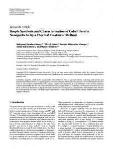
Baruwati et al. product was then dispersed in 10.0 mL of DMSO. To this, 20 mmol of DCC was added. After 10 min, 15 mmol of L-lysine methyl ester (produced by diazomethane protection of the acid group using (trimethylsilyl)diazomethane as the diazomethane source) was added to the solution. After 2 h, the reaction was stopped, and the product was collected by addition of ethanol and centrifugation. The acid group was then deprotected by following the standard procedure for methyl ester hydrolysis.31 Characterization of the Ferrite Nanoparticles. The phase of the as-synthesized ferrite nanoparticles was determined by X-ray diffraction in a MMS X-ray diffractometer with a Cu KR source in the 2θ range 10-65. The data were collected with a step of 0.5°/min. A few drops of the as-synthesized nanoparticles in hexane were added to a quartz plate and dried at room temperature before recording the X-ray pattern. TEM micrographs were recorded on a JEOL JSM-1200 II microscope at an operating voltage of 120 kV. A drop of the as-synthesized nanoparticles was loaded onto a carbon-coated copper grid and then allowed to dry at room temperature before recording the micrographs. FTIR spectra were recorded on a Perkin-Elmer SPETRUM 2000 FTIR instrument in the range 400-3700 cm-1 by drop-casting the nanoparticles onto a polystyrene film to confirm the presence of the surface-functionalized moieties on the surface of the nanoparticles. Elemental analyses of the assynthesized samples were performed on a Perkin-Elmer Optima 3300 DV inductively coupled atomic emission spectroscope (ICP-AES). Five milligrams of each sample was dissolved in 2.5 mL of concentrated HNO3, and the volume was adjusted to 25 mL in a volumetric flask. Five milliliters of this solution was then further diluted to 25 mL. This diluted sample was then used for the elemental analysis. Magnetic characterization was performed using a Quantum Design MPMS-XL SQUID magnetometer under field-cooled and zero-field conditions as well as at room temperature. Results and Discussions The main feature of the described synthetic protocol for the ferrite nanoparticles is their formation in a confined nanoscale organic matrix and the crystallization under hydrothermal condition.32 Under hydrothermal temperatures, the water and organic phases are much different from those at normal conditions because of the autogenous high pressure when the temperature exceeds the boiling points of water and toluene (110 °C). Under this condition, both of the liquids diffuse into each other, and the inorganic metal salts present in the water phase come into contact with the oleic acid present in the organic phase. This might facilitate the formation of an oleate complex and its subsequent decomposition to form the ferrite nanoparticles. Under the cooling condition, the two phases separate from each other and the nanoparticles remains in the organic phase. Figure 1a, b shows the typical TEM micrograph of the NiFe2O4 samples synthesized under conventional heating conditions at 250 °C for 1 h, with oleic acid to iron nitrate ratios of 3:1 and 6:1, respectively. In both cases, the as-synthesized particles were monodisperse with a uniform size of 9 ( 2 nm, as shown in Figure 1c. The corresponding selected area electron diffraction (SAED) pattern (inset of Figure 1a) established the highly crystalline nature of the product and can be indexed to a cubic system. There was no variation in the particle size distribution or particle morphology, even when the reaction time was increased to 4 h. This salient feature might be very desirable for large-scale production with size selectivity. For the MW method, the particles obtained at 160 °C for a reaction time of 1 h had a size of 4 ( 1 nm. The microwave
Synthesis of Monodisperse Ferrite Nanoparticles
J. Phys. Chem. C, Vol. 112, No. 47, 2008 18401
Figure 1. TEM micrograph of the NiF2O4 samples synthesized at the hydrothermal temperature 250 °C for 1 h with oleic acid to iron nitrate ratio (a) 3:1 and (b) 6:1. Inset shows the corresponding electron diffraction pattern. (c) Particle size distribution calculated on 100 particles. Error bar indicates 8% error.
Figure 2. TEM micrographs of the NiFe2O4 particles synthesized under microwave conditions with reaction times (a) 1 and (b) 2 h. (c, d) The corresponding particle size distribution calculated over 100 particles. Error bar indicates 10% error.
Figure 3. TEM micrographs for the (a) CoFe2O4 and (b) MnFe2O4 particles and (c) γ-Fe2O3. (d-f) Corresponding particle size distributions calculated over 100 particles. Error bar indicates 10% error.
18402 J. Phys. Chem. C, Vol. 112, No. 47, 2008
Baruwati et al.
TABLE 1: Summary of the Reaction Conditions, Morphology, and Crystallite Sizes Calculated from the Scherrer Formula and the Particle Sizes Calculated from TEM
sample
crystallite particle size (XRD) size (TEM) (nm) (nm)
reaction condition
NiFe2O4, CoFe2O4, MnFe2O4 conventional hydrothermal; 250 °C, 1 h; oleic acid to iron nitrate ratios of 3:1, 6:1 conventional hydrothermal; 250 °C, 2 h; oleic acid to iron nitrate ratio of 6:1 microwave, 160 °C, 1 h microwave, 160 °C, 2 h γ-Fe2O3 conventional hydrothermal; 120 °C, 1 h; oleic acid to iron nitrate ratio of 3:1 microwave; 120 °C, 1 h; oleic acid to iron nitrate ratio of 3:1
morphology
7(1
9(1
spherical
7(1
9(1
spherical
3.5 ( 1 6.5 ( 1 9(1
5(2 9(1 10 ( 1
4(1
5(1
spherical spherical spherical, some larger cubelike structures are also seen in the TEM images spherical
TABLE 2: Summary of the Magnetization Values Obtained for Different Samples coercivity (Oe) sample NiFe2O4 NiFe2O4 NiFe2O4 NiFe2O4 γ-Fe2O3
(iron nitrate to oleic acid ratio, 3:1) (iron nitrate to oleic acid ratio, 6:1) (iron nitrate to oleic acid ratio, 6:1; time, 4 h) (conventional)
saturation magnetization (emu/g)
5K
300 K
5K
300 K
blocking temp, K
53 55 280 210
11 12 11 12 11
22 20.1 25 53
24 22.8 22 42.2 45
10 10 25 60 105
reactions were explored at various temperatures, and 160 °C was selected because we were able to get single-phase ferrites at this minimum temperature under MW irradiation conditions. The smaller particle size obtained in this case is because of the low synthesis temperature and shorter reaction time. When the reaction time was increased to 2 h, the particles obtained had a size of 8 ( 1 nm. Figure 2a, b shows the particles synthesized under MW conditions with reaction times 1 and 2 h, respectively. Figure 2c, d shows the corresponding particle size distributions. Control experiments carried out at a similar temperature for 24 h under conventional reflux conditions did not yield any particle formation. The experiments conducted for CoFe2O4 and MnFe2O4 yielded nanoparticles with morphology similar to the NiFe2O4 nanoparticles under identical reaction conditions, which make the method general for the synthesis of a range of ferrite nanoparticles. In the case of γ-Fe2O3 nanoparticles, formation of a number of cubelike structures was observed from the TEM micrographs. When the temperature of the reaction was increased to 150 °C, the γ-Fe2O3 phase was converted to R-Fe2O3. Figure 3a-c and d-f show the TEM micrographs and particle size distributions for the CoFe2O4, MnFe2O4, and γ-Fe2O3 nanoparticles, respectively. X-ray diffraction confirmed the formation of single-phase cubic spinel nanostructures for all the ferrites synthesized. The crystallite sizes calculated from the Scherrer formula were found to be 7 ( 1 nm, which is comparable to the particle size calculated from TEM micrographs in the case of the NiFe2O4 nanoparticles. In the case of all other ferrites, the crystallite size was approximately equal to the particle size calculated from TEM. This implies that each particle is a single domain. Figure 4 shows the X-ray diffraction pattern for the as-synthesized ferrite samples. Table 1 summarizes the reaction conditions, morphology, and the crystallite sizes calculated from Scherrer formula and the particle sizes calculated from TEM micrographs for the various ferrite samples. ICP-AES studies confirmed that the ratios of Fe to Ni, Mn, and Co were approximately equal to 2. The values for the ratio of Fe to Ni, Mn, and Co are 1.95, 1.89, and 1.98 respectively. FTIR confirmed the presence of oleic acid moieties on the surface of the as-synthesized particles. The peaks that are
characteristics of oleic acid were also seen in the spectrum for as-synthesized nanoparticles. Figure 5 shows a representative FTIR spectrum of as-synthesized NiFe2O4 nanoparticles (5a) and oleic acid (5b). The peaks at 3009 and 3005 cm-1, which can be seen in Figure 5a and b, can be ascribed to the stretching frequency of the vinyl group. The peaks at 2927 and 2857 cm-1 in the case of Figure 5a and 2925 and 2852 cm-1 in the case of Figure 5b are due to the asymmetric and symmetric vibrations of the CH2 groups of oleic acid. The strong peak at 1710 cm-1 seen in both of the figures is due to the CdO stretching. The peaks at 1431 and 1300 cm-1 in Figure 5a and the peak at 1440 cm-1 in Figure 5b correspond to the C-H bending frequencies
Figure 4. The X-ray diffraction pattern for the as synthesized (a) CoFe2O4, (b) NiFe2O4, (c) MnFe2O4, and (d) γ-Fe2O3.
Synthesis of Monodisperse Ferrite Nanoparticles
J. Phys. Chem. C, Vol. 112, No. 47, 2008 18403
Figure 7. Zero-field-cooled and field-cooled plots for the NiFe2O4 sample synthesized at 250 °C for 4 h with an oleic acid-to-iron nitrate ratio of 6:1 under an applied field of 500 Oe.
Figure 5. Representative FTIR spectrum of (a) as-synthesized NiFe2O4 nanoparticles and (b) oleic acid.
Figure 8. TEM and photographic image of the surface functionalized nanoparticles with L-lysine.
Figure 6. Magnetization versus applied field plot for the NiFe2O4 sample synthesized at 250 °C for 4 h with an oleic acid-to-iron nitrate ratio 6:1 at room temperature as well as at 5 K. Inset shows the expanded view of the coercivities at the respective temperatures.
of CH2 group of oleic acid. The peaks at 920 cm-1 in Figure 5a and 923 cm-1 in Figure 5b are due to the O-H outplane vibrations. The strong peaks at 599 and 435 cm-1 that are present only in Figure 5a confirmed the presence of ferrite nanoparticles. The magnetization versus applied field plots for the assynthesized NiFe2O4 particles at various reaction conditions show that the particles are superparamagnetic at room temperature with negligible coercivities equal to 11-12 Oe. The
Figure 9. FTIR spectrum of the surface functionalized nanoparticles with L-lysine.
saturation magnetizations were found to be in the range of 20-25 emu/g, which is less than the theoretical value of 47 emu/g.33 The reduced saturation magnetization of ferrite nanocrystals is generally believed to be due to the decreased particles size and presence of a magnetic dead, or antiferromagnetic layer on the surface.34,35 The NiFe2O4 sample synthesized without oleic acid shows the saturation magnetization of 42 emu/g at room temperature and 53 emu/g at 5 K. These values are almost
18404 J. Phys. Chem. C, Vol. 112, No. 47, 2008 equal to the theoretical values for NiFe2O4 and, hence, confirm the presence of oleic acid on the surface that might be considered as a magnetically dead layer. The saturation magnetization at 5 K is almost equal to the saturation magnetization at room temperature. The coercivities at this temperature are in the range 55-280 Oe. Figure 6 shows the magnetization versus applied field plot for the NiFe2O4 sample synthesized at 250 °C for 4 h with an oleic acid-to-iron nitrate ratio of 6:1 at room temperature as well as at 5 K. Table 2 summarizes the different saturation magnetizations obtained for various samples. The zero-field-cooled (ZFC) and field-cooled (FC) plots under an applied field of 500 Oe gives the blocking temperature as in the range of 10-25 K for various samples. Figure 7 shows the zero-field-cooled and field-cooled plots for the NiFe2O4 sample synthesized at 250 °C for 4 h with an oleic acid-to-iron nitrate ratio of 6:1 under an applied field of 500 Oe. Surface functionalization renders the particles water-dispersible, and the water dispersions are highly stable up to 4 weeks. This makes the particles suitable for biological applications. Figure 8 shows the TEM micrographs and photographic image of the nanoparticles in water after surface functionalization with L-lysine. It can be seen from the TEM micrograph that the morphology and distribution of the particles remained unchanged, even after surface modification. Figure 9 shows the FTIR spectrum of the nanoparticles after surface functionalization with L-lysine. The broad pattern in the range 3300-2800 cm-1 corresponds to the presence of OH groups on the surface of the molecules. The peaks at 3336 and 1515 cm-1 correspond to the N-H stretching and bending vibrations of primary amides. The peak at 1568 cm-1 corresponds to the -COOgroups on the surface of the particles. The peaks at the range 1400-1300 are a result of the carboxylate anion stretching vibrations. They also correspond to the various O-H bending vibrations. Conclusions In conclusion, we have developed a facile method for the bulk synthesis of monodispersed spinel ferrite nanoparticles with size selectivity using readily available inorganic precursors via a water-organic interface. The method is equally feasible under both conventional hydrothermal and MW hydrothermal methods. The use of MW has the advantage of low temperature expedient synthesis. The as-synthesized particles were highly dispersible as well as stable in nonpolar organic solvents, which is useful in the applications such as ferrofluids and other magnetic applications. Surface functionalization of the as-synthesized particles with lysine made them water-dispersible for possible biological applications. Acknowledgment. BB is supported by the Postgraduate Research Program at the National Risk Management Research Laboratory administered by the Oak Ridge Institute for Science and Education through an interagency agreement between the U.S. Department of Energy and the U.S. Environmental Protection Agency. The authors are thankful to Dr. Jayappa
Baruwati et al. Manjanna, Faculty of Engineering, Iwate University, Japan for his help in magnetic measurements. References and Notes (1) Son, S.; Taheri, M.; Carpenter, E.; Harris, V. G.; McHenry, M. E. J. Appl. Phys. 2002, 91, 7589. (2) Gillot, B. Eur. Phys. J. Appl. 2002, 91, 10. (3) Jorgensen, F. The Complete Handbook of Magnetic Recording; McGraw-Hill: New York, 1995. (4) Alivisatos, A. P. Science 1996, 271, 933. (5) Berkovsky, M. V.; Medvedev, F. V.; Krakov, S. M. Magnetic Fluids Engineering Applications; Oxford University Press: Oxford, UK, 1993. (6) Manova, E.; Tsoncheva, T.; Estourne`s, Cl.; Paneva, D.; Tenchev, K.; Mitov, I.; Petrov, L. Appl. Catal. 2006, 2, 300. (7) Satyanarayana, L.; Reddy, K. M.; Manorama, S. V. Mater. Chem. Phys. 2003, 82, 21. (8) Reddy, K. M.; Satyanarayana, L.; Manorama, S. V.; Misra, D. K. Mater. Res. Bull. 2004, 39/10, 1491. (9) Tiefenauer, L. X.; Tscgirky, A.; Kuhne, G.; Andres, R. Y. Magn. Reson. Imaging 1996, 14, 391. (10) Tang, Z. X.; Sorensen, C. M.; Klabunde, K. J.; Hadjipanayis, G. C. Phys. ReV. Lett. 1991, 67, 3602. (11) Kulkarni, G.; Kannan., U.; Arunarkavalli, K. R. T.; Rao, C. N. R. Phys. ReV. B 1994, 49, 724. (12) Viroonchatapan, E.; Ueno, M.; Sato, H.; Adachi, I.; Nagae, H.; Tazawa, K.; Horikoshi, I. Pharm. Res. 1995, 12, 1176. (13) Gupta, P. K.; Hung, C. T. Life Sci. 1989, 44, 175. (14) Lu¨ders, U.; Barthe´le´my, A.; Bibes, M.; Bouzehouane, K.; Fufil, S.; Jacquet, E.; Contour, J.; Bobo, J. F.; Fontcuberta, J.; Fert, A. AdV. Mater. 2006, 18, 1733. (15) Sugimoto, T.; Shimotsuma, Y.; Itoh, H. Powder Technol. 1998, 96, 85. (16) Pannaparayil, T.; Marande, R.; Komarneni, S. J. Appl. Phys. 1991, 69, 5349. (17) Lopez Perez, A. J.; Lopez Quintela, A. M.; Mira, J.; Rivas, J.; Charles, W. S. J. Phys. Chem. B 1997, 101, 8045. (18) Shafi, M. P.V. K.; Gedanken, A.; Prozorov, R.; Balogh, J. Chem. Mater. 1998, 10, 3445. (19) Fatemi, J. D.; Harris, G. V.; Browning, M. V.; Kirkland, P. J. J. Appl. Phys. 1998, 83, 867. (20) Hyeon, T.; Chung, Y.; Park, J.; Lee, S. S.; Kim, Y.-W.; Park, B. H. J. Phys. Chem. B 2002, 106, 6831. (21) Sun, S.; Zeng, H. J. Am. Chem. Soc. 2002, 124, 8204. (22) Qing, S.; Zhang, Z. J. J. Am. Chem. Soc. 2004, 126, 6164. (23) Redl, F. X.; Black, C. T.; Papaefthymiou, G. C.; Sandstrom, R. L.; Yin, M.; Zeng, H.; Murray, C. B.; O′Brien, S. P. J. Am. Chem. Soc. 2004, 126, 14583. (24) Feng, S. H.; Xu, R. R. Acc. Chem. Res. 2001, 34, 239. (25) Siskin, M.; Katritzky, A. R. Science 1991, 254, 231. (26) (a) Komarneni, S. Curr. Sci. 2003, 85, 1730. (b) Celer, E. B.; Jaroniec, M. J. Am. Chem. Soc. 2006, 128, 14408. (27) Tsuji, M.; Hashimoto, M.; Nishizawa, Y.; Kubokawa, M.; Tsuji, T. Chem.sEur. J. 2005, 11, 440. (28) (a) Zhu, Y. J.; Wang, W. W.; Qi, R. J.; Hu, X. L. Angew. Chem., Int. Ed. 2004, 43, 1410. (b) Zhu, Y. J.; Hu, X. L. Chem. Lett. 2004, 33, 760. (c) Zhu, Y. J.; Hu, X. L. Chem. Lett. 2003, 32, 1140. (d) Zhu, Y. J.; Hu, X. L. Chem. Lett. 2003, 32, 732. (29) (a) Liao, X. H.; Zhu, J. M.; Zhu, J. J.; Xu, J. Z.; Chen, H. Y. Chem. Commun. 2001, 937. (30) Yong, K. T.; Roy, I.; Pudavar, H. E.; Bergey, J. E.; Tramposch, M. K.; Swihart, T. M.; Prasad, N. P. AdV. Mater. 2008, 20, 1412. (31) Corey, E. J.; Sze´kely, I.; Schiner, C. S. Tetrahedron Lett. 1977, 3529. (32) Tang, K.; Zhang, J.; Yan, W.; Li, Z.; Wang, Y.; Yang, W.; Xie, Z.; Sun, T.; Fuchs, H. J. Am. Chem. Soc. 2008, 130, 2676. (33) Geng, B. Y.; Ma, J. Z.; Liu, X. W.; Du, Q. B.; Kong, M. G.; Zhang, L. D. Appl. Phys. Lett. 2007, 90, 043120. (34) Zheng, M.; Wu, X. C.; Zou, B. S.; Wang, Y. J. J. Magn. Magn. Mater. 1998, 183, 152. (35) Pankhurst, Q. A.; Pollard, R. J. Phys. ReV. Lett. 1991, 67, 248.
JP807245G