Instructions: (PROVIDER) Use this form to assess the woman's eligibility for medication abortion. For open responses, write the response in space provided.
Study #3: Control, isotype-matched IgG. (0.3 mg/kg, 3 times/week). AngII-infused (1,000 ng/kg/min). S15 Fig. Page 2. #12: Died. 1.08 mm. 0.95 mm. 1.03 mm.
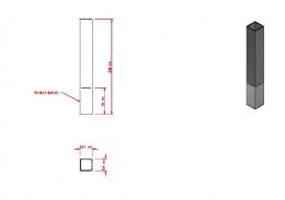
Jan 31, 2013 - chamfer and knife-edge finish lines. The interproximal areas wound up being critical due to thin RDT, potentially interfering with the structural ...
Body weights (g). Body composition (%). Total cholesterol (mM). Triglycerides (mM). Diet n w 0 w 8 w 16. LBM. FFM w 8 w 16 w 8 w 16. All: Herring. 9. 21.4±0.47.
12 Jul 2018 - ... Liu1, Guangbin Shi1, Man Liu1, Kenneth R. Boheler2, Samuel ...... Matlib MA, Zhou Z, Knight S, Ahmed S, Choi KM, Krause-Bauer J, et al.
Oct 31, 2018 - coral calcification are poorly understood (reviewed in [5]). ... proteins that likely supply dissolved inorganic carbon [5â7], as well as coral acidic ...
m o log 1/. Contains 1 FYVE-type zinc finger. Schizosaccharo m y ces po m b e ... 1516. ,. 8.9,. 0.0026. 1516. ,. 5.2,. 0.14. 90. ,. 83.3,. 1.5e-25 no hit no hit. 1494.
0.7 mm. 0.8 mm. 0.9 mm. 1.0 mm. 1.3 mm. 1.4 mm. A. B. C. 1.1 mm. 1.2 mm. Figure S1. Transmission electron microscopy images of ultra-thin sections of ...
Page 1. 56 mm. 200 mm. 25.4 mm. 25.4 mm. POLISHED SURFACE. Page 2. 56 mm. 200 mm. 25.4 mm. 25.4 mm. POLISHED SURFACE. R 2.5 mm.
on isotacticity control in propylene polymerization with silylene-bridged group 4 metallocenes. C2 symmetrical and asymmetrical catalysts, Organometallics 15,.
Sep 30, 2013 - such as MMFF94, Chemistry at Harvard Molecular Mechanics. (CHARMM), Assisted Model Building with Energy Refinement ..... In QuanPol, we implemented our latest QM/ ...... updated versions of these files can be directly used to perform .
Cougar. Montego. Mercury, Mark III &. Lincoln Continentai. 1970 MODELS AND
... TION CHART FOR 1970 MODELS 40-43. AUTOLITE ... 0 Quick ratio manual
steering gear ..... disconnecting the solenoid lead wire at the bullet connector.
S2. supplementary information. If extracellular (EC) [Ca2+] was elevated from 0 to 2 mM before the application of the calcium-ionophore ionomycin (Iono., 1 μM), ...
S2. supplementary information
2
0
current (nA)
0
30 Iono.
2
[Ca2+] 0 (mM) [K+] 2 (mM)
-0.5
-1.0 0
2
4
6 8 10 12 time (min)
If extracellular (EC) [Ca2+] was elevated from 0 to 2 mM before the application of the calcium-ionophore ionomycin (Iono., 1 µM), then TRESK current was activated by the elevation of [Ca2+] in some HEK293 cells, but only by ionomycin in the others. To circumvent this biological variability, we administered calcium and ionomycin together for reliable TRESK activation in further experiments. It is important to note, however, that in some cells the elevation of [Ca2+] alone did not influence TRESK current at all (blue arrow, see the representative recording above). This indicates that the elevation of EC [Ca2+] did not directly influence channel activity through a biophysical mechanism. For example, the “screening” effect of Ca2+ ions on the negatively charged phospholipid bilayer, which so profoundly affects the activation of voltage-gated channels, and the binding of the ion to the channel protein, could not be responsible for TRESK activation. This was also verified by the insensitivity of activated TRESK current to the reduction of EC [Ca2+] from 2 to 0 mM (not shown). Instead of a direct biophysical effect of EC calcium on the channel, the ion entered the cytoplasm and activated TRESK via calcineurin. It is also apparent in the figure shown above that TRESK current was small at the beginning of the measurement (less than 0.25 nA, when EC [K+] was increased from 2 to 30 mM), but the K+ current slowly increased in the calcium-free EC solution (green arrow). This activation was not negligible compared to that evoked by ionomycin (red arrow). We elevated EC [Ca2+] only when the K+ current stabilized (blue arrow). The initial activation of TRESK current in the calcium-free EC solution also supported the hypothesis that TRESK was preactivated by the experimental manipulations before the start of recording. The final phase of this preactivation could be detected at the beginning of the measurement. Because no EC Ca2+ was present at this time, calcium-dependent activation of the channel was possible through the release of the ion from intracellular stores. This brought ATP into suspicion as an agonist of Gq protein-coupled receptors, leaking from the patch pipette tip before seal formation.