Am J Physiol Cell Physiol 284: C562–C570, 2003. First published October 23, 2002; 10.1152/ajpcell.00336.2002.
Calcineurin transgenic mice have mitochondrial dysfunction and elevated superoxide production M. R. SAYEN,1 ÅSA B. GUSTAFSSON,1 MARK A. SUSSMAN,2 JEFFERY D. MOLKENTIN,2 AND ROBERTA A. GOTTLIEB1 1 The Scripps Research Institute, La Jolla, California 92037; and 2Children’s Hospital Medical Center, Cincinnati, Ohio 45229 Submitted 30 September 2002; accepted in final form 19 October 2002
Sayen, M. Richard, Åsa B. Gustafsson, Mark A. Sussman, Jeffery D. Molkentin, and Roberta A. Gottlieb. Calcineurin transgenic mice have mitochondrial dysfunction and elevated superoxide production. Am J Physiol Cell Physiol 284: C562–C570, 2003. First published October 23, 2002; 10.1152/ajpcell.00336.2002.—Introduction of the constitutively active calcineurin gene into neonatal rat cardiomyocytes by adenovirus resulted in decreased mitochondrial membrane potential (P ⬍ 0.05). Infection of H9c2 cells with calcineurin adenovirus resulted in increased superoxide production (P ⬍ 0.001). Transgenic mice with cardiac-specific expression of a constitutively active calcineurin cDNA (CalTG mice) exhibit a two- to threefold increase in heart size that progresses to heart failure. We prepared mitochondria enriched for the subsarcolemmal population from the hearts of CalTG mice and transgene negative littermates (control). Intact, well-coupled mitochondria prepared from one to two mouse hearts at a time yielded sufficient material for functional studies. Mitochondrial oxygen consumption was measured with a Clark-type oxygen electrode with substrates for mitochondrial complex II (succinate) and complex IV [tetramethylpentadecane (TMPD)/ascorbate]. CalTG mice exhibited a maximal rate of electron transfer in heart mitochondria that was reduced by ⬃50% (P ⬍ 0.002) without a loss of respiratory control. Mitochondrial respiration was unaffected in tropomodulin-overexpressing transgenic mice, another model of cardiomyopathy. Western blotting for mitochondrial electron transfer subunits from mitochondria of CalTG mice revealed a 20–30% reduction in subunit 3 of complex I (ND3) and subunits I and IV of cytochrome oxidase (CO-I, CO-IV) when normalized to total mitochondrial protein or to the adenine nucleotide transporter (ANT) and compared with littermate controls (P ⬍ 0.002). Impaired mitochondrial electron transport was associated with high levels of superoxide production in the CalTG mice. Taken together, these data indicate that calcineurin signaling affects mitochondrial energetics and superoxide production. The excessive production of superoxide may contribute to the development of cardiac failure.
EXPRESSION OF A CONSTITUTIVELY ACTIVE FORM of calcineurin in transgenic mice has been shown to result in
cardiac hypertrophy and progression to failure by 8–12 wk (26). Calcineurin signaling is recognized to contribute to the progression of disease in a number of models, as its inhibition has been shown to prevent cardiac hypertrophy (36). Cardiac function depends upon several factors, including adequate cell mass, intact contractile machinery, and adequate production of ATP. Sarcomeric dysfunction, as occurs in tropomodulinoverexpressing transgenic (TOT) mice, leads to dilated cardiomyopathy within 3–4 wk after birth. Loss of heart muscle through apoptosis leads to heart failure, as seen in the G␣q transgenic mice and in the FKBPcaspase-8 transgenic mice (1, 42). Mitochondrial dysfunction also leads to heart failure, as has been described in a variety of mitochondrial DNA deletion syndromes and in the targeted deletion of the adenine nucleotide translocator 1 (ANT1) (11, 41). The relationship between calcineurin signaling and these aspects of cardiac function are unclear, although calcineurin does not seem to promote apoptosis; in fact, activated calcineurin protected cultured cardiomyocytes from apoptosis mediated by 2-deoxyglucose or staurosporine and was associated with reduced infarct size after ischemia-reperfusion (I/R) (9). We hypothesized that calcineurin might affect mitochondrial function. This was suggested by the observation that calcineurin dephosphorylated the family of transcriptional regulators known as nuclear factor of activated T cells (NFAT), thereby permitting their translocation from cytosol to nucleus (31). NFAT has been shown to interact with GATA4 in the heart (26) and with MEF2c (4, 22, 43). These transcription factors have been shown to play a role in the regulation of expression of mitochondrial proteins (27, 44). To examine mitochondrial function by polarography, we adapted previously described techniques for isolating mitochondria from rat and rabbit hearts and optimized them for use with much smaller and more fragile mouse hearts (14, 17, 24, 29, 39). As we describe in this report, isolated mitochondria from transgenic mice expressing activated calcineurin exhibit impaired oxidative phosphorylation and increased superoxide production that may contribute to heart failure.
Address for reprint requests and other correspondence: R.A. Gottlieb, Dept. of Molecular and Experimental Medicine, The Scripps Research Institute MEM220, 10550 North Torrey Pines Road, La Jolla, CA 92037 (E-mail:
[email protected]).
The costs of publication of this article were defrayed in part by the payment of page charges. The article must therefore be hereby marked ‘‘advertisement’’ in accordance with 18 U.S.C. Section 1734 solely to indicate this fact.
mitochondria; bioenergetics; hypertrophy; genetic models of heart failure
C562
0363-6143/03 $5.00 Copyright © 2003 the American Physiological Society
http://www.ajpcell.org
MITOCHONDRIAL DYSFUNCTION IN CALCINEURIN TG MICE METHODS
Neonatal cardiomyocyte culture and analysis of mitochondrial membrane potential. Neonatal rat cardiomyocytes were prepared by collagenase digestion as previously described (16), with the following changes: 100 M bromodeoxyuridine (BrdU) was added to inhibit proliferation of nonmyocytic cells. Cells were plated on fibronectin-coated chamber slides at 24,000 cells/cm2. After 24 h, they were infected with adenovirus for activated calcineurin (8), Adgal, or empty vector AdSR and cultured for 48 h. Cells were loaded with rhodamine 123 (R123) (Molecular Probes, Eugene, OR) at 1 g/ml for 30 min and costained with Hoechst 33342 at 30 g/ml for 5 min for nuclear detection. Successive fields were imaged with a Nikon TE300 fluorescence microscope (Nikon, Tokyo, Japan) and digitally collected with a Spot2 digital camera (Diagnostic Instruments, Sterling Heights, MI) using identical parameters. Field selection was performed in bright-field illumination and was restricted to areas in which cell density was ⬍85 cells per field. This was necessary to ensure that cells in close proximity could be scored as individual cells. Mitochondrial membrane potential was assessed as the ratio of relative fluorescence intensity of R123 to the number of nuclei in each field using MetaMorph imaging software (Universal Imaging, Westchester, PA). To rule out loss of mitochondrial mass, parallel wells of cells were stained with MitoTracker Green (Molecular Probes), which stains mitochondria but is independent of mitochondrial membrane potential. Cells (500–1,000) were scored from each condition. Five fields from each slide were analyzed, and three to four slides were averaged from each experiment. Experiments were repeated in two independent myocyte preps with essentially identical results. Mice. Mice were generated and characterized as previously described (26, 37). All animal procedures were approved by the Animal Care and Use Committee of The Scripps Research Institute. Isolation of heart mitochondria. This protocol was based on several previously published methods (17, 24, 29, 39). Hearts were removed while still beating from mice anesthetized with ketamine/xylazine. Two mouse hearts were pooled and rapidly minced in ice-cold MSE buffer [in mmol/l: 220 mannitol, 70 sucrose, 2 EGTA, 5 MOPS (pH 7.4), and 2 taurine supplemented with 0.2% fatty acid-free bovine serum albumin (BSA)]. Heart tissue was homogenized in MSE buffer with a Polytron-type tissue grinder at 11,000 RPM for 2.5 s, followed by 2 quick strokes at 500 RPM with a loose fit PotterElvehjem tissue grinder. The homogenate was centrifuged at 500 g twice for 5 min and the supernatant was saved. Mitochondria were pelleted from the supernatant by centrifugation at 3,000 g twice, and the pellet was rinsed with MSE buffer. The supernatant was saved as crude cytosol. The final pellet was rinsed and resuspended in 50 l of incubation medium [in mmol/l: 220 mannitol, 70 sucrose, 1 EGTA, 5 MOPS (pH 7.4), 2 taurine, 10 MgCl2, and 5 KH2PO4, supplemented with 0.2% fatty acid-free BSA] (33). Mitochondria were incubated for 15 min on wet ice, and protein concentration was determined with BSA as a standard by a Bradford assay. All work was performed on wet ice at 0°C. Measurement of respiration in mitochondria from mouse or rat hearts. Oxygen consumption was measured at 30°C with a Clark-type oxygen electrode (Instech) in 600 l of KCl respiration buffer [in mmol/l: 140 KCl, 1 EGTA, 10 MOPS (pH 7.4), 10 MgCl2, and 5 KH2PO4, supplemented with 0.2% fatty acid-free BSA] (6, 23, 33). Complex II activity was measured using 200 g of mitochondria with 5 mM succinate as a substrate. Complex IV activity was measured using 150 AJP-Cell Physiol • VOL
C563
g of mitochondria with 0.4 mM TMPD and 1 mM ascorbate as a substrate. For each complex, the ADP-stimulated respiration rate (state 3) was measured after the addition of 2 mM ADP; the ADP-independent respiration rate, oligomycin-in-
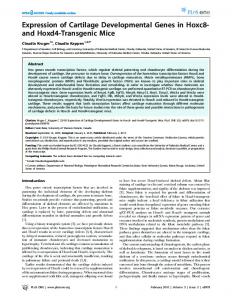
Fig. 1. Neonatal cardiomyocytes were infected with the activated calcineurin adenovirus (AdCal) and, after 48 h, were assessed for mitochondrial membrane potential (rhodamine 123 staining, green) and counterstained with Hoechst (blue nuclei). A: loss of mitochondrial membrane potential is seen in many of the AdCal cells, which demonstrate typical hypertrophic morphology when viewed in brightfield. Infection with adenovirus for -galactosidase (Adgal) or hypertrophic stimulation with phenylephrine (PE) does not affect mitochondrial membrane potential. B: rhodamine 123 fluorescence is shown as a ratio to the number of nuclei in the field, providing an average brightness per cell. Multiple fields were collected and analyzed with identical parameters. Error bars represent SD. (n ⫽ 4 separate slides, each representing 500–1,000 cells analyzed per condition.)
284 • FEBRUARY 2003 •
www.ajpcell.org
C564
MITOCHONDRIAL DYSFUNCTION IN CALCINEURIN TG MICE
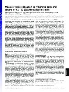
Fig. 2. Immunoblot of calcineurin. Arrowhead denotes the endogenous form of calcineurin, whereas the arrow indicates the activated calcineurin. Lane 1: cytosol from control, transgene negative hearts; lane 2: cytosol from calcineurin TG⫹ hearts; lanes 3 and 4: mitochondria from control hearts; lanes 5 and 6: mitochondria from calcineurin TG⫹ hearts.
sensitive (state 4), was measured after the addition of 2 M oligomycin, and the maximal respiration rate was measured after uncoupling the mitochondria with 2 M FCCP. Rates were calculated as nA O 䡠 min⫺1 䡠 mg⫺1 protein after subtracting the rate that was insensitive to the inhibitors, 1 M antimycin A for complex III and 1 mM KCN for complex IV. As a measure of mitochondrial integrity, the respiratory control ratio (RCR) state 3 divided by state 4 was calculated. The data reported herein represent five independent mitochondrial preparations comprising two hearts each from a total of ten control and ten transgenic mice. Comparison between calcineurin transgenic (CalTG) and wild-type (WT) mice was analyzed using Student’s t-test. Immunoblotting for mitochondrial proteins. Mitochondria (50 g) and crude cytosol (60 g) were resolved by SDSPAGE and transferred to polyvinylidene difluoride (PVDF) nylon membranes. Membranes were probed for ND3 and Rieske iron-sulfur protein with antibodies kindly provided by Dr. Akemi Matsuno-Yagi of the Scripps Research Institute for cytochrome oxidase subunits I and IV (Molecular Probes), ANT (Oncogene Research Products, Boston, MA), and calcineurin (Santa Cruz Biotechnology, Santa Cruz, CA). Detection was performed with enhanced chemiluminescence (ECL; Amersham, Piscataway, NJ). Nonsaturated autoradiographs were quantitated with Scion/NIH Image. Comparison between CalTG and WT mice was analyzed using Student’s t-test. Langendorff heart perfusions. All procedures were approved by the Animal Care and Use Committee at the Scripps Research Institute. In brief, mice were anesthetized with 150 g/kg pentothal, and hearts were removed and quickly cannulated onto the Langendorff perfusion apparatus. WT and transgenic hearts were prepared and hung in parallel on Langendorff setups with independent flow. Hearts were perfused with Krebs-Ringer buffer (in mM: 118.5 NaCl, 4.7 KCl, 1.18 KH2PO4, 1.18 MgSO4, 25 NaHCO3, 11.1 glucose, and 2.5 CaCl2) (Sigma) for 15 min before ischemia. No-flow ischemia was maintained for 30 min, and reperfusion was accomplished by restoring flow for 15 min. Measurement of superoxide generation. Superoxide generation was assessed via the conversion of dihydroethidium (DHE) to ethidium as previously described (25). DHE enters the cell and is oxidized primarily by superoxide to yield fluorescent ethidium, which intercalates into DNA and is thereby retained in the cell (3). Because the superoxide anion is short-lived, this method only detects the ongoing production of superoxide in the tissue. Although mitochondria are the major source of superoxide in the heart, other enzymatic sources of superoxide include xanthine oxidase, NADPH oxidase, and peroxisomes. In brief, hearts were frozen at ⫺20°C for 24 h. Hearts were then slightly thawed, sliced into 1-mmthick sections on a chilled template, and arranged in sepaAJP-Cell Physiol • VOL
rate wells of a 24-well plate in a solution consisting of 2 M DHE (Molecular Probes) in PBS. Plates were incubated in the dark at 37°C for 20 min. Sections were then placed on glass slides and imaged using an ultraviolet transilluminator (Fisher Scientific). All images were identically captured using a Kodak DC120 digital camera (Kodak) using Kodak Digital Science 1D software (Kodak). Images were treated identically and saved as TIFF files and analyzed using Adobe Photoshop 5.5. The percentage of superoxide production was quantified as the ratio of fluorescent (white) pixels to the total heart area. Statistical analysis was performed using ANOVA with Tukey-Kramer post test (InStat statistical software version 4.10; GraphPad, San Diego, CA). Measurement of superoxide production in cultured cells. H9c2 cells were plated in 12-well tissue culture plates at 5,000 cells per cm2 in DMEM supplemented with 10% FBS. At 24 and 48 h after being plated, cells were infected with replication deficient adenovirus expressing activated calcineurin or -galactosidase at a multiplicity of infection (MOI) of 10–20 plaque-forming units (PFU) per cell. At 24 h after the second infection, fresh medium containing 10 M DHE was added to wells (40). DHE fluorescence (excitation 518 nm/emission 605 nm) was measured at the indicated time with a Gemini fluorescence microplate reader (Molecular Devices). Images of cells were also captured to confirm intracellular fluorescence.
Fig. 3. Representative oxygen electrode tracings from calcineurin and control mice. Scale indicates nA O consumed per unit time. Each tracing represents mitochondria pooled from 2 mouse hearts. Inflections represent the following additions: a, mitochondria; b, substrate [succinate for complex II, tetramethylpentadecane (TMPD)/ascorbate for complex IV]; c, ADP; d, oligomycin; e, FCCP; f, inhibitor (antimycin A for complex II/III, KCN for complex IV).
284 • FEBRUARY 2003 •
www.ajpcell.org
MITOCHONDRIAL DYSFUNCTION IN CALCINEURIN TG MICE
Fig. 4. Respiration in 3- to 4-wk-old and from 9- to 10-wk-old calcineurin transgenic mice (CalTG) is impaired relative to control mice. Rates are normalized to mitochondrial protein. Filled bars are control, transgene negative animals, whereas open bars are CalTG⫹ mice. State 3 is defined as the rate obtained after the addition of 2 M ADP, whereas state 4 is the rate after addition of 2 M oligomycin. Maximal stimulated rate is obtained with the addition of 2 M FCCP, whereas the respiratory control ratio (RCR) is defined as the ratio of state 3 to state 4. Error bars represent SD (n ⫽ 6 independent analyses from 12 animals per genotype for all conditions, except for FCCP, where n ⫽ 4 analyses, 8 animals per genotype). P values between wild-type (WT) and CalTG are shown below each condition.
AJP-Cell Physiol • VOL
284 • FEBRUARY 2003 •
www.ajpcell.org
C565
C566
MITOCHONDRIAL DYSFUNCTION IN CALCINEURIN TG MICE
RESULTS
Adenoviral-mediated gene transfer of the activated calcineurin has been shown to induce neonatal cardiomyocyte hypertrophy and to upregulate expression of atrial natriuretic peptide 48 h after infection (9). Accordingly, we infected neonatal rat cardiomyocytes with the activated calcineurin adenovirus and examined mitochondrial membrane potential using rhodamine 123 (Fig. 1). We found that calcineurin-overexpressing myocytes consistently exhibited less rhodamine 123 fluorescence per cell than control or adenovirus-galactosidase-infected myocytes. Moreover, the diminished mitochondrial membrane potential was not merely a consequence of hypertrophy, because cells stimulated with phenylephrine possessed normal mitochondrial membrane potential. These results suggest that calcineurin regulates mitochondrial function. These results led us to examine the effects of calcineurin on mitochondrial function in the CalTG mice. To examine the possibility that calcineurin might directly regulate mitochondrial function through dephosphorylation, we isolated mitochondria from WT and CalTG hearts and performed subcellular fractionation.
However, we did not detect the activated calcineurin transgene in the mitochondrial fraction (Fig. 2). This finding suggests that the mechanism is indirect, possibly through transcriptional pathways. Mitochondria from the hearts of CalTG and transgene negative littermates were prepared and mitochondrial electron transfer capacity was analyzed. Representative tracings are shown in Fig. 3. Respiration in CalTG mitochondria was impaired when substrates for complex II (succinate) and complex IV (TMPD/ascorbate) were used. ADP stimulated respiration fourfold in CalTG and control mice when succinate was used as a substrate. The respiratory control ratio is used as an indication of mitochondrial integrity and demonstrates that the mitochondria isolated from the CalTG hearts did not sustain greater damage. We examined the maximal stimulated rate using FCCP to uncouple the mitochondria. We examined mice at 3–4 wk of age. We found that respiratory control ratios were similar to control mitochondria but that state 3, state 4, and FCCPuncoupled respiration was reduced by 29–37% in mitochondria prepared from CalTG hearts (Fig. 4).
Fig. 5. A: Western blotting demonstrates a decrease in the abundance of cytochrome oxidase (COX) subunits I and IV and a decrease in ND3 of complex I, with no change in the abundance of the Rieske iron-sulfur protein or the adenine nucleotide translocator (ANT). The first 4 lanes are from WT mice heart mitochondria, whereas the last 5 lanes are from the activated CalTG mice. Each lane represents material from 2 hearts. Equal amounts of mitochondrial protein were loaded in each lane. B: quantitation of mitochondrial proteins in control (n ⫽ 4 pairs of hearts) and CalTG (n ⫽ 5 pairs of hearts). After densitometry, samples were normalized to the nuclear-encoded Rieske iron-sulfur protein. Error bars represent SD. P values are shown below each comparison.
AJP-Cell Physiol • VOL
284 • FEBRUARY 2003 •
www.ajpcell.org
MITOCHONDRIAL DYSFUNCTION IN CALCINEURIN TG MICE
These studies were repeated on 9- to 10-wk-old mice when heart failure had set in (9). Overall, CalTG mitochondria demonstrated a statistically significant reduction by 50% in oxygen consumption per milligram of mitochon-
drial protein for state 3, state 4, and FCCP-uncoupled using succinate or TMPD/ascorbate as substrates. To further address the possibility that these changes in mitochondrial respiration were secondary to heart
Fig. 6. Superoxide production in hearts from WT and CalTG mice was measured by quantifying dihydroethidium (DHE) conversion. A shows representative stained heart slices from WT and CalTG mice at baseline and after ischemia-reperfusion (I/R). B shows mean ⫾ SE for 4 hearts for each condition. P values were determined by ANOVA. AJP-Cell Physiol • VOL
C567
284 • FEBRUARY 2003 •
www.ajpcell.org
C568
MITOCHONDRIAL DYSFUNCTION IN CALCINEURIN TG MICE
failure, we examined a model of dilated cardiomyopathy due to overexpression of the actin-capping molecule tropomodulin (37). Structural alterations are apparent as early as 1 wk of age (37). We examined respiration in heart mitochondria in 9- to 10-wk-old TOT mice and found that it did not differ from age-matched controls (data not shown). These results indicate that the mitochondrial alterations are directly associated with the activated calcineurin transgene rather than with hypertrophy or heart failure itself. To assess the basis for impaired mitochondrial respiration, we hypothesized that components of the electron transfer complexes might be altered. We used antibodies to both nuclear-encoded and mitochondrialencoded components of the respiratory chain. Westerns were loaded with equal amounts of mitochondrial protein, and loading was further normalized to the mitochondrial proteins ANT and Rieske iron-sulfur protein. Blots were probed for the proteins shown in Fig. 5A. We found that CalTG mitochondria exhibited similar amounts of ANT and Rieske iron-sulfur protein compared with control mitochondria but consistently showed less protein for complex I subunit 3 (ND3) and cytochrome oxidase subunits I and IV, summarized in Fig. 5B. Loss of these subunits is consistent with the impaired respiration we observed. In addition, we probed three samples of control and CalTG mitochondria for nuclear-encoded complex I subunits (75, 51, 49, 42, 39, 24, 18, and 9 kDa) and complex V subunits (␣, , b, and OCSP) and found no change in the abundance of these proteins relative to nontransgenic controls (data not shown). Many studies have shown that a limitation to electron flow can result in increased superoxide production via the ubisemiquinone. To determine whether the observed mitochondrial abnormalities resulted in increased superoxide production, we stained heart slices with DHE, which is converted to the fluorescent ethidium by superoxide anion (3). We found that su-
peroxide production was 4.5-fold greater in the CalTG hearts than WT (P ⬍ 0.001)(Fig. 6). Ischemia and reperfusion are also known to result in increased superoxide production. We assessed superoxide production in CalTG and WT hearts after 30 min ischemia and 15 min reperfusion (I/R). In WT hearts, superoxide production increased 3.4-fold over control levels after I/R (P ⬍ 0.01). In contrast, CalTG hearts increased superoxide production ⬍1.2-fold over their already high baseline level (P ⫽ NS) (Fig. 6). To determine whether the increased superoxide production was causally related to calcineurin activity, we infected H9c2 cells for 48 h with AdCal or Adgal and assessed superoxide production by DHE conversion. Activated calcineurin caused an increased rate of superoxide production (Fig. 7). No increase in apoptosis was observed over this time period. We conclude that calcineurin directs cellular alterations that include mitochondrial alterations and elevated superoxide production. DISCUSSION
We have developed methodology to isolate mitochondria from the hearts of transgenic mice for polarography studies. This technique will allow investigators to better characterize mitochondrial alterations in a variety of genetically modified mice. To maximize the quality of the mitochondria obtained, we chose to use only the mitochondria released by brief homogenization of the minced tissue. This results in an enrichment of subsarcolemmal mitochondria, with relatively poor recovery of interfibrillar mitochondria. Thus our conclusions are based on the properties of the subsarcolemmal population of mitochondria, and it should be noted that interfibrillar mitochondria might exhibit different properties, as reported by Hoppel (19). Optimal function of mitochondria in the heart is essential for ATP production and calcium homeostasis.
Fig. 7. Superoxide production in H9c2 cells infected with adenovirus for activated calcineurin or -galactosidase was measured by DHE conversion. Error bars denote SE for 3 experiments done in triplicate. The rate of superoxide production in the calcineurin expressing cells was significantly greater (P ⬍ 0.005).
AJP-Cell Physiol • VOL
284 • FEBRUARY 2003 •
www.ajpcell.org
MITOCHONDRIAL DYSFUNCTION IN CALCINEURIN TG MICE
Mitochondrial dysfunction may contribute to heart failure, as is well recognized in the mitochondrial deletion syndromes. Calcineurin is upregulated/activated in conditions that lead to cardiac hypertrophy, such as pressure overload and inhibition of calcineurin suppresses hypertrophy (7, 21). Although calcineurin is upregulated in the TOT mice, the level of activity is still considerably lower than that obtained in the activated calcineurin transgenic mice, which might explain why we do not observe mitochondrial dysfunction in the TOT mice. Overexpression of activated calcineurin leads to cardiac hypertrophy and eventual failure through a process that does not seem to involve apoptosis but likely involves new gene transcription, because the process can be recapitulated by NFAT3, a key target of calcineurin regulation in the heart (26). We have described mitochondrial alterations that arise as a result of overexpression of activated calcineurin in cell culture and the transgenic mouse model. Alterations in energy metabolism due to calcineurin have not been heretofore described. The impairment of oxidative phosphorylation is associated with a decrease in the amount of protein corresponding to components of the electron transfer complexes in the transgenic mouse. Both nuclear-encoded (cytochrome oxidase subunit IV) and mitochondrial-encoded (ND3, cytochrome oxidase subunit I) proteins appear to be downregulated. This may be mediated by increased degradation, a change in mitochondrial and/or nuclear transcription or translation. Proper assembly and stabilization of electron transfer components requires the coordinated synthesis of both nuclear and mitochondrial-encoded proteins (2, 28). Therefore, dysregulation of nuclear or mitochondrial transcription or translation could result in the destabilization of electron transfer complexes. Moreover, protein import into the mitochondria depends upon membrane potential. Diminished mitochondrial membrane potential, as we observed in the neonatal cardiomyocytes infected with adenovirus for activated calcineurin, could lead to impaired import of nuclear-encoded proteins. Further work will be required to establish the mechanism for the loss of electron transfer capacity mediated by calcineurin. Calcineurin is known to regulate the activity of transcription factors GATA4, NFAT3, and MEF2C (10, 20, 22). These transcription factors have been shown to regulate the expression of some nuclear genes of mitochondrial energy metabolism (18, 27, 44). Furthermore, it has recently been reported that calcineurin activates transcription of PGC-1, a transcriptional coactivator that regulates mitochondrial biogenesis (32). PGC-1 and the other transcription factors would be expected to increase mitochondrial biogenesis, a common feature of cardiac hypertrophy (30, 34, 45, 46). One would predict that PGC-1 overexpression would also lead to hypertrophy and elevated levels of superoxide production. The excessive superoxide production observed in the CalTG mice may explain several features of their phenotype. Superoxide at low levels has been shown to AJP-Cell Physiol • VOL
C569
stimulate hypertrophy (35), but chronic or high levels of superoxide will result in cumulative cellular damage and dilated cardiomyopathy (5, 15). Although dysregulated mitochondrial biogenesis may be responsible for excessive superoxide production and progression to heart failure, it is also possible that calcineurin activates superoxide production through other mechanisms, including upregulation of other enzyme systems that can generate superoxide, such as cytochrome P450 monooxygenases (12, 13, 38). Excessive superoxide production would lead to secondary mitochondrial damage and eventual progression to heart failure. This work was supported by National Heart, Lung, and Blood Institute Grant HL-61518 (to R. A. Gottlieb) and National Institute of Diabetes and Digestive and Kidney Diseases Training Grant DK-07022 (to Å. B. Gustafsson). REFERENCES 1. Adams JW, Sakata Y, Davis MG, Sah VP, Wang Y, Liggett SB, Chien KR, Brown JH, and Dorn GW. Enhanced G␣q signaling: a common pathway mediates cardiac hypertrophy and apoptotic heart failure. Proc Natl Acad Sci USA 95: 10140– 10145, 1998. 2. Bai Y and Attardi G. The mtDNA-encoded ND6 subunit of mitochondrial NADH dehydrogenase is essential for the assembly of the membrane arm and the respiratory function of the enzyme. EMBO J 17: 4848–4858, 1998. 3. Becker LB, van den Hoek TL, Shao ZH, Li CQ, and Schumacker PT. Generation of superoxide in cardiomyocytes during ischemia before reperfusion. Am J Physiol Heart Circ Physiol 277: H2240–H2246, 1999. 4. Blaeser F, Ho N, Prywes R, and Chatila TA. Ca2⫹-dependent gene expression mediated by MEF2 transcription factors. J Biol Chem 275: 197–209, 2000. 5. Cesselli D, Jakoniuk I, Barlucchi L, Beltrami AP, Hintze TH, Nadal-Ginard B, Kajstura J, Leri A, and Anversa P. Oxidative stress-mediated cardiac cell death is a major determinant of ventricular dysfunction and failure in dog dilated cardiomyopathy. Circ Res 89: 279–286, 2001. 6. Chance B and Higihara B. Direct spectroscopic measure measurements of interaction of components of the respiratory chain with ATP, ADP, phosphate, and uncoupling agents. In: Proceedings on Intracellular Respiration. Moscow: Congress Biochemistry, 1961, p. 3–26. 7. De Windt LJ, Lim HW, Bueno OF, Liang Q, Delling U, Braz JC, Glascock BJ, Kimball TF, del Monte F, Hajjar RJ, and Molkentin JD. Targeted inhibition of calcineurin attenuates cardiac hypertrophy in vivo. Proc Natl Acad Sci USA 98: 3322– 3327, 2001. 8. De Windt LJ, Lim HW, Haq S, Force T, and Molkentin JD. Calcineurin promotes protein kinase C and c-Jun NH2-terminal kinase activation in the heart. Cross-talk between cardiac hypertrophic signaling pathways. J Biol Chem 275: 13571–13579, 2000. 9. De Windt LJ, Lim HW, Taigen T, Wencker D, Condorelli G, Dorn GW, Kitsis RN, and Molkentin JD. Calcineurin-mediated hypertrophy protects cardiomyocytes from apoptosis in vitro and in vivo: an apoptosis-independent model of dilated heart failure. Circ Res 86: 255–263, 2000. 10. Delling U, Tureckova J, Lim HW, De Windt LJ, Rotwein P, and Molkentin JD. A calcineurin-NFATc3-dependent pathway regulates skeletal muscle differentiation and slow myosin heavychain expression. Mol Cell Biol 20: 6600–6611, 2000. 11. Esposito LA, Melov S, Panov A, Cottrell BA, and Wallace DC. Mitochondrial disease in mouse results in increased oxidative stress. Proc Natl Acad Sci USA 96: 4820–4825, 1999. 12. Fleming I, Michaelis UR, Bredenkotter D, Fisslthaler B, Dehghani F, Brandes RP, and Busse R. Endothelium-derived hyperpolarizing factor synthase (cytochrome P450 2C9) is
284 • FEBRUARY 2003 •
www.ajpcell.org
C570
13.
14.
15.
16.
17. 18.
19.
20.
21.
22. 23.
24. 25.
26.
27.
28.
29.
MITOCHONDRIAL DYSFUNCTION IN CALCINEURIN TG MICE
a functionally significant source of reactive oxygen species in coronary arteries. Circ Res 88: 44–51, 2001. Gergel D, Misik V, Riesz P, and Cederbaum AI. Inhibition of rat and human cytochrome P4502E1 catalytic activity and reactive oxygen radical formation by nitric oxide. Arch Biochem Biophys 337: 239–250, 1997. He H, Chen M, Scheffler NK, Gibson BW, Spremulli LL, and Gottlieb RA. Phosphorylation of mitochondrial elongation factor Tu in ischemic myocardium: basis for chloramphenicolmediated cardioprotection. Circ Res 89: 461–467, 2001. Ide T, Tsutsui H, Kinugawa S, Utsumi H, Kang D, Hattori N, Uchida K, Arimura K, Egashira K, and Takeshita A. Mitochondrial electron transport complex I is a potential source of oxygen free radicals in the failing myocardium. Circ Res 85: 357–363, 1999. Karwatowska-Prokopczuk E, Nordberg J, Li HL, Engler RL, and Gottlieb RA. Effect of the vacuolar proton ATPase on intracellular pH, calcium, and on apoptosis in neonatal cardiomyocytes during metabolic inhibition and recovery. Circ Res 82: 1139–1144, 1998. Kuo TH and Giacomelli FWJ. Oxidative metabolism of Polytron vs. Nagarse mitochondria in hearts of genetically diabetic mice. Biochim Biophys Acta 806: 9–15, 1985. Lenka N, Vijayasarathy C, Mullick J, and Avadhani NG. Structural organization and transcription regulation of nuclear genes encoding the mammalian cytochrome c oxidase complex. Prog Nucleic Acid Res Mol Biol 61: 309–344, 1998. Lesnefsky EJ, Tandler B, Ye J, Slabe TJ, Turkaly J, and Hoppel CL. Myocardial ischemia decreases oxidative phosphorylation through cytochrome oxidase in subsarcolemmal mitochondria. Am J Physiol Heart Circ Physiol 273: H1544–H1554, 1997. Liang Q, De Windt LJ, Witt SA, Kimball TR, Markham BE, and Molkentin JD. The transcription factors GATA4 and GATA6 regulate cardiomyocyte hypertrophy in vitro and in vivo. J Biol Chem 276: 30245–30253, 2001. Lim HW, De Windt LJ, Steinberg L, Taigen T, Witt SA, Kimball TR, and Molkentin JD. Calcineurin expression, activation, and function in cardiac pressure-overload hypertrophy. Circulation 101: 2431–2437, 2000. Mao Z and Wiedmann M. Calcineurin enhances MEF2 DNA binding activity in calcium-dependent survival of cerebellar granule neurons. J Biol Chem 274: 31102–31107, 1999. McKee EE, Grier BL, Thompson GS, and McCourt JD. Isolation and incubation conditions to study heart mitochondrial protein synthesis. Am J Physiol Endocrinol Metab 258: E492– E502, 1990. Meta L and Seitz C. Isolation of mitochondria with emphasis on heart mitochondria from small amounts of tissue. Methods Enzymol 55: 39–46, 1979. Miller FJJ, Gutterman DD, Rios CD, Heistad DD, and Davidson BL. Superoxide production in vascular smooth muscle contributes to oxidative stress and impaired relaxation in atherosclerosis. Circ Res 82: 1298–1305, 1998. Molkentin JD, Lu JR, Antos CL, Markham B, Richardson J, Robbins J, Grant SR, and Olson EN. A calcineurin-dependent transcriptional pathway for cardiac hypertrophy. Cell 93: 215–228, 1998. Moore ML, Wang GL, Belaguli NS, Schwartz RJ, and McMillin JB. GATA-4 and serum response factor regulate transcription of the muscle-specific carnitine palmitoyltransferase I  in rat heart. J Biol Chem 276: 1026–1033, 2001. Nijtmans LG, Henderson NS, Attardi G, and Holt IJ. Impaired ATP synthase assembly associated with a mutation in the human ATP synthase subunit 6 gene. J Biol Chem 276: 6755– 6762, 2001. Palmer JW, Tandler B, and Hoppel CL. Biochemical properties of subsarcolemmal and interfibrillar mitochondria isolated from rat cardiac muscle. J Biol Chem 252: 8731–8739, 1977.
AJP-Cell Physiol • VOL
30. Rajamanickam C, Merten S, Kwiatkowska-Patzer B, Chuang CH, Zak R, and Rabinowitz M. Changes in mitochondrial DNA in cardiac hypertrophy in the rat. Circ Res 45: 505–515, 1979. 31. Rao A, Luo C, and Hogan PG. Transcription factors of the NFAT family: regulation and function. Annu Rev Immunol 15: 707–747, 1997. 32. Schaeffer PJ, Vega R, Williams RS, and Kelly DP. Calcineurin signaling activates transcription of the PGC-1 gene: a potential mechanism for regulation of mitochondrial capacity. Circulation 104, Suppl: II-203, 2001. 33. Scholte HR, Yu Y, Ross JD, Oosterkamp II, Boonman AMC, and Busch HFM. Rapid isolation of muscle and heart mitochondria, the lability of oxidative phosphorylation and attempts to stabilize the process in vitro by taurine, carnitine and other compounds. Mol Cell Biochem 174: 61–66, 1997. 34. Sims JM, Patzer B, Kumudavalli-Reddy M, Martin AF, Rabinowitz M, and Zak R. The pathways of protein synthesis and degradation in normal heart and during development and regression of cardiac hypertrophy. Recent Adv Stud Card Struct Metab 12: 19–28, 1976. 35. Siwik DA, Tzortzis JD, Pimental DR, Chang DL, Pagano PJ, Singh K, Sawyer DB, and Colucci WS. Inhibition of copper-zinc superoxide dismutase induces cell growth, hypertrophic phenotype, and apoptosis in neonatal rat cardiac myocytes in vitro. Circ Res 85: 147–153, 1999. 36. Sussman MA, Lim HW, Gude N, Taigen T, Olson EN, Robbins J, Colbert MC, Gualberto A, Wieczorek DF, and Molkentin JD. Prevention of cardiac hypertrophy in mice by calcineurin inhibition. Science 281: 1690–1693, 1998. 37. Sussman MA, Welch S, Gude N, Khoury PR, Daniels SR, Kirkpatrick D, Walsh RA, Price RL, Lim HW, and Molkentin JD. Pathogenesis of dilated cardiomyopathy: molecular, structural, and population analyses in tropomodulin-overexpressing transgenic mice. Am J Pathol 155: 2101–2113, 1999. 38. Tan FL, Moravec CS, Li J, Apperson-Hansen C, McCarthy PM, Young JB, and Bond M. The gene expression fingerprint of human heart failure. Proc Natl Acad Sci USA 99: 11387– 11392, 2002. 39. Tyler DD and Gonze J. The preparation of heart mitochondria from laboratory animals. Methods Enzymol 10: 75–101, 1967. 40. Vanden Hoek T, Becker LB, Shao ZH, Li CQ, and Schumacker PT. Preconditioning in cardiomyocytes protects by attenuating oxidant stress at reperfusion. Circ Res 86: 541–548, 2000. 41. Wallace DC. Mouse models for mitochondrial disease. Am J Med Genet 106: 71–93, 2001. 42. Wencker D, Chandra M, Armstrong RC, Garantziotis S, Factor SM, Shirani J, and Kitsis RN. Rescue of dilated cardiomyopathy by caspase inhibition in FKBP-caspase-8 transgenic mice. Circulation 102, Suppl: II-28, 2000. 43. Wu H, Rothermel B, Kanatous S, Rosenberg P, Naya FJ, Shelton JM, Hutcheson KA, DiMaio JM, Olson EN, BasselDuby R, and Williams RS. Activation of MEF2 by muscle activity is mediated through a calcineurin-dependent pathway. EMBO J 20: 6414–6423, 2001. 44. Xia Y, McMillin JB, Lewis A, Moore M, Zhu WG, Williams RS, and Kellems RE. Electrical stimulation of neonatal cardiac myocytes activates the NFAT3 and GATA4 pathways and upregulates the adenylosuccinate synthetase 1 gene. J Biol Chem 275: 1855–1863, 2000. 45. Zak R and Rabinowitz M. Molecular aspects of cardiac hypertrophy. Annu Rev Physiol 41: 539–552, 1979. 46. Zak R, Rabinowitz M, Rajamanickam C, Merten S, and Kwiatkowska-Patzer B. Mitochondrial proliferation in cardiac hypertrophy. Basic Res Cardiol 75: 171–178, 1980.
284 • FEBRUARY 2003 •
www.ajpcell.org