Rev. Inst. Med. trop. S. Paulo 46(3):171-174, May-June, 2004
CASE REPORT CARRION´S DISEASE (BARTONELLOSIS BACILLIFORMIS) CONFIRMED BY HISTOPATHOLOGY IN THE HIGH FOREST OF PERU
Vicente MACO(1,2), Ciro MAGUIÑA(1,3), Antonio TIRADO(2), Vicente MACO C.(4) & José E. VIDAL(5)
SUMMARY Bartonellosis or Carrion´s disease is endemic in some regions of Peru, classically found in the inter-Andean valleys located between 500 and 3200 meters above sea level. We report the case of a 43 year-old male patient, farmer, who was born in the Pichanaki district (Chanchamayo, Junin), located in the High Forest of Peru. He presented with disseminated, raised, erythematous cutaneous lesions, some of which bled. The distribution of these lesions included the nasal mucosa and penile region. Additionally subcutaneous nodules were distributed over the trunk and extremities. Hematologic exams showed a moderate anemia. Serologic studies for HIV and Treponema pallidum were negative. The histopathologic results of two biopsies were compatible with Peruvian wart. Oral treatment with ciprofloxacin (500 mg BID) was begun. Over 10 days, the patient showed clinical improvement. This is the first report of a confirmed case of bartonellosis in the eruptive phase originating from the Peruvian High Forest, showing the geographical expansion of the Carrion´s disease. KEYWORDS: Bartonellosis; Bartonella bacilliformis; Carrion´s disease; Epidemiology; High Forest; Peru.
INTRODUCTION The infections caused by the pathogens of the genus Bartonella comprise a heterogeneous group of emerging and reemerging diseases2,12,13,20. At the present time, 20 species have been described, eight of which affect humans, particularly Bartonella bacilliformis, described in 1905 and responsible for Carrion´s disease. The Carrion´s disease is an reemerging illness native to Peru, present since the pre-Columbian period14,15, found in the inter-Andean valleys between 500 and 3,200 meters above sea level, between the 2º north latitude and 13º south latitude, where ecological conditions for the development for Lutzomyia verrucarum, the main vector involved in its transmission, are found6,16. It has been estimated that the population at risk is 1,686,236 inhabitants16 distributed in a surface of 144,496 km². There have also been cases described in rural areas of Ecuador and Colombia1,22. The illness develops through two phases: the acute hematic phase, or Oroya fever, and the eruptive phase or Peruvian wart, both separated by an asymptomatic period. The acute phase is characterized by fever, moderate to severe pallor, general discomfort and myalgias. Neurological complications (i.e. altered mental status, ataxia, seizure, agitation, coma), heart complications (i.e. congestive heart failure, pericardial effusions, myocarditis) and respiratory complications (i.e. adult respiratory distress syndrome) there
has been related in severe cases. Approximately one third of patients present with opportunistic infections due to non-typhoid Salmonellas (i.e. S. dublin, S. typhymurium, S. anatum), sepsis by Shigella dysenteriae, Enterobacter, Pseudomona aeruginosa, Staphylococcus aureus, pneumonia by Pneumocystis jirovecii or reactivation of tuberculosis, toxoplasmosis, and histoplasmosis. Fatality among untreated patients varies generally among 40 to 88%10. With prompt treatment, fatality is reduced to less than 9% of admitted patients13. A recent report after an outbreak of bartonellosis in a non-endemic area suggested that the illness seems to be significantly more benign than that frequently reported10. After weeks or months of the acute phase, less than 10% of the patients will progress to the eruptive phase13,22. This phase is characterized for the presence of diverse cutaneous lesions, in which the patient develops nodular dermal eruptions known as Peruvian wart, classified in three types: miliary (erythematous papules, less than 3 mm, frequently numerous and occasionally pruritic), mulaire (erythematous round lesion, over 5 mm, frequently eroded and sessile), and nodular (subdermal nodules)12. Characteristically, the lesions are painless and prone to bleed. In this study we report the first confirmed case of Carrion´s disease in the eruptive phase to originate in the High Forest of Peru, previously considered a non-endemic area of bartonellosis.
(1) Instituto de Medicina Tropical Alexander von Humboldt, Universidad Peruana Cayetano Heredia, Lima, Perú. (2) Hospital de Apoyo de Pichanaki, Chanchamayo, Junín, Perú. (3) Departamento de Enfermedades Transmisibles y Dermatológicas, Universidad Peruana Cayetano Heredia, Lima, Perú. (4) Departamento de Anatomia Patológica, Hospital Central de la Policia Nacional del Perú, Lima, Perú. (5) Instituto de Infectologia Emílio Ribas, São Paulo, Brazil. Correspondence to: Dr. Ciro Maguiña, Instituto de Medicina Tropical Alexander von Humboldt, Universidad Peruana Cayetano Heredia, Av. Honorio Delgado N° 460, Urb. Ingeniería, San Martín de Porres, Lima, Perú. E-mail:
[email protected]
MACO, V.; MAGUIÑA, C.; TIRADO, A.; MACO C., V. & VIDAL, J.E. - Carrion‘s disease (Bartonellosis bacilliformis) confirmed by histopathology in the High Forest of Peru. Rev. Inst. Med. trop. S. Paulo, 46(3):171-174, 2004.
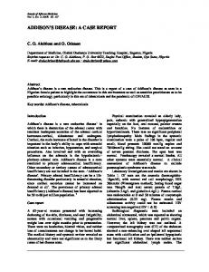
CASE REPORT A 43 year-old male farmer was admitted to the Pichanaki Hospital (Fig. 1) in June of the 2003, after 6 weeks of illness. The illness was first characterized by reddish cutaneous lesions approximately 2 mm diameter in the left forearm which were not painful. The lesions continuously enlarged in size over the following three weeks. They were removed by the patient and resulted in light bleeding. Four weeks before presentation, the patient noticed similar lesions in the upper and lower extremities and over the thorax, progressively increasing in number and size and spreading to the face, hair and nose. One week before arriving into the hospital, the patient went to another hospital, where he was prescribed oral dicloxacillin (500 mg QID), without improvement. He denied having lived or traveled to endemic areas of Carrion´s disease. Likewise, he denied previous illnesses, hospitalizations and prior surgical interventions.
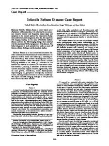
Fig. 2 - Miliary (left arrow) and mulaire (right arrow) lesions.
followed by the mulaire type (36.6%) and the nodular type (4.7%). The treatment was initiated with oral ciprofloxacin (500 mg BID) for 10 days. On the fifth day of treatment there was a decrease in the size of the miliary lesions. The patient was released and returned five days after completing treatment. At the time, the miliary warts had disappeared, including the penile lesion, and the size of the nodular lesions had decreased by approximately 50%.
Fig. 1 - Geographical location of Pichanaki district (*), in Junin, new reported endemic area of human bartonellosis in Peru.
Physical exam showed moderate pallor of skin and mucosa. Multiple cherry red papules and nodular lesions were seen, between 0.3 to 1 cm of diameter, located in the scalp, neck, face (Fig. 2), thorax, abdomen, upper and lower extremities, some of them eroded with signs of recent bleeding. Likewise, a 2 mm papular reddish wound was found in the penis and nodular subcutaneous wounds in the periumbilical and epitrochlear region, thigh and in nasal alae. The remainder of the exam was normal. Hematologic studies showed moderate anemia with a hematocrit of 34% and RBCs count of 5,700 cells/mm3. Serologic studies for human immunodeficiency virus and Treponema pallidum were negative. Biopsy of two lesions was performed. Microscopy study showed marked vascular proliferation with prominent endothelium and stromal angiosarcomal cells, arranged in nodules. In the interstitium was visualized the presence of mature lymphocytic inflammatory cells and evidence of ischemic necrosis with hemorrhage and dense infiltration of mixed inflammatory cells, lymphocytes and polymorphonuclear cells, without cellular typing (Fig. 3). They were identified as the mulaire and the nodular or subdermal types of the eruptive phase of the Carrion´s disease. Once the diagnosis was determined, the lesions were quantified and classified, and a total of 191 were identified. The majority were warts of the miliary type (58.6%), 172
Fig. 3 - Histopathology (H-E 400X). Biopsy of skin showing intense neovascularization and diffuse infiltrate of lymphocytes, accompanied by some histiocytes and extravasated red blood cells.
DISCUSSION This is the first histopathologically confirmed case of Carrion’s disease in the eruptive phase found in the Peruvian High Forest. Previous reports have documented isolated cases as well as an epidemic cluster of the hemolytic phase in this region. Nevertheless, no evidence of the presence of the complete cycle of the illness existed previously. The first report of a patient in the high forest was described by LOJA et al. in 1994. The case described the acute phase, in Quillabamba (Cuzco), found in a patient who lived at 1,047 meters above sea level11. Our patient was from the high forest in the Pichanaki district (Chanchamayo, Junin), located at 525 meters
MACO, V.; MAGUIÑA, C.; TIRADO, A.; MACO C., V. & VIDAL, J.E. - Carrion‘s disease (Bartonellosis bacilliformis) confirmed by histopathology in the High Forest of Peru. Rev. Inst. Med. trop. S. Paulo, 46(3):171-174, 2004.
above sea level. This area was previously known as a non-endemic area. The first epidemic cluster that showed the presence of the Carrion’s disease in the Peruvian forest occurred between October 1992 and April 1993 in the Aguarunas communities in the province of San Ignacio (Cajamarca). Seven patients were detected who had peripheral blood smears consistent with infection by B. bacilliformis16. No patient presented in the eruptive phase. Between 1996 and 1997 a bartonellosis cluster was found in the department of Amazonas, where several cases of Peruvian wart were confirmed with histopathology10. However, the cases originated more commonly from mountainous districts rather than the forest of Peru. Because of the lack of information about the exact origin of the wart cases, our report constitutes the first case confirmed in the Peruvian Forest. In recent years, Carrion’s disease clusters continued to be detected in Peru. Between April and June of 1998 there were 26 reported cases confirmed in the Sacred Valley of the Incas (Cuzco), the majority of which were children. These cases had a fatality rate of 23%19. On the other hand, between January and December of 1998 there were 71 reported cases of the acute phase and 121 in the eruptive phase in the province of Antonio Raymondi (Ancash)7. The recognition of a new endemic area of Carrion’s disease has an enormous epidemiological significance. In the district where our patient lives, there are approximately 3,700 families: 3,000 in urban zones (16,000 inhabitants), and 700 in rural areas (24,000 inhabitants). All this population is therefore at risk to acquire the illness. At the present time, there have been reported cases of Carrion’s disease clusters in 12 of the 24 departments of Peru. Ancash is the area with the highest number of cases in both acute hemolytic phase and the eruptive one, as well as the highest rates of incidence12,14,16,17. Five of these departments (Cajamarca, Junín, Huánuco, Amazonas, and Cuzco) have some districts located in the Peruvian Forest. The acute phase of Carrion’s disease has also been described in other non-endemic areas. Among them, the most severely affected districts are Chincha (Ica) and Pasamayo-Boza (Huaral, Lima)18. Both are coastal cities, out of the traditional ecological niches for bartonellosis. These findings present the possibility of the extension of Carrion’s disease not only to the forest of Peru, as it establishes of final form our report, but also to the Peruvian coast, in areas situated at less than 500 m above sea level, suggesting important epidemiological changes. Carrion’s disease evolves through several phases. Some studies show that to 60% of the population in endemic areas are asymptomatic and have no known history of bartonellosis, but have positive serology for B. bacilliformis8. On the other hand, the acute hemolytic phase does not represent a sine qua non condition for the presence of the eruptive phase. This partially explains why the complete cycle of the illness had not been described in the Peruvian forest. Finally, as confirmed in our report, the inhabitants of endemic areas can develop the eruptive phase as the only manifestation of the illness10,13.
The observation of mucosal lesions is also infrequent. MAGUIÑA, in a prospective series of 145 cases of Carrion’s disease, showed mucosal compromise in 4 (3%) patients17. The lesions were found on oral and nasal mucosa and on the ocular conjunctiva13,16,21,22. The diagnosis of Carrion’s disease requires clinical, epidemiological and laboratory information. The laboratory methods used frequently are peripheral blood smears and histopathologic studies. The final diagnosis in the acute phase uses the peripheral blood smears, which shows parasitized red cells with the bacillary forms of B. baciliformis seen in late phases some coccidial forms. The Romanovski, Giemsa and Wright stains facilitate the identification of the microorganisms2,12,13,20. The final diagnosis of the eruptive phase requires the demonstration of characteristic histopathological changes such as proliferation of endothelial cells, monocytes and macrophages, capillary proliferation, and in some cases, cellular atypia3,4,13. The Warthin-Starry stain confirms the diagnosis, allowing visualization of the bacteria. Other techniques include electron microscopy and immunohistochemistry5. The differential diagnosis of the eruptive phase of Carrion’s disease, depending on the type of lesion, includes bacillary angiomatosis, Kaposi’s sarcoma, pyogenic granuloma, hemangioma, fibrosarcoma, epithelial sarcoma, cutaneous lymphoma, histioid phase of Hansen’s disease and juvenile melanoma (Spitz tumor)2,13,20,22. The treatment of Carrion’s disease depends on the stage. In the acute phase, the treatment of choice is chloramphenicol for 10 to 14 days. Quinolones, especially ciprofloxacin are alternative drugs 12 . Chloramphenicol and ciprofloxacin have intracellular activity and antibacterial effect against Gram-negative germs like Salmonella typhi and non-typhoid species that usually complicate the course of the illness13. In the eruptive phase, the treatment of choice is rifampin for 14-21 days. Erythromycin, azithromycin, ciprofloxacin, and streptomycin have also been used (C. Maguiña, personal communication). As seen in our patient, ciprofloxacin represents a good alternative, for a period of 7-10 days13,16,17. Treatment during this phase produces a decrease in size of the lesions in 7 days (range: 4 -14 days) and complete disappearance in 27 days (range: 14-70 days)16. In spite of good therapeutic results, larger clinical trials are still necessary, which is often limited by the availability of patients in the eruptive phase. The lesions are benign and spontaneously remit without leaving a scar within 2-6 months. Our report shows extension of Carrion’s disease and its presence in the Peruvian forest, probably due to the continuous commerce carried out among that region with settlers from in adjacent endemic zones. On the other hand, it is also possible that the sandfly vector Lutzomyia verrucarum and other species of Lutzomyia, potential transmitters of the illness, have evolved. For these reasons, implementation of control measures for this disease are necessary in Peru, including strategies of entomological control, seroepidemiologic studies, and training of the technical personnel and health workers in these areas. RESUMO
The clinical manifestations of Carrion’s disease have been wellstudied2,12,13,20,22. Characteristically, the lesions of the eruptive phase are found predominantly on the upper and lower extremities and on the face17. The clinical presentation of our patient, with miliary, mulaire and nodule subdermal lesions, distributed in a disseminated form is unusual.
Doença de Carrión (Bartonelose baciliforme) confirmada por histopatologia na Selva Alta do Peru A bartonelose ou doença de Carrión é endêmica em algumas regiões 173
MACO, V.; MAGUIÑA, C.; TIRADO, A.; MACO C., V. & VIDAL, J.E. - Carrion‘s disease (Bartonellosis bacilliformis) confirmed by histopathology in the High Forest of Peru. Rev. Inst. Med. trop. S. Paulo, 46(3):171-174, 2004.
do Peru, descrevendo-se classicamente nos vales inter-andinos, entre 500 e 3.200 metros acima do nível do mar. Os autores relatam o caso de um paciente de 43 anos de idade, fazendeiro, natural do distrito de Pichanaki (Chanchamayo, Junín), localizado na Selva Alta do Peru. O paciente apresentou-se com lesões disseminadas, elevadas e eritematosas, algumas delas com sinais de sangramento. Distribuiam-se também na mucosa nasal e no penis. Foram também observados nódulos subcutâneos no tronco e nas extremidades. Os exames laboratorais evidenciaram anemia moderada. Os testes sorológicos para detectar anticorpos contra o HIV e Treponema pallidum foram negativos. Os resultados dos estudos histopatológicos de duas biópsias de pele foram compatíveis com verruga peruana. Iniciou-se antibioticoterapia com ciprofloxacina (500 mg duas vezes ao dia). Após dez dias de tratamento, o paciente apresentou melhora clínica importante. Este relato representa o primeiro caso autóctone confirmado da fase eruptiva da bartonelose em um paciente da Selva Alta do Peru, sugestiva da expansão geográfica da doença. ACKNOWLEDGEMENTS The authors are thankful to Dr. José Espinoza for his assistance in preparation of the manuscript. REFERENCES 1. AMANO, Y.; RUMBEA, J.; KNOBLOCH, J.; OLSON, J. & KRON, M. - Bartonellosis in Ecuador: serosurvey and current status of cutaneous verrucous disease. Amer. J. trop. Med. Hyg., 57: 174-179, 1997. 2. ANDERSON, B.E. & NEUMAN, M.A. - Bartonella spp. as emerging human pathogens. Clin. Microbiol. Rev., 10: 203-219, 1997.
9. GRAY, G.C.; JOHNSON, A.; THORNTON, S.A. et al. - An epidemic of Oroya fever in the Peruvian Andes. Amer. J. trop. Med. Hyg., 42: 215-221, 1990. 10. KOSEK, M.; LAVARELLO, R.; GILMAN, R.H. et al. – Natural history of infection with Bartonella bacilliformis in a nonendemic population. J. infect. Dis., 182: 865872, 2000. 11. LOJA, D.; ORMEA, A.; RICSE, R. & VILCA, M. - Bartonelosis: reporte de un caso procedente de zona no endémica y presentación inusual. Bol. Soc. peru. Med. interna, 7: 24-27, 1994. 12. MAGUIÑA, C.; GARCIA, P.J.; GOTUZZO, E.; CORDERO, L. & SPACH, D.H. – Bartonellosis (Carrion’s disease) in the modern era. Clin. infect. Dis., 33: 772-779, 2001. 13. MAGUIÑA, C. & GOTUZZO, E. - Bartonellosis new and old. Infect. Dis. Clin. N. Amer., 14: 1-22, 2000. 14. MAGUIÑA, C. & MAGUIÑA, T. - Nuevos aportes sobre la historia de la verruga peruana o enfermedad de Carrión. Diagnóstico, 13: 47-51, 1984. 15. MAGUIÑA, C.; MAGUIÑA, T.; ARRESE, J. & PIERARD, G.E. - La verruga peruana chez les Huaylas. Rev. Europ. Derm. MTS., 2: 594-596, 1990. 16. MAGUIÑA, C. - Bartonellosis o enfermedad de Carrión. Nuevos aspectos de una vieja enfermedad. Lima, AFA, 1998. 17. MAGUIÑA, C. - Bartonellosis humana en el Perú: estudio de 145 casos de Bartonellosis en el Hospital Nacional Cayetano Heredia. Lima, 1993. (Tesis Universidad Peruana Cayetano Heredia). 18. MAGUIÑA, C.; SÁNCHEZ-VERGARAY, E.; GOTUZZO, E. et al. - Estudios de nuevas zonas endémicas de bartonellosis humana o enfermedad de Carrión en el Perú. Acta méd. peru., 18: 22-27, 2001.
3. ARIAS-STELLA, J. & ARIAS-STELLA, C.J. - Formas histológicas de la verruga peruana. Folia Derm. peru., 8: 15-20, 1997.
19. MONTOYA, M.; MAGUIÑA, C.; VIGO, B. et al. - Brote epidémico de la enfermedad de Carrión en el valle sagrado de los Incas (Cuzco). Bol. Soc. peru. Med. interna, 11: 170-176, 1998.
4. ARIAS-STELLA, J. & ARIAS-STELLA, C.J. - Identificación de la Bartonella bacilliformis a la microscopía de luz en la verruga peruana. Folia Derm. peru., 9: 16-21, 1998.
20. SPACH, D.H. & KOEHLER, J.E. - Bartonella-associated infections. Infect. Dis. Clin. N. Amer., 18: 137-155, 1998.
5. ARIAS-STELLA Jr., J. & ARIAS-STELLA, J. - Warthin-Starry stain identifies Bartonella bacilliformis in verruga peruana. Mod. Path., 10: 41a, 1997.
21. TORRES, R.; BALLONA, R.; CACERES, H.; CASTRO, R.; KIKUSHIMA, I. & PAZ, E. - Verruga peruana: compromiso de mucosas. Revisión de la literatura. Folia Derm. peru., 7: 55-58, 1996.
6. CACERES, A.G.; QUATE, L.; TROYES, L. et al. - Bartonellosis humana en Amazonas, Perú. Aspectos entomológicos. Folia Derm. peru., 9: 33-35, 1998. 7. CHAVEZ, M.; ZORRILLA, L.; CASAS, G. & MAGUIÑA, C. - Brote de la enfermedad de Carrión en la provincia Antonio Raymondi, Ancash-Perú. Rev. peru. Enferm. infec. trop., 1: 38-41, 2001. 8. GONZALES, F.; MINAYA, P.; ROMERO, S. et al. - Seroprevalencia de bartonellosis en localidades rurales de Carhuaz y Huaraz, Ancash. In: CONGRESO PERUANO DE ENFERMEDADES INFECCIOSAS Y TROPICALES, 3, Lima, 1993. Resúmenes.
174
22. WALKER, D.H.; GUERRA, H. & MAGUIÑA, C. - Bartonellosis. In: GUERRANT, R.; WALKER, D. & WELLER, P. Tropical infectious diseases: principles, pathogens and practice. Philadelphia, Churchill Livingstone, 1999. p. 494-497. Received: 22 January 2004 Accepted: 27 April 2004