Case Report
Successful complete regression of isolated intramedullary spinal cord metastases from epithelial ovarian carcinoma with chemotherapy and radiotherapy Bakshi A, Biswas G, Deshmukh C, Prasad N, Nair R, Parikh PM Department of Medical Oncology, Tata Memorial Hospital, Parel, Mumbai, India
m o fr d ns a lo tio Abstract n w lica o d ub e rf e w P m). r o o o f kn .c le ed ow b la M dkn i a by e v a si ted w.m The patient was asymptomatic at the time of Introduction presentation to us and clinical examination was Fpresentsosas advancedw Epithelial ovarian cancer usually unremarkable except for the well-healed scar of prior D h (w surgery. Ultrasonography of the abdomen and pelvis stage disease with widespread intra-abdominal P e t residual right ovarian tumor. The Serum CAmetastases. The liver and lung are the commonest sites s i i s system metastases revealed 125 level was within normal range. The patient received of distant metastases. h Central nervous are rare in epithelialTovarian a cancer and usually occur as three cycles of chemotherapy with paclitaxel and a part of widespread systemic dissemination. Isolated carboplatin. At the end of three cycles, ultrasound of Correspondence to: Dr. Purvish Parikh, E-mail:
[email protected]
Advances in the management of ovarian cancer by use of aggressive surgery and effective platinum-based
chemotherapy have prolonged survival; this may have resulted in an alteration of the metastatic pattern of the disease and spread to unusual sites (e.g., CNS) has become more common. Also, with the availability of more
sensitive imaging techniques, these tumors are being diagnosed with increasing frequency. Intramedullary spinal cord metastasis is rare. We report one such case treated successfully with chemotherapy and radiotherapy with long-term survival.
Key words: Intramedullary metastases, ovarian cancer
metastasis to the intramedullary cavity of spinal cord is very rare. We report one such case from eastern India treated successfully with local radiotherapy and oral chemotherapy with prolonged survival. Case Report A 40-year-old lady presented with acute abdominal pain in June 2000, when she underwent exploratory laparotomy elsewhere. She was found to have a rightsided ovarian tumor, which was partially resected. There was no ascites or intra-abdominal lymphadenopathy. Histologically, the tumor was found to be serous papillary adenocarcinoma grade III.
136 136 CMYK
abdomen and pelvis was normal. She then underwent a total abdominal hysterectomy with bilateral salpingooophorectomy and omentectomy. Histologically, the right ovary revealed a residual serous papillary adenocarcinoma, while the omentum and other tissues were free of disease. Postoperatively, she received another three cycles of paclitaxel and carboplatin. Subsequently, she remained under observation. In August 2002, she presented with backache, progressive weakness in both lower limbs and loss of bowel and bladder control. On clinical examination, there was grade IV power in both lower limbs and the deep tendon reflexes and plantar reflexes were absent.
Indian Journal of Cancer | July–September 2006 | Volume 43 | Issue 3
Bakshi, et al.: Spinal cord metastases from epithelial ovarian carcinoma
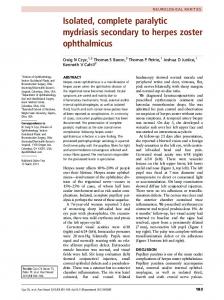
MRI of the spine revealed an intramedullary 1.2 x 4.5 x 1.5 cm lesion [Figures 1A and B] in conus medullaris and cauda equina. There was diffuse edema of the cord from the level of C4 vertebra to conus medullaris. CT scan of thorax, abdomen and pelvis was normal. Serum CA-125 level was elevated (63.2 iu/ml). The cerebrospinal fluid (CSF) examination showed proteins 143 mg/dl, sugar 50 mg/dl, chlorides 116 mEq/l, LDH 43 iu/l and CA-125 of 384 iu/ml. However, no malignant cells were detected on CSF cytology. The patient received dexamethasone and had symptomatic improvement. Subsequently, she received local radiotherapy to the spine and oral Etoposide (50 mg OD, D1-15, every 21 days). MRI of the spine at the end of six cycles of chemotherapy showed complete regression of the metastatic lesion. CT scan of thorax, abdomen and pelvis continued to remain normal. At last follow up (May 2005), patient continued to be in complete remission and has an excellent quality of life.
chemotherapy have lengthened survival, which may have changed the metastatic pattern of the disease (e.g., spread to unusual sites has become more common).[2] Also, with the availability of more sensitive imaging techniques, these tumors are being diagnosed with increasing frequency.[3] The common sites of CNS involvement are the brain parenchyma (cerebrum, cerebellum and pons), leptomeninges and very rarely the spinal cord.[4] Median survival from diagnosis of CNS metastases was 5 months. [4] Our patient has survived for 33 months without any evidence of disease.
m o r Intramedullary spinal cord involvement in 8·5% f to the CNSoccurs of all patients with metastasis and d ns of the lungin is2·1% of all cases of cancer. aCarcinoma the o of intramedullary o most common source metastases, l ti melanoma, renal-cell n followed by breast carcinoma, a w ltumors, carcinoma, colorectal ic cervical carcinoma and o Discussion lymphoma.d b u e P of).spinal cord involvement might Three e patterns Neurologic manifestations in a patient with malignancy r distinct f suggest the w spread of tumor to the cord by different may be due to paraneoplastic syndrome, metastases, m r infection, vascular event or an adverse effect of the routes. Most likely, it o fospread.kn .co is a result of hematogenous treatment received. e ed ow l In stage IV epithelial ovarian cancer, liver and lung are other cases, there is direct extension of b Inleptomeningeal n metastatic tumor across the pia into the a the commonest sites of metastases besides widespread Mparenchyma l k i d of the cord or the tumor may involve the intra-abdominal disease. Involvement of theacentral y e b nervous system by metastatic epithelial ovarianvcancer is medulla a part .m along the perineural sheets. rare. Whenever it occurs, these lesions are usually a d s cancer w Patients with spinal cord involvement can present with of widespread systemic dissemination. i Ovariante s w radicular pain, weakness, paresthesia and bowel and accounted for only 1% of the cases.F o involvement. Sometimes, they can be subtle and h by(wuse bladder PDovarian nonspecific. A high index of suspicion is essential in Advances in the management of cancer e of aggressive surgery andiseffectiveit platinum-based order to prevent permanent neurologic deficit. s Th a MRI is a sensitive modality for the detection of nervous [5]
[6]
[7]
[1,2]
[3]
A
B
system involvement. In epithelial ovarian cancer, measurement of CA-125 levels in the cerebrospinal fluid may be contributory to establishing the diagnosis. In our patient, the CSF CA-125 levels were elevated at the time of diagnosis of spinal cord metastases and declined after the treatment completion.
Figure 1: A. MRI (saggital) shows intramedullary ovoid mass at the level of L1 and L2 (T2 W). B. MRI (coronal) shows intramedullary enhancing lesion at the level of L1 and L2 post contrast (T1W)
Neurosurgical resection may be of benefit in selected patients if the tumor is well encapsulated, the systemic disease is under control and the tumor type is radio resistant. The role of chemotherapy in the management of CNS involvement from ovarian carcinoma remains unclear. Recent reports have demonstrated objective response of CNS metastases from ovarian cancer treated with platinum-based chemotherapy. It has been suggested that diffusion of systemically administered
Indian Journal of Cancer | July–September 2006 | Volume 43 | Issue 3
137 CMYK137
Bakshi, et al.: Spinal cord metastases from epithelial ovarian carcinoma
cytotoxic drugs into the CNS might be related to an alteration of the blood-brain barrier by intracranial lesions as well as radiotherapy. It is of interest that in the reported patient, carboplatin had good activity in the spinal cord metastasis.[7] To our knowledge, only four patients with isolated intramedullary spinal cord involvement from epithelial ovarian carcinoma have been reported. First reported case received steroids and radiotherapy. [2] Second patient survived for 10 months after the diagnosis of intramedullary metastases and received steroids, surgical resection and carboplatin.[7] The third patient underwent excision only and there was no disease recurrence till 24 months. [8] Fourth one, reported recently, underwent surgery and postoperative radiotherapy with a disease-free follow-up of 16 months. [6] However, our patient with isolated intramedullary spinal cord metastasis from epithelial ovarian cancer has responded very well to radiotherapy and chemotherapy and is in complete remission for the last 33 months. References 1.
138 CMYK
3.
4.
5.
m o fr d ns a lo tio n w lica o d ub e rf e w P m). r o o o f kn .c le ed ow b la M dkn i a by e v a si ted w.m F os w D P te h (w is si h T a
Rose PG, Piver MS, Tsukada Y, Lau TS. Metastatic patterns in
138
2.
histologic variants of ovarian cancer: An autopsy study. Cancer 1989;64:1508-13. Thomas AW, Simon SR, Evans C. Intramedullary spinal cord metastases from epithelial ovarian carcinoma. Gynec Oncol 1992;44:195-7. Connolly ES Jr, Winfree CJ, McCormick PC, Cruz M, Stein BM. Intramedullary spinal cord metastases and review of the literature. Surg Neurol 1996;46:329-38. Cormio G, Maneo A, Parma G, Pittelli MR, Miceli DM, Bonazzi C. Central nervous system metastases in patients with ovarian carcinoma. A report of 23 cases and a literature review. Ann Oncol 1995;6:571-4. Costigan DA, Winkelman MD. Intramedullar y spinal cord metastasis: A clinicopathological study of 13 cases. J Neurosurg 1985;62:227-33. Rastelli F, Benedetti G, Di Tommaso L, Mazzoli M, Calbucci F, Crino L. Intramedullary spinal metastasis from ovarian cancer. Lancet Oncol 2005;6:123-5. Cormio G, Di Vagno G, Di Fazio F, Loverro G, Selvaggi L. Intramedullary spinal cord metastasis from ovarian carcinoma. Gynec Oncol 2001;81:506-8. Isoya E, Saruhash Y, Katsuura A, Takahashi S, Matsusue Y, Hukuda S. Intramedullary spinal cord metastasis of ovarian tumor. Spinal Cord 2004;42:485-7.
6.
7.
8.
Source of Support: Nil, Conflict of Interest: None declared.
Indian Journal of Cancer | July–September 2006 | Volume 43 | Issue 3