Received: 2.2.2006
Accepted: 1.8.2006
Case Report Tuberculous cerebritis and tuberculoma in a patient with AIDS: Literature review and case report. Saeed Abrishamkar* Abstract Tuberculous brain cerebritis, abscess and tuberculoma in AIDS patients are considered as rare conditions and only few cases have been reported in the literature. The present case is a 28-year-old man with AIDS and previous systemic tuberculosis, denied by him and his family. He was admitted to our department due to headache, hemiparesis and seizures. A brain computed tomography (CT) scan disclosed a frontal hypodense lesion with a non-homogenous contrast enhancement that was reported as a high grade glioma. Magnetic resonance imaging (MRI) showed a diffuse hypointense lesion in right frontal area on T1-weighted, and hyperintense on T2-weighted and flair view, but there was a small paraventricular region with hypointensity on both T1, T2 and flair series, which was also reported to be a high grade glioma. Because of clinical course and imaging findings, the patient was a candidate for operation. After operation, the results of pathology and laboratory examination confirmed the diagnosis of tuberculous brain cerebritis and tuberculoma with positive Acquired Immune Virus (HIV) serology. Thus, tuberculous cerebritis, tuberculoma and abscesses should be considered in the differential diagnosis of focal brain lesions in AIDS patients, but AIDS should also be considered in every patient with an uncommon cerebral lesion who is not cooperative with medical healthcare providers. Surgical excision or biopsy and anti-tuberculous treatment are the mainstay in management of these lesions in patients with AIDS. KEY WORDS:
AIDS, tuberculoma, tuberculosis cerebritis. JRMS 2006; 11(4): 273-277
H
uman immunodeficiency virus (HIV) infection and acquired immunodeficiency syndrome (AIDS) are known risk factors in exacerbating tuberculosis in individuals previously infected with Mycobacterium Tuberculosis 1. The HIV epidemic rendered tuberculosis more prevalent and increased mortality, while promoting multi-drug resistance and increasing extra-pulmonary and disseminated forms of the disease 2-4. The central nervous system (CNS) is involved in 10-20% of cases in whom HIV infection coexists with tuberculosis 2,3. While the main clinical syndrome is tuberculous meningitis 2,3, focal tuberculous lesions are infrequent and may appear as tuberculomas and rarely as abscesses 5. This is a case report of a brain lesion, which according to clinical examination
and imaging reports seemed to be a high-grade glioma, but was later found to be cerebral tuberculosis in AIDS. Because of social problems, on the day of admission, he and his family concealed his HIV status and previous history of tuberculosis.
Case Report A 28-years-old man was admitted to our service in November 2005 because of holocranial headache, two episodes of generalized tonicclonic seizures and a month-long progressive left hemiparesis. The imaging findings and reports of MRI and CT scan were consistent with high grade glioma and therefore the patient was a candidate for craniotomy and tumor resection. At the first visit, he and his family denied his past history of intravenous drug
* Assistant Professor, Department of Neurosurgery, Isfahan University of Medical Sciences. Isfahan, Iran. e-mail:
[email protected]
Journal of Research in Medical Sciences July & Aug. 2006; Vol 11, No 4.
273
A case of tuberculous cerebritis in AIDS
Abrishamkar
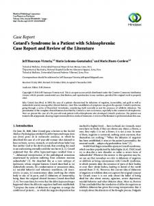
abuse, but declared after his operation that he was a drug abuser and two years before his admission, was hospitalized and treated with the diagnosis of systemic tuberculosis. Laboratory results revealed moderate anemia (hemoglobin of 10 g/dL), but other examinations including chest radiography were normal. After receiving confirmation of cerebral tuberculosis by pathologist, other laboratory examinations were requested. The absolute Cluster Designation Four Plus (CD4+) count was less than 100 cells/µL, and tuberculin skin test using Purified Protein Derivative (PPD) was negative. Brain computed tomography (CT) scan showed a hypodense frontal lesion with significant associated edema. Magnetic resonance imaging (MRI) showed a diffuse hypointense right frontal lesion on T1- weighted view, which was hyperintense on T2-weighted and flair view. Meanwhile, there was a small central region with hypointensity in both T1/T2 and flair series (figure 1). In light of the pathology report, clinical findings, and lack of knowledge as to the patient's history of TB or AIDS, the first diagnosis was high-grade
glioma; hence craniotomy was administered on the day following his admission. Intraoperative findings showed loose and firm abnormal tissues and central hard matrix that seemed to be calcification of tumor. After operation, the specimen was sent for pathological examination and the patient was transferred to intensive care unit. The next day, he was awake with his previous neurological deficits. The patient was discharged from hospital three days later with dexamethasone and phenytoin. One week later, he was referred to the Emergency Room because of progressive neurological deficits and his control CT scan showed a large cerebral edema in the right frontal lobe. On the same day, pathology confirmed cerebral tuberculosis and the ZiehlNeelsen staining showed abundant acid-fast bacilli (AFB). After additional consultation with Department of Infectious Diseases, isoniazid, rifampicin, pyrazinamide and ethambutol were started and the requested HIV test confirmed the diagnosis of AIDS. At this time, the patient's family acknowledged that he was an intravenous drug abuser and had a history of systemic tuberculosis two years before.
Figure 1. Tuberculoma and surrounding cerebritis in T1/T2 and flair series; the central hypointense region in all series is tuberculoma.
274
Journal of Research in Medical Sciences July & Aug. 2006; Vol 11, No 4.
A case of tuberculous cerebritis in AIDS
Discussion Tuberculous brain cerebritis, tuberculoma and abscess are infrequently described in the literatures, but in the past two decades the frequency of Mycobacterium Tuberculosis infection has increased 6. The histopathological features of cerebral tuberculosis are different from other kinds of infection and the definitive diagnosis depends on microscopic evidence of acute inflammatory alterations in the tuberculoma and the presence of AFB 6,7. Tuberculoma generally develops as a single lesion in CNS 8-11 and evolves gradually 12. Tuberculomas can develop through four pathophysiological mechanisms: invasion of bacilli from CSF (TB meningitis), as a consequence of a disseminated TB, paradoxical reaction in patients with antituberculous treatment with or without antiretrovirals, and local reactivation of latent foci 2,11. Cerebral TB infection may produce local areas of cerebritis with formation of tuberculomas. It is unknown why an abscess is produced in some cases with or without medical therapy, and tuberculoma develops in some others. Farrar believes that if the quantity of bacilli is high enough or the immunity is depressed, a focal cerebritis may progress to an abscess 12. On the other hand, most authors consider abscesses to be the result of liquefaction of tuberculomas 10,11,13. In the AIDS era, cerebral tuberculosis or tuberculoma is usually a subacute illness and the most frequent clinical manifestations are seizure 12,14, consciousness alterations 11, paresis 9,11,15 and headache 8,11,16. Other findings are paresthesis 9,11, cerebellar signs 8,16, facial palsy 16, and intracranial hypertension 8. Before the AIDS era, Whitener 6 reported that patients with cerebral tuberculosis usually had an acute illness, and the most common findings were focal neurological deficits (71%), headache (47%), fever (46%), seizures (35%) and consciousness alterations (24%). The present case is in accordance with other reports of AIDS patients. Formerly, Whitener 6 reported that patients with tuberculomas frequently presented epidemiological, historical or laboratory evidence of extra-cranial tubercu-
Abrishamkar
losis, but chest X-ray consistent with tuberculosis was uncommon. The principal cause of intracranial focal lesions coexistent with CD4+ values below 100 cells/µL 14 is toxoplasmosis. CD4+ values in cerebral tuberculosis without other CNS infections are reported to be between 112-270 cells/µL 9,16. This important finding would be misleading to diagnostic reasoning in some cases. This finding and the history of toxoplasmosis encephalitis, plus the lack of secondary prophylaxis for this opportunistic infection could have more retarded the definite diagnosis and consequently delayed treatment. CT scan findings of cerebral tuberculosis could be similar to those related to other causes of expanding lesions in AIDS patients 12; i.e. a hyperdense lesion and moderate contrast enhancement. But, the presence of a single lesion, a thick capsule, ring enhancement, and a lobular contour make the diagnosis of tuberculous abscess more likely 9,11,17,18. On the other hand, toxoplasmic encephalitis lesions are generally multiple, not multilobulated, and their capsules are not thick. The differential diagnosis of cerebral tuberculosis is wide, including toxoplasmic encephalitis (the most frequent cause of intracranial mass in AIDS patients) and primary CNS lymphoma 19-21. Other rare causes include cryptococcal abscess and cryptococcoma21, non-tuberculous mycobacterial lesion 22, pyogenic abscess 9, syphilitic gumma 21, aspergilloma 23, Chagas Disease 24, intracranial mass due to cytomegalovirus 25 and intra-axial primary tumors. The AIDS patients with focal brain lesions, IgG antibodies against t. gondii and CD4+ cell count below 200 cells/µL should receive antitoxoplasma treatment. If no clinical or imaging improvement is seen after two weeks of treatment, a stereotaxic brain biopsy will be indicated 17. The availability of imaging techniques such as CT scan and MRI with Gadolinium (Gd) can improve the chance of correct diagnosis. However, until larger validation studies are com-
Journal of Research in Medical Sciences July & Aug. 2006; Vol 11, No 4.
275
A case of tuberculous cerebritis in AIDS
Abrishamkar
pleted, brain biopsy will continue to be the only definitive diagnostic modality 14. Operation and early medical treatment of cerebral tuberculosis in AIDS patients yielded a good outcome in the majority of reported cases 8,9,15. Furthermore, none of these patients had other associated CNS diseases and all deaths which occurred were linked to other causes rather than the tuberculoma themselves 10,16,22. Interestingly, before the AIDS era, Whitener 6 reported longer survival in all cases that received anti-tuberculous chemotherapy and surgical intervention.
AIDS should be considered in any patient with uncommon cerebral lesion who is not cooperative enough with medical healthcare providers. Many patients and their families deny their condition out of fear of social stigma. Considering the present case, cerebral tuberculosis should be included in the differential diagnosis of focal lesions in AIDS patients. A high index of suspicion would prompt an earlier surgical excision or stereotaxic biopsy, which are considered to be the gold standard for the diagnosis and treatment of cerebral tuberculosis.
References 1. Sepkowitz KA, Raffalli J, Riley L, Kiehn TE, Armstrong D. Tuberculosis in the AIDS era. Clin Microbiol Rev 1995; 8(2):180-199. 2. Iseman MD. Treatment of multidrug-resistant tuberculosis. N Engl J Med 1993; 329(11):784-791. 3. Slutsker L, Castro KG, Ward JW, Dooley SW, Jr. Epidemiology of extrapulmonary tuberculosis among persons with AIDS in the United States. Clin Infect Dis 1993; 16(4):513-518. 4. Small PM, Schecter GF, Goodman PC, Sande MA, Chaisson RE, Hopewell PC. Treatment of tuberculosis in patients with advanced human immunodeficiency virus infection. N Engl J Med 1991; 324(5):289-294. 5. Zuger A, Lowy FD. Tuberculosis. In: Scheld WM, Whitley RJ, Durack DT, editors. Infections of the central nervous system. Philadelphia: Lippincott-Raven, 1997: 417-443. 6. Whitener DR. Tuberculous brain abscess. Report of a case and review of the literature. Arch Neurol 1978; 35(3):148-155. 7. Tyson G, Newman P, Strachan WE. Tuberculous brain abscess. Surg Neurol 1978; 10(5):323-325. 8. Dechambenoit G, Boni NG, Santini JJ, Ba Z, V, Beaumel A, Kakou M. [Tuberculous abscess of the cerebellum]. Neurochirurgie 1993; 39(5):326-329. 9. Farrar DJ, Flanigan TP, Gordon NM, Gold RL, Rich JD. Tuberculous brain abscess in a patient with HIV infection: case report and review. Am J Med 1997; 102(3):297-301. 10. Fischl MA, Pitchenik AE, Spira TJ. Tuberculous brain abscess and Toxoplasma encephalitis in a patient with the acquired immunodeficiency syndrome. JAMA 1985; 253(23):3428-3430. 11. Machado DM, Marchiori E, Praxedes MC, Malheiros NR, Santos AA. Abscesso cerebral tuberculoso na SIDA: relato de um caso. Radiol bras 1995; 28:343-346. 12. Whiteman M, Espinoza L, Post MJ, Bell MD, Falcone S. Central nervous system tuberculosis in HIV-infected patients: clinical and radiographic findings. AJNR Am J Neuroradiol 1995; 16(6):1319-1327. 13. Yang PJ, Reger KM, Seeger JF, Carmody RF, Iacono RP. Brain abscess: an atypical CT appearance of CNS tuberculosis. AJNR Am J Neuroradiol 1987; 8(5):919-920. 14. Skiest DJ. Focal neurological disease in patients with acquired immunodeficiency syndrome. Clin Infect Dis 2002; 34(1):103-115. 15. Gettler JF, Garner BF. Long-term survival of an AIDS patient with a tuberculous cerebral abscess. J Natl Med Assoc 1996; 88(9):605-606. 16. Velasco-Martinez JJ, Guerrero-Espejo A, Gomez-Mampaso E, Navas-Elorza E, Garcia-Ribas G. Tuberculous brain abscess should be considered in HIV/AIDS patients. AIDS 1995; 9(10):1197-1199. 17. Reichenthal E, Cohen ML, Schujman CB, Eynan N, Shalit M. Tuberculous brain abscess and its appearance on computerized tomography. Case report. J Neurosurg 1982; 56(4):597-600. 18. Sheller JR, Des Prez RM. CNS tuberculosis. Neurol Clin 1986; 4(1):143-158. 19. Mamidi A, DeSimone JA, Pomerantz RJ. Central nervous system infections in individuals with HIV-1 infection. J Neurovirol 2002; 8(3):158-167.
276
Journal of Research in Medical Sciences July & Aug. 2006; Vol 11, No 4.
A case of tuberculous cerebritis in AIDS
Abrishamkar
20. Price RW, Worley JM. Management of neurologic complications of HIV-1 infection and AIDS. In: Sande MA, Volpering Pa, editors. The medical management of AIDS. Philadelphia: W.B. Saunders, 1995: 261-288. 21. Evaluation and management of intracranial mass lesions in AIDS. Report of the Quality Standards Subcommittee of the American Academy of Neurology. Neurology 1998; 50(1):21-26. 22. Trilla A , Graus F, Ananos G, Navarro M. Abceso cerebral tuberculoso syndrome da inmunodeficiencia adquirida. Med clin (Barcelona) 1989; 93:236. 23. Mylonakis E, Paliou M, Sax PE, Skolnik PR, Baron MJ, Rich JD. Central nervous system aspergillosis in patients with human immunodeficiency virus infection. Report of 6 cases and review. Medicine (Baltimore) 2000; 79(4):269-280. 24. Corti M. AIDS and Chagas' disease. AIDS Patient Care STDS 2000; 14(11):581-588. 25. Bassil HF, William DC. Cytomegalovirus encephalitis in an HIV positive patient presenting with a cerebral mass lesion. AIDS Patient Care STDS 1997; 11(5):319-321.
Journal of Research in Medical Sciences July & Aug. 2006; Vol 11, No 4.
277