nutrients Article
Catechol Groups Enable Reactive Oxygen Species Scavenging-Mediated Suppression of PKD-NFkappaB-IL-8 Signaling Pathway by Chlorogenic and Caffeic Acids in Human Intestinal Cells Hee Soon Shin 1,2,3 , Hideo Satsu 1,4, *, Min-Jung Bae 1,5 , Mamoru Totsuka 1 and Makoto Shimizu 1,6 1
2 3 4 5 6
*
Department of Applied Biological Chemistry, Graduate School of Agricultural and Life Sciences, The University of Tokyo, Tokyo 113-8657, Japan;
[email protected] (H.S.S.);
[email protected] (M.-J.B.);
[email protected] (M.T.);
[email protected] (M.S.) Division of Nutrition and Metabolism Research, Korea Food Research Institute, Seongnam-si 13539, Korea Department of Food Biotechnology, University of Science and Technology (UST), Daejeon 34113, Korea Department of Biotechnology, Faculty of Engineering, Maebashi Institute of Technology, Gunma 371-0816, Japan Institutes of Entrepreneurial BioConvergence, Seoul National University, Seoul 08826, Korea Department of Nutritional Science, Tokyo University of Agriculture, Tokyo 156-8502, Japan Correspondence:
[email protected]; Tel.: +81-27-265-7374
Received: 30 December 2016; Accepted: 13 February 2017; Published: 20 February 2017
Abstract: Chlorogenic acid (CHA) and caffeic acid (CA) are phenolic compounds found in coffee, which inhibit oxidative stress-induced interleukin (IL)-8 production in intestinal epithelial cells, thereby suppressing serious cellular injury and inflammatory intestinal diseases. Therefore, we investigated the anti-inflammatory mechanism of CHA and CA, both of which inhibited hydrogen peroxide (H2 O2 )-induced IL-8 transcriptional activity. They also significantly suppressed nuclear factor kappa-light-chain-enhancer of activated B cells (NF-κB) transcriptional activity, nuclear translocation of the p65 subunit, and phosphorylation of IκB kinase (IKK). Additionally, upstream of IKK, protein kinase D (PKD) was also suppressed. Finally, we found that they scavenged H2 O2 -induced reactive oxygen species (ROS) and the functional moiety responsible for the anti-inflammatory effects of CHA and CA was the catechol group. Therefore, we conclude that the presence of catechol groups in CHA and CA allows scavenging of intracellular ROS, thereby inhibiting H2 O2 -induced IL-8 production via suppression of PKD-NF-κB signaling in human intestinal epithelial cells. Keywords: chlorogenic acid; caffeic acid; interleukine-8; nuclear factor κB; protein kinase D; reactive oxygen species; catechol group
1. Introduction Chlorogenic acid (CHA) is one of the most widely consumed polyphenols because it occurs abundantly in numerous foods, especially coffee. CHA has antioxidative [1], anti-inflammatory [2], anti-obesity [3], anti-diabetic [4], and anti-cancer activities [5] and owing to its biological effects, CHA has been used in various functional foods and as supplemental agents. Caffeic acid (CA) is one of the CHA metabolites hydrolyzed by esterase and gut microflora in the gastrointestinal tract. However, CA is a phenolic acid that also occurs naturally in numerous agricultural products such as fruits, vegetables, wine, olive oil, and coffee. Similar to CHA, CA has been reported to have antioxidant, Nutrients 2017, 9, 165; doi:10.3390/nu9020165
www.mdpi.com/journal/nutrients
Nutrients 2017, 9, 165
2 of 12
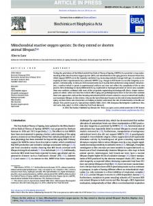
anti-tumor, and anti-inflammatory activities [6–8]. In a previous study, we reported that oxidative Nutrients 2017, 9, 165 2 of 12 stress-induced interleukin-8 (IL-8) secretion and mRNA expression were markedly inhibited by CHA. Furthermore, its metabolite CA, suppressed production but not thatmarkedly of quinicinhibited acid (another stress‐induced interleukin‐8 (IL‐8) secretion IL-8 and mRNA expression were by CHA. Furthermore, its metabolite CA, suppressed IL‐8 production but not that of quinic acid (another metabolite of CHA) in Caco-2 cells [9]. Recently, it was reported that CHA and CA reduced the mRNA metabolite CHA) in Caco‐2 cells [9]. protein Recently, it was reported CHA and reduced the expression of of macrophage inflammatory 2 (MIP-2, a mousethat homologue ofCA IL-8) in a dextran mRNA expression of macrophage inflammatory protein 2 (MIP‐2, a mouse homologue of IL‐8) in a sulfate sodium-induced colitis model [10]. However, the inhibitory mechanism of CHA and CA on IL-8dextran sulfate sodium‐induced colitis model [10]. However, the inhibitory mechanism of CHA and production has not yet been fully elucidated. CA on IL‐8 production has not yet been fully elucidated. Oxidative stress can trigger toxicity, damage, and inflammation in cells. In general, reactive general, reactive – Oxidative stress can trigger toxicity, damage, and inflammation in cells. 1 oxygen species (ROS) including singlet oxygen ( O2 ), hydrogen peroxide (H2 OIn 2 ), superoxide (O2 ), oxygen species (ROS) including singlet oxygen (1O2), hydrogen peroxide (H2O2), superoxide (O2–), and hydroxyl radical (HO•) develop antioxidant defenses by enhancing the expression of superoxide and hydroxyl radical (HO•) develop antioxidant defenses by enhancing the expression of superoxide dismutase, catalase, glutathione peroxidase, and peroxiredoxins, which maintain the redox balance [11]. dismutase, catalase, glutathione peroxidase, and peroxiredoxins, which maintain the redox balance However, when cellular ROS production overwhelms the antioxidant capacity, the ROS causes [11]. However, when cellular ROS production overwhelms the antioxidant capacity, the ROS causes oxidative stress-induced serious cell damage via production of toxic chemical compounds and oxidative stress‐induced serious cell damage via production of toxic chemical compounds and cytokines/chemokines, that results in the pathogenesis of several diseases. For example, oxidative cytokines/chemokines, that results in the pathogenesis of several diseases. For example, oxidative stress promotes the production of inflammatory cytokines in intestinal epithelial cells. Among the stress promotes the production of inflammatory cytokines in intestinal epithelial cells. Among the inflammatory cytokines, IL-8 in particular, plays a key rolerole in the of inflammatory bowel inflammatory cytokines, IL‐8 in particular, plays a key in pathogenesis the pathogenesis of inflammatory diseases which include Crohn’s disease and ulcerative colitis [12].colitis Therefore, the inhibition bowel (IBDs), diseases (IBDs), which include Crohn’s disease and ulcerative [12]. Therefore, the of IL-8inhibition of IL‐8 as a therapeutic target is essential in controlling inflammatory diseases like IBDs. as a therapeutic target is essential in controlling inflammatory diseases like IBDs. Nevertheless, theNevertheless, the mechanism underlying the regulation of IL‐8 has not yet been fully elucidated. mechanism underlying the regulation of IL-8 has not yet been fully elucidated. Therefore, we investigated the inhibitory mechanisms of CHA and CA against oxidative stress‐ Therefore, we investigated the inhibitory mechanisms of CHA and CA against oxidative induced IL‐8 production in human intestinal epithelial Caco‐2 cells. Furthermore, we elucidated the stress-induced IL-8 production in human intestinal epithelial Caco-2 cells. Furthermore, we elucidated thefunctional moieties mediating these anti‐inflammatory effects using structural analogs (Figure 1) of functional moieties mediating these anti-inflammatory effects using structural analogs (Figure 1) CHA and CA. of CHA and CA.
Figure 1. Chemical structures of analogues derived from chlorogenic acid (CHA) andand caffeic acid (CA). Figure 1. Chemical structures of analogues derived from chlorogenic acid (CHA) caffeic acid (CA). Chemical structure of CHA (A); CA (B); cinnamic acid (C); p‐coumaric acid (D); m‐coumaric Chemical structure of CHA (A); CA (B); cinnamic acid (C); p-coumaric acid (D); m-coumaric acid (E); acid (E); protocatechuic acid (F); and dihydrocaffeic acid (G). protocatechuic acid (F); and dihydrocaffeic acid (G).
Nutrients 2017, 9, 165
3 of 12
2. Materials and Methods 2.1. Reagents The Caco-2 cell lines were obtained from the American Type Culture Collection (Rockville, MD, USA). Dulbecco’s modified Eagle’s medium (DMEM) was purchased from Wako Pure Chemicals (Osaka, Japan). Fetal bovine serum (FBS), penicillin-streptomycin and non-essential amino acids (NEAA) were purchased from Gibco (Gaithersburg, MD, USA). CHA (PubChem CID: 1794427), CA (PubChem CID: 689043), p-coumaric acid (PubChem CID: 637542), and cinnamic acid (PubChem CID: 444539) were purchased from Sigma (St. Louis, MO, USA). m-coumaric acid (PubChem CID: 637541), protocatechuic acid (PubChem CID: 72), and dihydrocaffeic acid (PubChem CID: 348154) were purchased from Wako (Osaka, Japan). The dual-luciferase reporter assay system was obtained from Promega (Madison, WI, USA). The anti-human nuclear factor kappa-light-chain-enhancer of activated B cells (NFκB) p65 and IκB kinase (IKK) α/β (p-IKKα/β) antibodies were purchased from Santa Cruz Biotechnology (Santa Cruz, CA, USA). Protein kinase D (PKD) and p-PKD antibodies were purchased from Cell Signaling Technology (Beverly, MA, USA), and horseradish peroxidase-linked goat anti-rabbit IgG was from ICN Biomedicals (Aurora, OH, USA). ECL Plus Western blotting detection reagents were obtained from GE Healthcare Amersham Bio-Sciences (Buckinghamshire, UK). 20 ,70 -dichlorodihydrofluorescein diacetate (H2 DCFDA) was purchased from Invitrogen (Carlsbad, CA, USA). 2.2. Cell Culture Caco-2 cells were cultured at 37 ◦ C in humidified air containing 5% CO2 . The cells were maintained in a 100-mm dish with DMEM containing 1000 mg/L of glucose and supplemented with 10% FBS, 1% NEAA, 200 U/mL of penicillin and 200 µg/mL of streptomycin. The cells were subcultured weekly by trypsinization (0.1% trypsin, 0.5-mM EDTA) and seeded at a density of 2 × 10⁵ cells/well on a 24-well plate. The cells were allowed to grow for 2 weeks for the experiments, and the medium was changed every 2–3 days. The Caco-2 cells were used between passages 40 and 60. 2.3. Transient Transfection and Luciferase Assay The IL-8 reporter vector was constructed by inserting the human IL-8 promoter region containing the glucocorticoid-responsive element, transcriptional factor activator protein 1 (AP-1), CCAAT/enhancer-binding protein (C/EBP), and NF-κB consensus element into the pGL3-basic vector (Promega, Madison, WI, USA). The NF-κB reporter vector was constructed by inserting four NF-κB consensus elements (TGGAATTTCC) into the pGL3-promoter vector (Promega, Madison, WI, USA). Caco-2 cells were seeded at 1 × 10⁵ cells/well on 12-well tissue culture plates 1 day prior to transient transfection. At 70% confluency, cells were co-transfected with pRL-CMV (the internal control plasmid) and the pGL3-IL-8 or NF-κB vector by using the LipofectAmine Plus reagent (Invitrogen, Carlsbad, CA, USA) according to the manufacturer’s protocol. The transfected cells were allowed to recover overnight at 37 ◦ C and subsequently exposed to 1 mmol/L H2 O2 for 24 h. At the same time, we treated the cells with CHA or CA. The luciferase activity was measured by a Luminometer using the dual-luciferase reporter assay system (Promega, Madison, WI, USA) according to the manufacturer’s instructions. 2.4. Western Blot Analysis In order to analyze the nuclear translocation of p65, the cells were suspended in buffer A (10 mmol/L HEPES, 10 mmol/L KCl, 0.1 mmol/L EDTA, 0.1 mmol/L EGTA, 10% NP-40, 1 mmol/L DTT, 0.5 mmol/L PMSF, and 1 µg/mL of a protease inhibitor cocktail from Sigma, at pH 7.9). The cells were vigorously vortexed and centrifuged. After removing the supernatant, the cell pellet was suspended in buffer C (20 mmol/L HEPES, 25% v/v glycerol, 0.4 M NaCl, 1 mmol/L EDTA, 1 mmol/L EGTA, 1 mmol/L DTT, 1 mmol/L PMSF, and 1 µL/mL of a protease inhibitor cocktail from Sigma,
Nutrients 2017, 9, 165
4 of 12
at pH 7.9). After vortexing and centrifuging, the supernatant was used as the nuclear extract. Using 20 µg of nuclear proteins, the nuclear translocation of NF-κB p65 was detected by electrophoresis and antibody response. The analysis was carried out with a chemiluminescent substrate (ECL; Amersham Biosciences). To analyze the phosphorylated and total protein level of IKK and PKD, the cells were suspended in lysis buffer (10 mmol/L Tris-HCl, 150 mmol/L NaCl, 1% Nonidet P-40, 1 mmol/L EDTA, 20 mmol/L NaF, 100 µmol/L sodium orthovanadate, 20 µmol/L PMSF, and 100 µg/mL of a calpain inhibitor, at pH 7.8). The cells were vigorously vortexed and centrifuged, and the supernatant was used as the whole cell extract. Using 20 µg of cytosolic proteins, the phosphorylation of IKK and PKD were detected by electrophoresis and antibody response as above. 2.5. Measurement of Intracellular ROS Generation The intracellular ROS level was detected by using H2 DCFDA. H2 DCFDA is a cell-permeable indicator for ROS that is non-fluorescent until the acetate groups are removed by intracellular esterases, which allows oxidation to occur within the cell, causing irreversible conversion to the fluorescent form, DCF. For ROS measurements, Caco-2 cells were incubated for 5 min with HBSS containing 10 µmol/L H2 DCFDA after pre-treatment with CHA or CA. The cells were washed with HBSS for removal of extracellular H2 DCFDA, and then treated with 2 mmol/L H2 O2 for 1 h. The fluorescence was measured by a plate reader with an excitation wavelength of 485 nm and an emission wavelength of 538 nm. 2.6. ROS-Scavenging Activity The scavenging activities were measured by monitoring the capacity to reduce superoxide, hydrogen peroxide, and hydroxyl radical generation. These ROS were detected by using 2-methyl-6p-methoxyphenylethinylimidazopyrazinone (MPEC; ATTO, Tokyo, Japan) and xylenol orange (Sigma, and a radical catch kit (ALOKA, Tokyo, Japan). The scavenging activity (%) was determined using the following formula: Scavenging activity = [(activity of control) − (activity of sample)/(activity of control)] × 100
(1)
2.7. Enzyme-Linked Immunosorbent Assay Confluent monolayers of Caco-2 cells on the 24-well plate were exposed to a culture medium containing 2 mmol/L H2 O2 and CA derivatives. After incubation, the level of IL-8 in the cell supernatant was determined using commercially available enzyme-linked immunosorbent assay kits (Pierce, Rockford, IL, USA) according to the manufacturer’s instructions. 2.8. RNA Isolation and Real-Time PCR Total RNA was isolated from the cells by using Isogen (Nippon Gene, Tokyo, Japan) according to the manufacturer’s instructions. Reverse transcription of the RNA was performed by using an ExScript RT reagent kit (Takara, Otsu, Japan). First-strand cDNA was prepared from 1 µg of the total RNA. The real-time PCR reaction was performed in a 10 µL volume containing 1 µg of cDNA, 0.5 µmol/L of each primer, and 2 × SYBR Premix Ex Taq™. Each sample was denatured at 95 ◦ C for 15 min and subsequently subjected to 40 cycles of 15 s at 95 ◦ C for denaturation, 15 s at the appropriate annealing temperature (56–57 ◦ C), and 10 s at 72 ◦ C for elongation. The primer sequences were as follows: human IL-8, 50 -AGAGTGATTGAGAGTGGACC-30 (forward) and 50 -ACTTCTCCACAACCCTCTG-30 (reverse); and β-actin, 50 -CCACGAAACTACCTTCAAC-30 (forward) and 50 -GATCTTCATTGTGCTGGG-30 (reverse). The real-time PCR reactions were run on a Lightcycler (Roche Applied Science, Penzberg, Germany). The gene expression levels were normalized to the level of the housekeeping gene (β-actin). Relative gene expression changes are reported as number-fold changes compared to the control samples.
Nutrients 2017, 9, 165
5 of 12
2.9. Statistical Analysis Each result is expressed as the mean ± the standard error (SE). The data were tested with Tukey’s multiple‐range test when significant differences (p