Wong etal. (11) reported the cloning ofa4-kb mRNA from a human stromalcell line, and Ladner et al. (12) published the sequence of a 2.3-kb M-CSF mRNA from ...
Proc. Natl. Acad. Sci. USA Vol. 85, pp. 6706-6710, September 1988 Biochemistry
cDNA cloning and expression of murine macrophage colony-stimulating factor from L929 cells (alternative splicing/cDNA sequence/COS cell expression/hematopoiesis/lymphokine)
MARTHA B. LADNER*t, GEORGE A. MARTIN*, JANELLE A. NOBLE*, VAUGHAN P. WITTMANt, M. KIM WARREN§, MICHAEL MCGROGAN*¶, AND E. RICHARD STANLEYII** Department of *Molecular Biology, tMicrobial Genetics, and §Cell Biology, Cetus Corp., 1400 Fifty-third Street, Emeryville, CA 94608; and Departments of I'Microbiology and Immunology and of **Cell Biology, Albert Einstein College of Medicine, 1300 Morris Park Avenue, Bronx, NY 10461
Communicated by Harry Eagle, June 10, 1988
A 4-kilobase and a 2-kilobase cDNA clone ABSTRACT encoding a murine macrophage colony-stimulating factor have been isolated. Except for 2 amino acid residue differences, these two clones encode the same 520 amino acid residue protein, which is preceded by a 32-amino acid residue signal peptide. The two clones, whose molecular masses correspond to the two transcripts observed in murine L929 fibroblasts, contain 3' untlated regions that are markedly different in sequence and length. Both clones can be expressed in COS cells and the recombinant protein is active in a mouse bone marrow colony assay.
medium (DMEM) with 10% fetal callf serum and RNA was isolated as described (10). Poly(A)+ RNA was selected by two passages through an oligo(dT) column and fractionated on 5-25% sucrose gradients for 16 hr at 30,000 rpm. One-fifth of each fraction was injected into Xenopus oocytes (15) and assayed 48 hr later essentially as described (16). RNA Blot Analysis of M-CSF Message. L929 cell mRNA (5 ,ug) was electrophoresed on a 1% agarose gel containing 2.2 M formaldehyde (17) and blotted onto nitrocellulose. A 1500-base-pair (bp) Ava II/Bgl I fragment from the human cDNA pcCSF-17 clone (10) was subcloned into M13mpl8, and a single-stranded DNA probe was made by extending with [32P]dCTP using the universal primer. The blots were hybridized at 420C using 5 x SSC/5 x Denhardt's solution/ 0.1% NaDodSO4/50%o formamide/106 cpm of probe per ml (1 x SSC = 0.15 M NaCl/0.015 M sodium citrate; 1 x Denhardt's solution = 0.02% bovine serum albumin/0.02% Ficoll/0.02% polyvinylpyrrolidone). The washes were 2 x SSC/0.1% NaDodSO4 at room temperature followed by 0.2 x SSC/0.1% NaDodSO4 at 550C. cDNA Synthesis. Fractions from sucrose gradients were used to synthesize double-stranded DNA as described (18). After EcoRI linker addition, the cDNA was electrophoresed on a 4% polyacrylamide gel, and the cDNA 800 bp and above was isolated and ligated into AgtlO (19). Candidate clones were found by screening plaque lifts with a 32P-labeled pcCSF-17 probe. Insert DNA was digested with various restriction enzymes and subcloned into M13mpl8 and M13mpl9 (20), and both strands were sequenced completely by the dideoxy chain-termination method (21). Transcription of M-CSF Message from the SP6 Promoter. The phage containing the 4-kb and 2-kb inserts were digested with EcoRI and the cDNA insert was ligated into EcoRIdigested pGEM2 (Promega Biotec, Madison, WI). Clones in the proper orientation were linearized with either Xba I, which cleaves in the pGEM polylinker, or BamHI, which cuts within the insert. Capped mRNA was prepared by transcribing 2 pug oflinearized plasmid in 40 mM Tris-HCl, pH 7.5/5.6 mM MgCl2/2 mM spermidine/0.5 mM ATP, GTP, and UTP/10 mM dithiothreitol/1 mM m7GpppG (Pharmacia)/ RNasin (1 unit/,&l) (Promega Biotec)/50 ,M GTP/1 GCi [3H]UTP (1 Ci = 37 GBq)/18 units ofSP6polymerase (Promega Biotec). Reticulocyte lysate translations were done with a translation kit according to the manufacturer's instructions (Promega Biotec).
Colony-stimulating factors (CSFs) are defined by their ability to promote the clonal growth and differentiation of hematopoietic progenitor cells. Four distinct CSF molecules have been identified (1). Based on the colonies formed in response to these factors, they have been designated granulocyte CSF, macrophage CSF (M-CSF), granulocyte-macrophage CSF, and multi-CSF (or interleukin 3). Murine M-CSF was initially purified to homogeneity from L-cell-conditioned serum-free medium (2) and was found to be a glycosylated disulfidelinked dimer of =70 kDa. The unglycosylated monomeric size of the protein was estimated to be 15 kDa (3). In vitro, purified L-cell M-CSF stimulates the survival, proliferation, and differentiation of mononuclear phagocytes (4). Murine M-CSF can stimulate macrophages to kill tumor cells (5), to resist vesicular stomatitis virus infection (6), and to produce granulocyte CSF (7, 8). In mice, M-CSF enhances mononuclear phagocyte production and increases the number of monocytes, neutrophils, and multipotent cells (9). Kawasaki et al. (10) initially reported the amino-terminal amino acid sequence of murine and human M-CSF and the cDNA cloning of a 1.6-kilobase (kb) human M-CSF molecule from a human pancreatic tumor cell line, MIA PaCa-2. Sequences that encode a longer M-CSF protein have also been published. Wong etal. (11) reported the cloning ofa4-kb mRNA from a human stromal cell line, and Ladner et al. (12) published the sequence of a 2.3-kb M-CSF mRNA from MIA PaCa cells. In the murine system, Rajavashisth et al. (13) published the sequence of a partial cDNA clone and DeLamarter et aL (14) reported the sequence of a 2-kb clone from L929 cells. To characterize fully the transcripts from L929 cells, we have isolated two full-length M-CSF cDNA clones that correspond to the two M-CSF transcripts we have observed and studied the expression of these two clones in COS cells.tt
MATERIALS AND METHODS Isolation and Sucrose Gradient Separation of RNA. Cells were grown to confluency in Dulbecco's modified Eagle's
Abbreviations: CSF, colony-stimulating factor; M-CSF, macrophage CSF. tTo whom reprint requests should be addressed. VPresent address: Invitron Corp., San Carlos, CA 94070. ttThe sequence reported in this paper is being deposited in the EMBL/GenBank data base (IntelliGenetics, Mountain View, CA, and Eur. Mol. Biol. Lab., Heidelberg) (accession no. J03862).
The publication costs of this article were defrayed in part by page charge payment. This article must therefore be hereby marked "advertisement" in accordance with 18 U.S.C. §1734 solely to indicate this fact. 6706
Biochemistry: Ladner et al.
Proc. Natl. Acad. Sci. USA 85 (1988)
6707
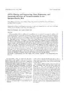
20 E 15
CO)
z < 10 T-
C#) CO)
5
0
10
15
20
25
FIG. 1. Sucrose gradient fractionation of L929 cell mRNA. Heavy line, concentration of M-CSF measured by an RIA of homogenates of Xenopus oocytes injected with fractionated mRNA. Ordinate, fraction number. (Inset) RNA blot analysis of murine M-CSF from L929 cells. The probe was 32P-labeled pcCSF-17 (10).
COS Cell Transfections. The murine cDNAs were ligated into the EcoRI site of a polylinker that had previously been inserted directly downstream of the simian virus 40 promoter in the Okayama-Berg expression vector (22). Plasmid DNAs were purified on CsCl gradients and transfected into COS A2 cells by a DEAE-dextran method (23) with the addition of chloroquine (24). The media were removed 72 hr later and assayed by a murine bone marrow colony assay (25, 26). Recombinant M-CSF Immunoprecipitation. COS A2 cells were transfected as described above. After 48 hr, transfected monolayers were rinsed in phosphate-buffered saline and incubated in cysteine- and methionine-free DMEM without serum for 2 hr. Approximately 25 gCi of [35S]methionine and [35S]cysteine per ml (-1000 Ci/mmol) were added, and the cells were grown overnight before harvesting the supernatant. The M-CSF protein was immunoprecipitated and electrophoresed on a NaDodSO4/polyacrylamide gel.
were able to direct the translation of a 60- to 70-kDa protein (Fig. 2). The size of the polypeptide indicated that these cDNAs must encode a protein in the same size range as the large human M-CSF protein. The slight shift in mobility between the proteins produced from the 2-kb clone and the 4-kb clone probably reflects the two amino acid differences between the clones; these amino acid differences are discussed in more detail below. This result also suggested that the difference in the two murine cDNAs resides in either the 5' or the 3' untranslated regions of the clones rather than in the coding region, since both clones encode proteins of the
A
B
C
D E
G
200 -
97.4-
RESULTS Cloning and Sequencing. RNA was isolated from mouse L cells that were grown to confluence in DMEM with 10% fetal calf serum. After poly(A) + selection on oligo(dT) columns, the mRNA was fractionated on 5-25% sucrose gradients. A portion of each fraction was injected into Xenopus oocytes, and the supernatants were tested for M-CSF activity by radioimmunoassay (16). Two active peaks of M-CSF were detected, one at 20 S and one at 25 S (Fig. 1). These activity peaks correspond in size to the transcripts detected with a human M-CSF cDNA probe (Fig. 1 Inset). A cDNA library was constructed in AgtlO (19) with the sucrose gradient fractions that contained M-CSF activity. The library was screened with a human M-CSF cDNA probe. Fourteen plaques were chosen for further analysis from -500,000 screened. After plaque purification and DNA isolation, the two largest inserts, which were 4 and 2 kb long, were subcloned into both M13 (20) and a vector containing the SP6 promoter (27) for further analysis. To determine the size of the protein encoded by these cDNAs, RNA was transcribed from the inserts in the pGEM vectors (Promega Biotec) and tested for translational activity in a reticulocyte lysate assay. Both the 4- and the 2-kb insert
F
-110 97
68- M.4.. 40 IN* 43-- 35
27.5
-
- 20
18.4 14.3 FIG. 2. NaDodSO4/polyacrylamide gel of reticulocyte lysate translations of M-CSF RNA transcribed from the SP6 promoter. Translations of uncapped mRNA from 4-kb insert digested with Xba I (lane A), capped mRNA from 4-kb insert digested with Xba I (lane B), capped mRNA from 4-kb insert digested with BamHI (lane C), capped mRNA from 2-kb insert digested with Xba I (lane D), capped mRNA from 2-kb insert digested with BamHI (lane E), no mRNA (lane F), brome mosaic virus RNA (a positive control) (lane G). Numbers are kDa.
Biochemistry: Ladner et al.
6708
Proc. Natl. Acad. Sci. USA 85 (1988)
same size. DNA sequencing of the 4- and the 2-kb clones confirmed that they both contain the same long open reading frame (Fig. 3). The protein begins with a 32-amino acid leader sequence followed by the mature M-CSF coding sequence, as determined by the following two criteria: first, the 181 amino acid residues following the initiating methionine at nucleotide 160 are 81% similar to the human clone pcCSF-17; second, the coding sequence beginning after the putative leader peptide (at nucleotide 256) is the same as that determined for A.
the amino terminus of purified M-CSF protein from murine L cells (10). The large size difference between the two clones is due to differences in the 3' untranslated sequences. The 2-kb clone has a 235-bp 3' untranslated region that is homologous to the human clone pcCSF-17. This clone contains the polyadenylylation signal ATTAAA 18 bp before the polyadenylylation site. The 4-kb clone diverges from the 2-kb clone 16 bp after the last amino acid residue and contains a 2.1-kb 3' untrans-
,
TGAAAGTTTGCCTCGGTGCTCTCGGTGTCGCTGCGGCTC
40
TCTGCATCCCAGGACAGCGGCGTGGCCCTCGACCGGGGCGCGGGCTCTTCAGCCACTAGCGAGCAAGGGAGCGAGCGAACCAGGGCGGCCAACACGCCGTGCCGGGACCCAGCTGCCCGT
160
ATGACCGCGCGGGGCGCCGCGGGGCGCTGCCCTTCTTCGACATGGCTGGGCTCCCGGCTGCTGCTGGTCTGTCTCCTCATGAGCAGGAGTATTGCCAAGGAGGTGTCAGAACACTGTAGC
METThrAlaArgGlyAlaAlaGlyArgCysProSerSerThrTrpLeuGlySerArgLeuLeuLeuValCysLeuLeuMETSerArgSerIleAlaLysGluValSerGluHisCysSer 1 280
CACATGATTGGGAATGGACACCTGAAGGTCCTGCAGCAGTTGATCGACAGTCAAATGGAGACTTCATGCCAGATTGCCTTTGAATTTGTAGACCAGGAACAGCTGGATGATCCTGTTTGC
HisMETIleGlyAsnGlyHisLeuLysValLeuGlnGlnLeuIleAspSerGlnMETGluThrSerCysGlnIleAlaPheGluPheValAspGlnGluGlnLeuAspAspProValCys 20
400
40
TACCTAAAGAAGGCCTTTTTTCTGGTACAAGACATAATAGATGAGACCATGCGCTTTAAAGACAACACCCCCAATGCTAACGCCACCGAGAGGCTCCAGGAACTCTCCAATAACCTGAAC
TyrLeuLysLysAlaPhePheLeuValG4nAspIleIleAspGluThrMETArgPheLysAspAsnThrProAsnAlaAsnAlaThrGluArgLeuGlnGluLeuSerAsnAsnLeuAsn 60
520
80
AGCTGCTTCACCAAGGACTATGAGGAGCAGAACAAGGCCTGTGTCCGAACTTTCCATGAGACTCCTCTCCAGCTGCTGGAGAAGATCAAGAACTTCTTTAATGAAACAAAGAATCTCCTT
SerCysPheThrLysAspTyrGluGluGlnAsnLysAlaCysValArgThrPheHisGluThrProLeuGlnLeuLeuGluLysIleLysAsnPhePheAsnGluThrLysAsnLeuLeu 100
640
120
GAAAAGGACTGGAACATTTTTACCAAGAACTGCAACAACAGCTTTGCTAAGTGCTCTAGCCGAGATGTGGTGACCAAGCCTGATTGCAACTGCCTGTACCCTAAAGCCACCCCTAGCAGT
GluLysAspTrpAsnIlePheThrLysAsnCysAsnAsnSerPheAlaLysCysSerSerArgAspValValThrLysiroAspCysAsnCysLeuTyrProLysAlaT'hrProSerSer 140
160
760
AspProAlaSerAlaSerProHis~nr~ol~oeMTl~oe~al~ul~ps~pe~nr~rl~ye~re~ur~rl~ur 180 200
880
1000
ProSerAlaGlyGlyProValProGly~ll~pl~ul~re~ulyh~nr~le~ul~ae~yl~ae~ul~ee~rl~ul Gly
1120
C
280
AAGTTTTCCCCCTCCACGCCTGTAGGGGGCAGCATCCAGGCAGAGACTGACAGACCCAGGGCCCTCTCAGCATCTCCATTCCCTAAATCAACAGAGGACCAAAAGCCAGTGGATATAACA LysPheSerroe hrr al ylye le nla uhrs rgr rg aeue lae roh roy erh lus lny roa sp eg300
Pro
320
1240
AspArgProLeuThrGluValAsnProMETArgProIleGlyGlnThrGln~snAsnThrProGluLysThrAspGlyThrSerThrLeuArgGluAspHisGlnGluProGlySerPro 340 360
1360
HisIleAlaThrProsn 1480
ol
CTCTTGGGGCATTGTGCTGCCCCTTGGGGAGCTTGAGGGCAAG
r
a
e
sn rl h r a la nee380u400 380 400
AGAGTACAGGATGAAGAGCCCCAGGCTGAAGAGATCGCAGTAGGGGGCAGCCAGGCCTGTGGCCCGTTTTAATTCCATTCCTTTGACTGACACAGGCCATGTGGAG
ir Arg~er~r~rg~s~rg~rger~ro~a~lu~e~lu~lyly~er~a~er~l~ly~lala~rg~o~al~l~rg~hesn~er~e~ro~e~hrysp ly aslal 420
l
440
1600
CAGCATGAGG-GATCCTCTGACCCCCAGATCCCTGAGTCTGTCTTCCACCTGCTGGTGCCGGGCATCATCCTAGTCTTGCTGACTGTTGGGGGCCTCCTGTTCTACAAGTGGAAGTGGAGG GlnHisGluGlyS rS
1720
r sp
ol~l~oluSerValPheHisLeuLeuValProGlyIleIleLeuValLeuLeuThrValGlyGlyL 460
uL uPheTy LysTrpLysTrpArg
480
AGCCATCGAGACCCTCAGACATTGGATTCTTCTGTGGGGCGACCAGAGGACAGCTCCCTGACCCAGGATGAGGACAGACAGGTGGAACTGCCAGTATAGAAAGGATTC
SerHisArgAspProGlnThrLeuAspSerSerValGlyArgProGluAspSerSerLeuThrGlnAspGluAspArgGlnValGluLeuProVal 500
520
1840
ACAGGACTATCTCTTTATGGAAGGAGACATATGGGAACATCCACCACTACCCTCTCCTACCATCTTCCTGGGAATGTGGCCTACCACTACCAGAGCTCCTGCCTACCAAGACTGGATGAA
AGAAGCAGCTTTGATGGGGTCTTTCCATCCTCACCCTTAGACTCTCAACCAAAGAGAAAGGGCTGGAGGATGCCCCCCACATACTGCCACTATTTATTGTGGGCCCTGGAGGCTCCCTGC ATTGGAGGAAGGGCAGCTCAGCAGCTCAGGACCCTTTCCCTTAGGGGCTGCTTCCTCCCCTCAAAACCAGAACCTGGCAAGGGACTCACTAGCCTGGATGGCCCATGGGAGACCAGGACA GATGAGAAGGAGCAGAAGAGCCCTGTGCCCAGAAGACCCAACTGGTGCCAAGGAATCCCAGCATGGACAGGCAGGGACCTGTTTCCCAAGAAGAGAGCCTGATATTCAAAGGGTGGGACA 2320 GCATCTGCCCGACTTCCCGTAAAGGCATAAAGGCACGCAGCCCAAAAGACGGGAAGAGGAGGCCTTTGGCTGCTTGTGTTGACAGCTTAAAGGGGTCTACACCCTCAACTTGCTTAAGTG CCCTCTGCTGATAGCCAGGAAGGAGGGAGACCAGCCCTGCCCCTCAGGACCTGACCTGGCTCATGATGCCAAGAGGAAGACAGAGCTCTAGCCTCGTCTTCTCCTGCCCACAGCCCCTGC 2560 CP.GAGTTCTTTTGCCCAGCAGAGGCACCCCTCATGAAGGAAGCCATTGCACTGTGAATACTGAACCTGCCTGCTGAACAGCCTGTCCCATCCATCCCTATGAGTGACCATCCGTCCGAAT GTTCTCCCACTTCCTTCAGCCTCTCCTCGGCTTCTTGCACTGAGCTGGCCTCACGTGTTGACTGAGGGAGCCCCTGAGCCCCAACCTTCCCCTGCCTCAGCCTTTGATTGTCCAGGGTGA 2800O AGCTGTGGGAGAACCGCCTGGGCTACCAGTCAGAGCTGGTCTTTGGGCTGTGTTCCTTGCCCAGGTTTCTGCATCTTGCACTTTGACATTCCCAGGAGGGAAGTGACTAGTGGAAGGGAG AGAGGAAGGGGAGGCAGAGACAAAGGCCACAGGCAGAGCTATGAATGAGAATGGGTCTTGAAAATATGTGTGCACCCCTAAGCTTGAAATTGATCTCTATACTCTAGCCCCTCAGCCAGC 3040 CT'CC'lTCCI'GTTGTCTGAAACCTGGAGCTAAGCAGGTTGTCCTGTCACAAGCTCTGGGGACTGAGCTCCATGCTCCAACCCCACCCTCTTCTGACCTTTGTTCTCCAGACCTGACCCAGG 2080
3280
3520 3760
TAGGCAAGGGTACCCTCCCAGTCTCACCTACCATACTGTGCCATCTCTAGCCAAGCAAGCCAGGTTTAGAGAAGGGTCAAAAAAAAAAAAAAAGGGTTGTTTACTTCCAACTTGTTCTGA 1'GCCCTCTGTTTCCCAGGCCAGGCTTGTCTGTGGTGACCTGGGCATGGGTGACAGGGCTCTCATTTGCCCCTTGGTCTCTTTATGCTGCTGAGTCCCCCTTTCCTGCCCTCCCTGGCTAC TGGGTCAA.TAATCTTTCAGGCCATGAATCTGGGAGGAGAGTGGTCTGTAAGCTCCATCAGCCCTGTCCTGAGACAGCAGGGGGGAAGGACACTGGAGACTTTCTTGTGGGGCTTACTTAG CCTTCTGGTTACAGACTATTTCCATGCTAGAAAATACATATTTTAAAATAGAAGGAAAAACACAGAAACAAAACAAAACAAGGCATTCTCTACCCCTCCACCTTAAACATATATTATTAA AGACAGAA.GAGAAAATCCAACCCATTGGCAAGAAGCTCTTTGTGGGTGCCTGGTTACATCGGAGCAGGGGAGCCTCAAATCCACCTTTGGAGCCGCCCCTGTGTGCATTAGGAACCCTTC
TCTCCTCTGAGAAAGCTCAGAGGGAGCACTGCCTCACAAACTGTGAGACTGCGTTTTTTATACTTGGAAGTGGTGAATTATTTTATATAAGGTCATTTAAATATCTATTTAAAAAATAGG AAGCTGCTTTTATATTTAATAATAAAAGAAGTGCACAAGCTGCAAAAAAAAAA
D.
1831 1951
4
GACCCCTCACCATCCTGGACACACTCGTTTGTCAATGTCCCTCTGAAAATGTGGCGCCCAGCCCTGGACACAGTACTCCAGATGTTGTCTGACCAGCTCAGAGTACAGTGGGACGGTTGT CTTCCTTGATCTGGACAGTACTCTTCTACTCGT'GCAGATTAAGATCACATTAGTTTTAACAGCTGCATCATATATTGTCATATGTTGAGCTTGTAGTCTATTAAAAACCCCAGTTCTAAA
FIG. 3. DNA sequence and deduced amino acid sequence of the 4-kb and the 2-kb murine M-CSF cDNA clones. (A) Coding sequence for the 4-kb and 2-kb clones and the 3' untranslated sequence of the 4-kb clone. (B) Alternative 3' sequence found in the 2-kb clone. The 5' end of the 2-kb sequence is indicated by a small triangle and the two nucleotide differences found in the 2-kb clone are shown above the sequence. The start of the mature M-CSF protein is designated as amino acid 1. The coding sequence not found in the human clone pcCSF-17 is underlined. The three potential N-linked glycosylation sites are underlined. Arrows indicate the point where the two sequences diverge. Hydrophobic region extends from amino acid residues 460-483.
Biochemistry: Ladner et al. lated region, which is not related to the 2-kb clone. The 4-kb clone has a polyadenylylation signal (AATAAT) (28) 23 bp before the polyadenylylation site, as well as the sequence TGCACAAGCTGC immediately preceding the poly(A) tails. The latter sequence is similar to the consensus sequence TTCACTGC that has been found to precede poly(A) tails in a number of mRNAs (29). It has been reported that a conserved AU sequence in the 3' untranslated region of some mRNAs renders them more unstable than mRNAs that do not contain this sequence (30). The longest sequence conserved between several mRNAs cited is AUUUA. The 3' end of the murine M-CSF 4-kb clone contains this motif three times within 58 bp. The presence of guanine and cytosine bases between the AU sequences may modify the effect of the AUUUA sequence, however. The 2-kb clone contains no such AU-rich sequences within its 3' end. Although no difference has yet been shown- in the mRNA stability between the short clone and the long clone, the possibility exists that the different 3' untranslated regions can have an effect on the half-life of the two M-CSF mRNAs and, ultimately, on the amount of protein produced. Two nucleotide differences in the coding sequence were found between the two clones. In the 4-kb clone, there is an A at position 1034 and a T at position 1192, which are a G and a C in the corresponding positions in the 2-kb clone. Both changes are reflected in changes at the amino acid level: in the first instance, the long clone encodes an aspartic acid, while the short clone encodes a glycine; in the second position the long clone encodes a serine and the short clone encodes a proline. The 2-kb sequence obtained by DeLamarter et al. (14), published while this manuscript was in preparation, encodes an aspartic acid and a seine at these positions. The two amino acid differences in our 2-kb clone could be due to polymorphism in the genomic sequence or reverse transcriptase errors. Expression of Murine M-CSF. To ascertain whether these sequences are capable of directing the translation of active M-CSF protein, both cDNAs were subcloned into an Okayama-Berg type expression vector (22) that had been modified by the addition of a polylinker immediately downstream of the simian virus 40 promoter sequence. CsCl gradientpurified DNA was transfected into COS A2 cells and the supernatants were tested 72 hr later for M-CSF activity by a murine bone marrow colony assay as described (25, 26). The assay shows that both clones direct the expression of active M-CSF (Table 1). The murine protein contains three N-linked glycosylation sites (Asn-X-Ser or Thr) and may be quite large in its native dimeric glycosylated form. In studies of the biosynthesis of M-CSF in mouse L cells, two major polydisperse species of M-CSF (=-75 kDa and 180-410 kDa) have been shown to be released into the medium (K. Price and E.R.S., unpublished observations). As shown in Fig. 4, a similar pattern was found Table 1. Mouse bone marrow colony assay Sample Experiment Units/ml 2 kb 1 7,733 4 kb 1,067 Vector alone 20 Mock transfection 0 2 kb 2 11,533 5,033 4 kb 0 Mock transfection 2 kb 3 1,500 4 kb 6,000 pcCSF-17 2,500 Expression of the 4-kb and the 2-kb cDNA clones in COS A2 cells. Vector alone is pcDB without insert, and pcCSF-17 is the original human M-CSF recombinant clone in the Okayama-Berg vector.
Proc. Natl. Acad. Sci. USA 85 (1988)
6709
A BC
200
1
116 97 -
66 -
43-
31-
FIG. 4. Gradient (5-15%) NaDodSO4/polyacrylamide gel electrophoresis of recombinant murine M-CSF protein immunoprecipitated (32) from [35S]methionine- and cysteine-labeled medium from COS A2 cells transfected with the 4-kb clone (lane A), the 2-kb clone (lane B), and the vector alone (control) (lane C).
for the M-CSF produced by either cDNA clone in COS A2 cells. The medium from CHO cells expressing the human 4-kb clone also contained two M-CSF species, one of 70-90 kDa and one >150 kDa (11). It is likely that some heterogeneity arises from differential glycosylation of the M-CSF protein. The larger (180-410 kDa) L-cell species appears to be very highly glycosylated and has not been studied in detail (K. Price and E.R.S., unpublished observations). The size reported for the native biologically active glycosylated dimeric protein isolated from L-cell medium is =70 kDa (2). The molecular mass of the entire polypeptide predicted from the DNA sequence is 60.5 kDa. This difference in size between the mature -70-kDa species and the predicted polypeptide subunit precursor implies that some form of posttranslational processing is occurring. The murine cDNAs contain a sequence that encodes a very hydrophobic region (residues 460-483), followed by five positively charged amino acids. This region, which is conserved in the human M-CSF sequence, has the features of a transmembrane domain (31). As suggested previously (10), M-CSF may exist in a membrane-bound form. Cleavage of the extracellular domain' by proteolytic enzymes accounts for the smaller M-CSF polypeptides detected in cell culture media (32) and serum.
DISCUSSION We have shown that murine L cells produce a 4-kb and a 2-kb M-CSF transcript that differ in their 3' sequences. The 2-kb clone is essentially the same as the clone described by DeLamarter et al. (14) except for the presence of nine additional bases before the poly(A) tail in the latter clone. Messages of both sizes have also been detected in mouse lung, heart, brain, and kidney (13). In a separate study we have identified a human 2.3-kb M-CSF cDNA clone from the MIA PaCa cell line, which encodes a 522-amino acid M-CSF protein (12). The coding region of this clone corresponds to the coding region of a 4-kb M-CSF cDNA isolated from another human cell line, TPA301(11). The murine DNA coding sequence is 81% identical to this human sequence. The human 2.3- and 4-kb cDNA clones differ in their 3' untranslated sequences (12), as do the two murine cDNAs. The 3' untranslated sequence of the 2-kb murine clone is 90% identical to the 3' untranslated sequence of the human 2.3-kb clone. The 3' untranslated sequence of the murine and human 4-kb clones are more divergent,
6710
Biochemistry: Ladner et al.
although the last 110 bp are homologous (12). Both the murine clones and the human clones diverge 13 bp after the TAG stop codon. We know that this divergence occurs at an intronexon boundary in the human genomic sequence (12) and assume that an intron occurs at the same position in the munne genomic sequence. The occurrence of two different 3' untranslated sequences on both murine and human M-CSF transcripts and the conservation of sequence between the species suggest that the 3' untranslated sequence, or portions of it, are important and could be involved in transcript stabilization (30). Different cell types could synthesize M-CSF transcripts that would vary in their turnover rates, depending on which 3' sequence was predominantly used. This differential turnover would provide a means of controlling the amount of M-CSF protein synthesized in cells in which the rate of M-CSF transcription remains constant. Beginning at amino acid 150 of the mature protein, the murine clones and the human M-CSF cDNA clones contain 295 additional amino acids not encoded in the human 1.6-kb pcCSF-17 cDNA clone (10). The 1.6-kb human transcript is generated by an alternative splicing event, which deletes a large portion of exon 6. Despite this deletion, the 1.6-kb pcCSF-17 clone encodes a protein that contains the putative membrane spanning region and is the membrane precursor from which a mature biologically active M-CSF is proteolytically cleaved in transfected cells (32). Our data, along with the data of others, show that both murine and human cells transcribe an array of M-CSF mRNAs. In both species, these messages have been shown to contain 3' untranslated sequences that are vastly different in size and sequence. In addition, we know that human M-CSF transcripts can encode two proteins; one of 224 amino acids and one of 522 amino acids (12). A 1.4-kb M-CSF transcript, which could encode the murine equivalent of the 224-amino acid form of M-CSF, has been reported by other investigators to be present at high levels in mouse liver and in small amounts in other tissues and L cells (13). Since a 224-amino acid form of murine M-CSF has not yet been reported, a sequence analysis of the 1.4-kb transcript would be very interesting. Further studies should be directed at elucidating the relationship of the various M-CSF transcripts to the size of the M-CSF protein produced by the cells and why different tissues appear to transcribe different M-CSF mRNAs. The variety of M-CSF transcripts and their wide distribution in different cell types suggest that M-CSF may have additional functions that have not been defined. We thank Mei-Ting Lee and Adam Sampson-Johannes for M-CSF assays, James Devlin for pCDB, Michelle Manos for COS A2 cells, Eric A. Ladner and Tim Culp for graphics, and Deborah Jackson for typing the manuscript. 1. Metcalf, D. (1984) The Haemopoietic Colony Stimulating Factors (Elsevier, Amsterdam). 2. Stanley, E. R. & Heard, P. M. (1977) J. Biol. Chem. 252,43054312. 3. Das, S. K. & Stanley, E. R. (1982) J. Biol. Chem. 257, 1367913684.
Proc. Natl. Acad. Sci. USA 85 (1988) 4. Tushinski, R. J., Oliver, I., Guilbert, L., Tynan, P., Warner, J. & Stanley, E. R. (1982) Cell 28, 71-81. 5. Ralph, P. & Nakoinz, I. (1987) Cell. Immunol. 105, 270-279. 6. Lee, M.-T. & Warren, M. K. (1987) J. Immunol. 138, 30193022. 7. Metcalf, D. & Nicola, A. (1985) Leuk. Res. 9, 35-50. 8. Warren, M. K. & Ralph, P. (1986) J. Immunol. 137, 2281-2285. 9. Broxmeyer, H. E., Williams, D. E., Cooper, S., Shadduck, R. K., Gillis, S., Waheed, A. & Urdal, D. (1986) Blood 66, Suppl. 1, 146a (abstr.). 10. Kawasaki, E. S., Ladner, M. B., Wang, A. M., Van Arsdell, J., Warren, M. K., Coyne, M. Y., Schweickart, V. L., Lee, M.-T., Wilson, K. J., Boosman, A., Stanley, E. R., Ralph, P. & Mark, D. (1985) Science 230, 291-2%. 11. Wong, G. G., Temple, P. A., Leary, A. C., Witek-Giannotti, J. S., Yang, Y.-C., Ciarletta, A. B., Chung, M., Murtha, P., Kriz, R., Kaufman, R. J., Ferenz, C. R., Sibley, B. S., Turner, K. J., Hewick, R. M., Clark, S. C., Yanai, N., Yokota, H., Yamada, M., Saito, M., Motoyoshi, K. & Takaku, F. (1987) Science 235, 1504-1508. 12. Ladner, M. B., Martin, G. A., Noble, J. A., Nikoloff, D. M., Tal, R., Kawasaki, E. S. & White, T. J. (1987) EMBO J. 6, 2693-2698. 13. Rajavashisth, T. B., Eng, R., Shadduck, R, K., Waheed, A., Ben-Avram, C. M., Shively, J. E. & Lusis, A. J. (1987) Proc. Natl. Acad. Sci. USA 84, 1157-1161. 14. DeLamarter, J. F., Hession, C., Semon, D., Gough, N. M., Rothenbuhler, R. & Mermod, J.-J. (1987) Nucleic Acids Res. 15, 2389-2390. 15. Gurdon, J. B., Lane, C. D., Woodland, H. R. & Marbaix, G. (1971) Nature (London) 233, 177-182. 16. Stanley, E. R. (1979) Proc. Natl. Acad. Sci. USA 76, 29692973. 17. Lehrach, H., Diamond, D., Wozney, J. M. & Boedtker, H. (1977) Biochemistry 16, 4743-4751. 18. Gubler, U. & Hoffman, B. J. (1983) Gene 25, 263-269. 19. Huynh, T. V., Young, R. A. & Davis, R. W. (1985) in DNA Cloning: A Practical Approach, ed. Glover, D. M. (IRL, Oxford), Vol. 1, pp. 49-78. 20. Yanisch-Perron, C., Vieira, J. & Messing, J. (1985) Gene 33, 103-119. 21. Sanger, F., Nicklen, S. & Coulson, A. R. (1977) Proc. Natl. Acad. Sci. USA 74, 5463-5467. 22. Okayama, H. & Berg, P. (1982) Mol. Cell. Biol. 2, 161-170. 23. Sompayrac, L. M. & Dana, K. J. (1981) Proc. Natl. Acad. Sci. USA 78, 7575-7578. 24. Luthman, H. & Magnusson, G. (1983) Nucleic Acids Res. 11, 1295-1308. 25. Moore, R. N. & Rouse, B. T. (1983) J. Immunol. 131, 23742378. 26. Prystowsky, M. B., Naujokas, M. F., Ihle, J. N., Goldwasser, E. & Fitch, F. W. (1984) Am. J. Pathol. 114, 149-156. 27. Melton, D. A., Kreig, P. A., Rebagliati, M. R., Maniatis, T., Zinn, K. & Green, M. R. (1984) Nucleic Acids Res. 12, 70357056. 28. Wickens, M. & Stephenson, P. (1984) Science 226, 1045-1051. 29. Benoist, C., O'Hare, K., Breathnach, R. & Chambon, P. (1980) Nucleic Acids Res. 8, 127-142. 30. Shaw, G. & Kamen, R. (1986) Cell 46, 659-667. 31. Sabatini, D. D., Kreibich, G., Morimoto, T. & Adesnick, M. (1982) J. Cell Biol. 91, 1-22. 32. Rettenmier, C. W., Roussel, M. F., Ashmun, R. A., Ralph, P., Price, K. & Sherr, C. J. (1987) Mol. Cell. Biol. 7, 23782387.