Original Article
SECOND OPINION IN PATHOLOGY OF LYMPHOID LESIONS – AN AUDIT Syed Naeem Raza Hamdani1, Muhammad Ashraf Sharif2, Sajid Mushtaq3, Nadira Mamoon4, Muhammad Tahir Khadim5 ABSTRACT Objective: Errors in medical practice remain under intense scrutiny because of their human costs in terms of disability and suffering. Second opinion in histopathology is a safeguard against misdiagnosis before instituting major therapeutic endeavor. Lymphoid lesions are peculiar for their diagnostic evaluation as they require ancillary techniques and expertise in most cases. This review was undertaken to assess the magnitude of diagnostic discrepancies in lymph node lesions of surgical pathology. Methodology: Forty cases of lymph node lesions initially reported from different centres and referred for second opinion during the year 2005 were included in the study. Special stains and immunohistochemistry were applied and initial diagnosis was compared with the review diagnosis. The discrepancies were divided into different categories keeping in view the implications on patient management. Results: Of the 40 cases included in the study, there was agreement with the initial diagnosis on review in 11 cases. There was discrepancy between the initial and review diagnosis in 29 cases. Conclusions: Review is a sensitive and effective method for quality control and to identify areas of disagreement. Mandatory second opinion is a good policy in patient management where crucial therapeutic decisions are involved. It is highly suggested in cases where the histpathologist initially reporting the case does not have requisite ancillary techniques like immunohistochemistry. KEY WORDS: Audit, Surgical pathology, Immunohistochemistry, Medical error, Second opinion. Pak J Med Sci
October - December 2008 (Part-II)
Vol. 24
No. 6
798-802
How to cite this article: Hamdani SNR, Sharif MA, Mushtaq S, Mamoon N, Khadim MT. Second opinion in pathology of lymphoid lesions– An audit. Pak J Med Sci 2008;24(6):798-802.
INTRODUCTION Diagnostic errors in histopathology are important and their human cost in terms of disability, suffering and death is tremendous.1,2 Lymphoid lesions are peculiar in surgical pathology. They require basic paraffin section Correspondence Dr. Muhammad Ashraf Sharif, House 107, Street - 110, Sector G-11/3, Islamabad - Pakistan. Email:
[email protected]
* Received for Publication:
March 26, 2008
* Accepted:
September 26, 2008
798 Pak J Med Sci 2008 Vol. 24 No. 6
www.pjms.com.pk
of high quality as well as ancillary techniques such as immunohistochemistry. At the same time expertise of the histopathologist can not be overemphasized. Misdiagnosis represents an underemphasized and understudied area of patient safety.3,4 They are challenging to detect and evaluate.5 It is often difficult to agree whether an error has occurred, and even harder to determine with certainty its causes and consequences.6 Institute of Medicine in 1999 published a report citing medical error as a cause of death for some Forty four thousand to ninety eight thousand Americans each year and ever
Pathology of lymphoid lesions
since then, the medical practices that affect patient safety have been under intense scrutiny.7 Efforts are being made to pinpoint mistake-prone practices within a complex health care system.8 The methodology of diagnoses has also come under audit. Diagnostic failure is regarded as one of the most common causes of medical malpractice claims against hospitals.9 Studying cases of “missed” diagnosis have a central role in medical education, research and quality assurance.10,11 Several different methods have been used to identify areas of disagreement, including second opinions and blinded reviews.12,13 Each of these method is subject to their own particular bias. Second opinion pathology is a patient safety practice whereby pathology material from one institution is reviewed at the treating institution before the initiation of any major therapeutic endeavour. Erroneous pathological diagnosis may be uncovered and inappropriate unnecessary therapeutic endeavors can be avoided. This study was carried to assess the magnitude of erroneous pathological diagnosis in lymphoid lesions in our country where there is an absence of external quality assurance programme and most of the pathology laboratories lack ancillary techniques. METHODOLOGY The study was carried out at Armed Forces Institute of Pathology Rawalpindi (AFIP), which is a referral laboratory for armed forces of Pakistan and also receives specimens from Northern areas and upper Punjab in addition to the local hospitals of the adjoining cities. It has an average annual biopsy workload of 28,000 specimens. Forty cases of lymphoid lesions referred to AFIP Histopathology department during the year 2005, for second opinion after being reported upon elsewhere were included in the study. The cases were reviewed in a systematic manner. Initially the cases were seen independently by a senior registrar and then by the consultant histpathologist. Ancillary techniques such as special stains for periodic acid Schiff (PAS),
reticulin and immunohistochemistry were applied where required. Epithelial markers like pancytokeratin (CK AE1/AE3), epithelial membrane antigen (EMA) and lymphoid markers like LCA, CD-3, CD-20, CD-45RO, CD-15 and CD-30 were applied using antigenantibody immunoperoxidase technique for immunohistochemistry as per requirement of each case. Intradepartmental consultation was done in a few cases where diagnostic difficulties had arisen due to any reason. After evaluation and reaching a conclusive diagnosis, the cases were categorized keeping in view the implications on patient management. The different categories were: 1. Category A: There was concurrence in the review and initial diagnosis. 2. Category B: The cases which required modification and sub-classification of their diagnosis according to the WHO classifications or ICD-O system. 3. Category C: Those lesions where the category of benign or malignant diagnosis remained the same but there was disagreement in the specific diagnostic entity. 4. Category D: The cases in which the initial diagnosis of malignancy was changed to a benign lesion. 5. Category E: The initial benign diagnosis was changed to malignant on review. RESULTS Forty cases were included in the study. Eleven cases (28%) showed concurrence of diagnosis and were typed as category A. These cases included six cases of metastatic carcinoma, four cases of malignant Non-Hodgkin Lymphoma and one case of reactive hyperplasia of lymph node. Twelve cases were placed in category B as there was discrepancy in the classification and sub-classification which was either absent or different. Four cases of Hodgkin lymphoma had an absence of sub-categorization or different from the previous diagnosis. Sub-classification of Non-Hodgkin lymphoma was not done in three cases. Likewise three cases Pak J Med Sci 2008 Vol. 24 No. 6
www.pjms.com.pk
799
M. Ashraf Sharif et al.
Table-I: Comparison of review and initial diagnosis in category ‘C’ Serial 1. 2. 3. 4. 5. 6. 7. 8. 9. 10. 11. 12. 13.
Initial diagnosis
Review diagnosis
Metastatic small cell carcinoma Poorly differentiated neoplasm Metastatic carcinoma Malignant NHL (Immunoblastic) Chronic granulomatous lymphadenitis Chronic granulomatous lymphadenitis Poorly differentiated lymphoma Poorly differentiated lymphoma Differential diagnosisLymphoma vs Carcinoma Hodgkin lymphoma Hodgkin lymphoma Suggestive of malignant NHL Malignant NHL (small lymphocytic)
Metastatic squamous cell carcinoma Malignant *NHL (DLBCL) Malignant NHL (DLBCL) Metastatic carcinoma Chronic caseating granulomatous lymphadenitis Chronic caseating granulomatous lymphadenitis Mixed cellularity Hodgkin lymphoma Malignant NHL (DLBCL) Malignant NHL (DLBCL) Rebiopsy Rebiopsy Rebiopsy Rebiopsy
*NHL Non-Hodgkin lymphoma DLBCL Diffuse large B-cell lymphoma
reported as lymphoproliferative disorder were categorized in to Hodgkin and Non-Hodgkin lymphoma. One case of metastatic carcinoma was type specified as metastatic squamous cell carcinoma and like wise a case of Castleman disease was reassigned a diagnosis of reactive hyperplasia. Thirteen (36%) cases were placed in category “C” (Table-I). Four cases were unfit for opinion either due to processing and fixation artifacts or fragmented nature of the biopsy. (Rebiopsy may be needed in some cases for a definite diagnosis). This was done after extensive deliberations in the form of deeper cuts or manual staining. A diagnosis of lymphoma was changed to carcinoma and vice versa after application of immunohistochemical stains in 2 cases (Fig-1). A differential diagnosis between carcinoma and lymphoma in two cases was resolved after application of immunohis-
tochemical stains for LCA and CK AE1/AE3. A case of metastatic small cell carcinoma was reassigned a diagnosis of metastatic squamous cell carcinoma. Two cases reported initially as malignant lymphoma were categorized in to B and T cell subtype after application of markers for CD-3, CD-45R0 and CD-20. Two cases of granulomatous lymphadenitis showed caseation necrosis at review. A change in diagnosis from benign to malignant and malignant to benign was seen in four cases which were in category “D” & “E” (Table-II). DISCUSSION The results of the study showed an agreement in diagnosis between the review and the initial diagnosis in eleven (28%) of the cases. The histological diagnosis of overt metastatic carcinoma and Non-Hodgkin lymphoma
Table-II: Comparison of cases with a benign and malignant diagnosis at review in category ‘D’ & ‘E’ Category D D D E
Initial diagnosis
Review diagnosis
B- cell lymphoma with plasmacytic differentiation Malignant neoplasmD/D carcinoma vs lymphoma Squamous cell carcinoma pharynx Atypical lymphoid hyperplasia
800 Pak J Med Sci 2008 Vol. 24 No. 6
www.pjms.com.pk
Colonic ulcer with necrotic slough Reactive hyperplasia lymph node Reactive lymphoid hyperplasia Malignant NHL (Follicular lymphoma with DLBCL areas)
Pathology of lymphoid lesions
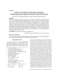
Fig-1: Photomicrographs showing a malignant non-Hodgkin large B-cell lymphoma which was reported as metastatic carcinoma on H &E stain (a) but negative for epithelial markers EMA and CK AE1/AE3 and positive for LCA & CD-20(b) on immunohistochemistry using immunoperoxidase streptavidin diaminobenzidine technique. (x400 magnification)
(diffuse large B-cell type) were in agreement in eleven of the cases. Non-Hodgkin lymphomas must be sub-categorized in to B and T cell types and further according to WHO/REAL classification of lymphoid malignancies since specific treatment protocols and drug regimens are used for B and T cell neoplasms.14 Even in B cell neoplasms the treatment of Follicular lymphoma is different from Burkitt’s lymphoma. Therefore it is mandatory to designate lymphomas into specific subtypes for prognostic and treatment purposes. However, this practice was not followed in twelve (30%) of category “B” lesions. Efforts should be made to classify metastatic poorly differentiated carcinomas in to specific subtypes wherever possible as it may be useful to determine the primary. A diagnosis of lymphoproliferative disorder on histopathology is of little benefit to the patient and treating clinician. All efforts must be made to stratify it into Hodgkin and Non-Hodgkin lymphoma. Immunohistochemistry is essential in all such cases. Adequate processing and fixation with thin sections of a lymph node are essential to avoid a misdiagnosis.15 Inexpertise of the technicians may put the pathologist under undue pressure. In such conditions, a rebiopsy should be suggested. Undifferentiated carcinomas and
lymphomas pose a diagnostic challenge on routine H&E sections which highlights the importance of immunohistochemistry to differentiate between them. It is the need of the hour that all relevant markers must be applied before giving a diagnosis relying on histology alone. Four cases in our study were also resolved by application of CK AE1/AE3 and LCA. Having said that, this does not undermine the importance of routine H&E stained sections as immunohistochemistry only acts as an adjunct to reach a final diagnosis. A case of metastatic keratinizing squamous cell carcinoma was misdiagnosed as a metastaic small small carcinoma. Although keratinisation was identified and a diagnosis was made on H&E stained sections but negativity for synaptophysin, chromogranin and neuron specific enolase excluded the diagnosis made initially before review. Categorisation of a benign lesion into malignant and vice versa is a major diagnostic error with drastic bearings on patient management. It not only shakes the confidence of the clinician but also puts the patient and family under undue stress. This also has medico-legal and malpractice implications. This further highlights the importance of continuing medical education, quality assurance programmes and surgical audits to put under check such practices. Pak J Med Sci 2008 Vol. 24 No. 6
www.pjms.com.pk 801
M. Ashraf Sharif et al.
Several international studies have reported major diagnostic changes in a small but meaningful number of cases. Abt et al16 found that 45 of 777 cases reviewed had a change in pathological diagnosis that was significant clinically. Kronz et al17 found that second opinion pathology resulted in a changed diagnosis in 1.4% of cases. Similarly Tsung18 noted major diagnostic disagreement in 35 of 673 (5.2%) consecutive cases. In each of these studies the vast majority of these changes reflected a conversion between benign and malignant or a significant modification of tumor classification. But surprisingly an alarming 72% change in diagnosis in any form needs critical analysis for the underlying factors and remedies to overcome this. The probable factors resulting in disagreement in our study are: a) Communication gap between clinician and histopathologist b) Lack of surgical audit and quality assurance programmes at local or national level c) Absence of trend of an intradepartmental consultation amongst histopathologists at places d) Lack of uniform availability of ancillary techniques such as immunohistochemistry at all laboratories. e) Lack of availability of quality assurance regulatory body to monitor and overlook the professional competence of practicing surgical pathologists. f) Overworked surgical pathologists without per person workload limitations. g) Poor processing and staining of tissue specimens
1. 2. 3. 4. 5. 6. 7. 8. 9. 10. 11. 12.
13. 14.
15.
16. 17. 18.
Second opinion in surgical pathology results in major diagnostic changes in a meaningful number of patients. It is strongly encouraged in cases reported from centers where ancillary techniques such as immunohistochemistry are not available before taking any major therapeutic intervention. www.pjms.com.pk
Baker GR, Norton P. Patient safety and healthcare error in the Canadian healthcare system. Ottawa, Canada: Health Canada: 2002;1-167. Leape L, Brennan T, Laird N. The nature of adverse events in hospitalized patients: Results of the Harvard medical practice study II. N Engl J Med 1991;324:377-84. Ramsay A. Errors in histopathology reporting: Detection and avoidance. Histopathology 1999;34:481-90. Edelman D. Outpatient diagnostic errors: unrecognized hyperglycemia. Eff Clin Pract 2002;5:11-6. Garber M, Gordon R, Franklin N. Reducing diagnostic errors in medicine: what’s the goal? Acad Med 2002;77:981-92. Kuhn G. Diagnostic errors. Acad Emerg Med 2002;9:740-50 Kohn LT, Corrigan JM, Donaldson MS. To err is human: building a safer health system. Washington, DC: National Academy Press 1999. Landrigan CP, Rothschild JM, Cronin JW. Effect of reducing interns’ work hours on serious medical errors in intensive care units. N Engl J Med 2004;351:1838-48. Glabman M. The top ten malpractice claims (and how to minimize them). Hosp Health Netw 2004;78:60-6. Gruver R, Fries E. A study of diagnostic errors. Ann Intern Med 1957;47:108-20 Kirch W, Schafii C. Misdiagosis at a university hospital in 4 medical eras. Medicine 1996;75:29-40. Jacques SM, Qureshi F, Munkarah A, Lawrence WD. Value of second opinion pathology review of endometrial cancer diagnosed on uterine curettings and biopsies. Mod Pathol 1997;10:103A. Hahm GK, Niemann H, Lucas JG, Frankel WL. The value of second opinion in gastrointestinal and liver pathology. Arch Pathol Lab Med 2001;125:736-9. Jaffe ES, Harris NL, Stein H, Vardiman JW, editors. In: World Health Organisation classification of tumors. Pathology and genetics of tumors of heamatopeitic and lymphoid tissues. IARC Press: Lyon (France);2001. Cousar JB, Casey TT, Macon WR, McCurley TL, Swerdlow SH. Lymph nodes. In: Mills SE, Carter D, Greenson JK, Oberman HA, Reuter V, Stroler MH, editors. Stermberg’s diagnostic surgical pathology. 4th ed. Philadelphia (USA): Lippincot Williams & Wilkins; 2004;777-848. Abt AB, Abt LG, Olt GJ. The effect of interinstitution anatomic pathology consultation on patient care. Arch Pathol Lab Med 1995;119:514-7. Kronz JD, Westra WH, Epstein JI. Mandatory second opinion surgical pathology at a large referral hospital. Cancer 1999;86:2426-35. Tsung JS. Institutional pathology consultation. Am J Surg Pathol 2004;28:399-402.
Authors: 1.
CONCLUSIONS
802 Pak J Med Sci 2008 Vol. 24 No. 6
REFERENCES
Dr. Syed Naeem Raza Hamdani, MBBS, FCPS Registrar Histopathology, 2. Dr. Muhammad Ashraf Sharif, MBBS, FCPS Registrar Histopathology, 3. Brig. Dr. Sajid Mushtaq, MBBS, FCPS, FRC Path Head Department of Histopathology, AFIP 4. Lt Col Dr. Nadira Mamoon, MBBS, FCPS Consultant Histopathologist, 5. Colonel Dr. Muhammad Tahir Khadim, MBBS, FCPS Consultant Histopathologist, 1-5: Armed Forces Institute of Pathology, Rawalpindi - Pakistan.