2482
J. Agric. Food Chem. 2007, 55, 2482−2488
Celiac-Related Properties of Chemically and Enzymatically Modified Gluten Proteins CRISTIANA BERTI,†,‡ LEDA RONCORONI,§,| MARIA LETIZIA FALINI,†,⊥ ROSITA CARAMANICO,†,# ERSILIA DOLFINI,§ MARIA TERESA BARDELLA,|,∇ LUCA ELLI,|,∇ CLAUDIA TERRANI,∇ AND FABIO FORLANI*,† Dipartimento di Scienze Molecolari Agroalimentari, Dipartimento di Biologia e Genetica per le Scienze Mediche, and Dipartimento di Scienze Mediche, Universita` di Milano, Milan 20133, Italy, and Centre for the Prevention and Diagnosis of Celiac Disease, Fondazione IRCCS, Ospedale Maggiore Policlinico, Mangiagalli e Regina Elena, Milan 20133, Italy
The effects of chemical (acid-heating treatment) and enzymatic (microbial transglutaminase, TGase) modification (deamidation) of gluten proteins on their physicochemical and celiac disease-related properties were studied. Ammonia release, sodium dodecyl sulfate-polyacrylamide gel electrophoresis, and sample solubility analyses were employed to check the extent of gluten modification. Among different treatments achieved, the acid-heating treatment performed at 90 °C for 3 h induced gluten deamidation, paralleling an increase of gluten solubility without relevant proteolysis. Changes in the immunoreactivity of celiac IgA anti-gliadin antibodies (AGAs) to modified gluten proteins were detected by using a competitive indirect enzyme-linked immunosorbent assay method. Chemical deamidation by acid-heating treatment of gluten lowered IgA-AGA immunoreactivity. IgA-AGA immunoreactivity to gliadins was increased when they were submitted to TGase-catalyzed deamidation. The acid-heating treatment of gluten reduced its cytotoxic activity on human colon adenocarcinoma LoVo cell line. These results showed that chemical deamidation of gluten may be envisaged as a way to lower the potential risk for celiac people due to widespread use of gluten as a food additive. KEYWORDS: Gluten; deamidation; IgA anti-gliadin antibody; immunoreactivity; LoVo cell line; cytotoxicity; celiac disease
INTRODUCTION
Given the unique inherent physical properties of its storage proteins (gliadins and glutenins) (1), it is not surprising that wheat gluten has been the subject of intense attention by the food industry (2). The most common usage of gluten continues to be in baked goods of various types and pasta production. However, an increasing awareness of gluten structural and functional properties, such as solubility, fat emulsification, gelation, and foaming (2-5), has caused an expanding diversity of applications. On occasion, manufacturers have been stimulated to explore ways of converting gluten into products with different properties. Deamidation is one of the methods used * To whom correspondence should be addressed. Tel: +390250316820. Fax: +390250316801. E-mail:
[email protected]. † Dipartimento di Scienze Molecolari Agroalimentari. ‡ Present address: Dipartimento di Scienze e Tecnologie Alimentari e Microbiologiche, Sez. Nutrizione, Universita` di Milano. § Dipartimento di Biologia e Genetica per le Scienze Mediche. | Dipartimento di Scienze Mediche. ⊥ Present address: Dipartimento di Scienze Mediche, Universita ` di Milano. # Present address: Dipartimento di Scienze e Tecnologie Alimentari e Microbiologiche, Sez. Industrie Agrarie, Universita` di Milano. ∇ Centre for the Prevention and Diagnosis of Celiac Disease.
for this purpose (6-12): Removal of the glutamine γ-amide and/or asparagine δ-amide groups to form carboxylic groups improves some of the additional functional properties of gluten proteins such as solubility, foam expansion, and emulsion capacity. The ingestion of wheat gliadins (prolamins) induces, in genetically susceptible individuals, celiac disease (CD), an inflammatory disease of the small intestine with an autoimmune component (T cell-mediated disease) (13-16). CD seems to manifest itself mainly in patients who carry HLA class II molecules DQ2 or DQ8. Adaptive immune responses are controlled by a preceding activation of the innate immunity mechanisms (17). The gluten prolamins are Gln- and Pro-rich proteins (18), and their CD-triggering sequences are characterized by the presence of multiple Pro and Gln residues. Most of these T cell epitopes are the preferred substrates of the tissue transglutaminase enzyme (tTGase; TG2), identified as the major autoantigen in CD (19). TG2 selectively deamidates certain Gln residues of gluten increasing the binding affinity of the epitopes to HLA-DQ2 or -DQ8 molecules, thereby enhancing their T cell stimulatory potential (20). In particular, a 33-mer peptide from the 266 amino acid R-gliadin, strongly resistant to
10.1021/jf062623n CCC: $37.00 © 2007 American Chemical Society Published on Web 02/21/2007
Celiac-Related Properties of Gluten Proteins gastrointestinal breakdown, was suggested as the primary initiator of the inflammatory response (21). CD is perhaps the most common human genetic disorder; thus, there is a strong need to find a way to detoxify “wheat” (20). For example, it may be possible to enzimatically modify gluten in vivo or during manufacturing processes in such a way that the epitopes are no longer recognized by the immune system (15, 21-23). On occasion, any consideration of new uses of modified gluten, particularly in noncereal-based products, should include the impact of the application on people with CD. As to this point, in an explorative study, minor immunoreactivity of anti-gliadin antibodies (AGAs) and cytotoxic effects on LoVo human cells were evidenced in acid-modified gluten proteins (24). This work aimed to deeply study the effects of a controlled chemical and enzymatic modification of gliadin and gluten proteins on their physicochemical and, mainly, CD-related properties, with the background idea of decreasing the potential risk associated with a general use of gluten as a food additive. MATERIALS AND METHODS Materials. Commercial gluten (pool of “hard red spring and winter” cultivars) (CG), 2-mercapto-ethylamine (cysteamine), pepsin, and trypsin were purchased from Sigma-Aldrich Co. (St. Louis, MO). Pancreatin was from Merck (Darmstadt, Germany). Gluten of bread wheat flour (Hereward cultivar) (HG) was prepared by the AACC standard method of hand washing, number 38-10 (25), and stored freezedried. Gliadins were extracted from bread wheat flour (Hereward cultivar) according to Capelli et al. (26). The ethanol extract containing gliadins was lyophilized and stored at 4 °C in a dry atmosphere. Activa TGase (EC. 2.3.2.13; TGase) from StreptoVerticillium sp. 8112 was purchased from Ajinomoto Foods Deutschland GmbH (Hamburg, Germany). Cell culture media were all purchased from Gibco (Milan, Italy), and plastics were from Corning (Milan, Italy). The employed cell line, LoVo, was obtained from human intestinal carcinoma and purchased from ATCC. Acid-Heating Treatment of Gluten. Gluten suspensions (5%) in 8.75 M acetic acid (AcG), in sealed glass tubes, were treated at 50 and 90 °C (0.75, 3, and 19 h) in a hybridization oven equipped with a rotating rack. After incubation, samples were exhaustively dialyzed (membrane cutoff, 3000 Da) against water, freeze-dried, and stored at 4 °C in a dry atmosphere. Gluten suspensions (5%) in water were processed either as above (wG) or omitting the heating step (untreated gluten, uG) and used as control samples. Treatment of Gliadins with TGase. TGase-catalyzed reaction was carried out on gliadins according to a modification of the method of Mamone et al. (27). Where it is stated, gliadins were tryptic-digested before the TGase treatment: Gliadins, suspended in 100 mM TrisHCl, pH 8.0, were hydrolyzed by trypsin (E:S ratio 1:30), and after 3 h at 37 °C, digestion was stopped by heating the reaction mixture at 100 °C for 5 min. For TGase treatment, 0.5 mg/mL gliadin sample suspensions in 70 mM Tris-HCl, pH 8.0, and 1 mM dithiothreitol were incubated with TGase (0.3 U/mg gliadins) for 2 h at 43 °C, in the presence or in the absence of 50 mM cysteamine. The TGase reaction was stopped by cooling at 4 °C and immediately diafiltered against 100 mM Tris-HCl, pH 8, using a Centricon device (Amicon, Danvers, MA) equipped with a 10000 Da cutoff membrane (or 3000 Da cutoff membrane when reactions were performed on tryptic-digested gliadins). Control experiments were prepared as above, omitting TGase. Quantification of the Ammonia Release. The ammonia amount in gliadin and gluten samples was determined before dialysis or diafiltration steps using an ammonia kit (Boehringer Mannheim GmbH, Darmstadt, Germany) according to the manufacturer’s instructions. Thiol Groups Determination. Thiol groups determination was carried out using the Ellman’s reagent, 5,5′-dithiobis-(2-nitrobenzoate) (28). Sodium Dodecyl Sulfate-Polyacrylamide Gel Electrophoresis (SDS-PAGE). Lyophilized uGs and AcGs were dissolved in the
J. Agric. Food Chem., Vol. 55, No. 6, 2007
2483
electrophoresis denaturing sample buffer and subjected to SDS-PAGE under reducing conditions according to Laemmli (29). Determination of Gluten Sample Solubility. Different buffers at different pH values were used as follows: 50 mM CH3COONa (pH 3, 4, and 5); 50 mM KH2PO4 (pH 6 and 7); 50 mM Tris-HCl (pH 8); and 50 mM 2-amino-2-methyl-1,3-propanediol (AMP, pH 9 and 10). Samples were dispersed in the different buffers (0.5 mg/mL) and stirred with a magnetic bar for 1 h and then centrifuged (10000g, 4 °C, 15 min). The absorbance of the supernatants was measured at 280 nm and used as a solubility parameter. Proteolytic Digestion. Proteolytic digestion was performed according to the method described in Dolfini et al. (30). Protein suspensions (2.5 mg/mL) in 0.05 M acetic acid, pH 3.05, were incubated with pepsin (E:S ratio 1:30) at 37 °C under continuous stirring for 24 h. The peptic digestion was stopped by adjusting the pH to 8.0 using 3 M Tris-HCl. The samples were then digested with pancreatin (E:S ratio 1:30) at 37 °C for 3 h, freeze-dried, and stored at 4 °C in a dry atmosphere. Determination of IgA AGA Immunoreactivity. Immunoreactivity of untreated celiac patient IgA AGAs to proteolitically digested acid heating-treated gluten and to TGase-treated gliadins was evaluated by the means of the competitive indirect enzyme-linked immunosorbent assay (ELISA), based on bread wheat flour gliadin as the coating antigen, human serum (diluted 1/150) as the primary antibody, goat anti-human IgA-peroxidase conjugate (Sigma, A0295) as the secondary antibody, and gliadin or proteolitically digested gluten samples (0.031000 µg/mL) as the competitor (31). Human serum was obtained from adults with CD diagnosed by means of intestinal biopsies according to standard criteria (32), selecting and pooling those with the highest AGA titers, using a commercial diagnostic kit (ORGenTec Diagnostika GmbH, Mainz, Germany). Control serum was obtained from healthy adults. Each experiment was performed in triplicate and repeated at least twice. The I50 value is expressed as the competitor concentration (µg/mL) yielding 50% ∆OD. Immunoreactivity for a tested protein was calculated as follows: (I50, gliadin or proteolized gluten/I50, tested protein) × 100. Cell Cultures. LoVo cells were maintained in a humidified 37 °C, 5% carbon dioxide incubator. Cells underwent serial steps as monolayers (split 1:5 every 5 days) in Ham’s F-12 medium supplemented with 10% foetal bovine serum, 1% MEM vitamin solution (100× stock solution), and 3% L-glutamine (200 mM stock solution). Mycoplasma contamination was regularly searched for and excluded using the Hoechst method (33). Multicellular tumor spheroids (MCTS) were performed by seeding 4 × 10⁵ cells/mL in 25 mL of complete IMDM medium supplemented with penicillin and streptomycin in Erlenmeyer flasks and incubated in a gyratory rotation incubator (60 rpm) at 37 °C in air. Homotypical aggregations are visible after 4 days of culture, and the MCTSs are usually complete within 7 days [average diameter ( standard deviation (SD), 370 ( 48.5 µm]. Two-dimensional cell cultures were treated for 48 h with different concentrations of proteolized gluten samples (125-750 µg/mL). MCTSs were treated with 750 µg/mL proteolized gluten samples from the seventh to the twelfth day of culture. At the end of the treatments, two-dimensional cell culture growth was evaluated and morphologically analyzed by phase contrast microscopy; the trypan blue exclusion method was used to evaluate cell viability. MCTSs were morphologically evaluated by means of phase contrast microscopy and scanning electron microscopy (SEM). MCTSs were washed twice in PBS and then fixed in 2.5% glutaraldehyde in phosphate buffer for 24 h at 4 °C. At the time of analysis, a representative sample of spheroids was recovered, immediately placed on a paper filter, and observed in low vacuum modality at a high voltage of 10 kV. The SEM analysis was performed using a Philips Scanning Electron Microscope (model XL20) (34). Statistical Analyses. Each of the measurements described above was carried out in at least three replicate experiments, and the results are reported as the mean and SD. Statistical analyses were performed using the Statistica software (Statsoft Inc., Tulsa, OK). The data from the ammonia release were analyzed by means of two-way analysis of variance (ANOVA) with temperature and time as dependent factors. Differences between the two types of gluten (HG and CG) were
2484
J. Agric. Food Chem., Vol. 55, No. 6, 2007
Berti et al.
Table 1. Ammonia Released during Treatment of CG, Suspended (5%) in Acetic Acid (AcG), at 50 and 90 °C for Different Time Intervals temperature (°C)
reaction time (h)
ammonia release (%)a
50
0.75 3.00 19.00 0.75 3.00 19.00
0.7 ± 0.4 1.1 ± 0.3 b 2.9 ± 0.7 c 3.3 ± 0.5 d 13.2 ± 0.6 e 67.3 ± 3.7 f
90
a Percent ratio (mean values ± SD, n ) 3) of ammonia released from gluten during treatment to that of completely hydrolyzed gluten (hydrolysis in vacuo for 3 h at 110 °C in 6 N HCl). Means within a column with different letters are significantly different at P < 0.001 when the letter is b and at P < 0.0001 when the letter is c. For each temperature, estimates with different letters (letters d−f) are significantly different (P < 0.0001).
analyzed by means of two-way ANOVA using the type of gluten as the independent condition. A two-way ANOVA with temperature and time as factors was used to test statistical significance of celiac IgAAGA immunoreactivity to gluten submitted to acid-heating treatment. Following a significant main effect in the ANOVA, individual means were compared using the LSD multiple range test. The data from cell cultures were analyzed using two-tailed Student’s t test. For all of the experiments, P < 0.05 was considered statistically significant. RESULTS AND DISCUSSION
Changes in Chemical Properties by Acid-Heating Treatment of Gluten. Gluten in its natural form has very low solubility due to the high concentration of amino acids residues such as Gln and Asn. In fact, the amide groups in the side of these amino acids play an important role in promoting the association between gliadin and glutenin molecules. However, in recent years, there is an increased interest in expanding the commercial use of gluten toward various food preparations beyond breadmaking and pasta production (2). The mild deamidation by acid is assumed to be a convenient and effective way to improve the functional properties of food proteins (4, 7, 9). In fact, the conversion of protein amide groups to carboxylic groups, with concomitant release of ammonia, induces conformational changes by imparting additional negative charges that decrease the isoelectric point of the protein. In addition, acidic deamidation may cause mild peptide hydrolysis leading to the formation of lower molecular weight polypeptides, which in turn enhances the solubility. We achieved an acid-heating treatment of gluten proteins under different conditions of temperature and time in order to identify the optimum levels of deamidation and hydrolysis. In other words, we investigate the conditions for deamidation of gluten having minimal changes in peptide molecular size (4, 6, 7). To this aim, ammonia release, electrophoretic patterns, and solubility of the modified gluten samples were assessed. Data about ammonia release of gluten suspensions, in acetic acid (AcGs), which underwent heating at 50 and 90 °C at different time conditions, are reported in Table 1. On the whole, no significant differences between CG samples and hand-washed gluten samples (HGs) were observed in all of the acid-heating treatment conditions. Our results indicate that the presence of acetic acid is a key factor in the release of ammonia. In fact, in all of the heattreated gluten water suspensions (wGs), there was little or no release of ammonia (data not shown), when heating was applied as well (on average 0.6% at 50 °C and 4.6% at 90 °C, for 19
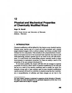
Figure 1. SDS-PAGE of gluten samples after acid-heating treatment at 50 (A) and 90 °C (B). CG was used. The Mr values of molecular weight markers (m) are indicated on the left-side. Lanes: 1, uG; 2−4, AcGs heat-treated for 0.75, 3, and 19 h, respectively.
h). A relevant role of temperature (P < 0.0005) on AcGs was assessed as follows: The higher the treatment temperature is, the stronger the effects are. In fact, the ammonia release of gluten was higher by the heat treatment at 90 °C as compared to that at 50 °C (significantly at 3 and 19 h). This effect was strengthened (P < 0.0005) by the extent of treatment times (significantly at 90 °C). Presumably, the ammonia release from gluten undergone heating at 50 °C depended on the hydrolysis of few accessible Gln γ-amide (and Asn δ-amide) groups to form carboxylic groups (deamidation). On the contrary, the high ammonia release of gluten that underwent heating at 90 °C, enhanced by the prolonged treatment, is caused by an unmasking of the hidden amide groups that become more accessible for further deamidation, also as a consequence of peptide bond cleavage (proteolysis). SDS-PAGE analysis was carried out in order to examine any change in size of the treated gluten proteins, in other words to verify the extent of proteolysis. The electrophoretic patterns of CGs and HGs were comparable under all of the conditions considered (data not shown). To evaluate the effect of different acid-heating treatment conditions on molecular size, the electrophoretic patterns of treated gluten samples were compared to those of untreated (uG) ones (Figure 1). Temperature exploited a strong effect on electrophoretic migration properties of the gluten. In fact, the electrophoretic patterns of AcGs obtained by heating at 50 °C (Figure 1A) were different with respect to those obtained at 90 °C (Figure 1B). The bands included in the 43000-67000 Mr range (see * in Figure 1A), observable in the uGs, disappeared in the AcGs heated at 50 °C for 19 h. These bands were not evident in the AcGs treated at 90 °C for 0.75 h and higher times. These results suggest that the heating step at 50 °C induces lower proteolysis with regard to that at 90 °C. In addition, the electrophoretic patterns revealed that, in AcGs produced by heating at 90 °C for 3 h, the intensity
J. Agric. Food Chem., Vol. 55, No. 6, 2007
Celiac-Related Properties of Gluten Proteins
2485
Table 2. Celiac IgA-AGA Immunoreactivity to Gluten Submitted to Acid-Heating Treatmenta sample
treatment temperature (°C)
treatment time (h)
immunoreactivity (%)
uG AcG
50
0.75 3.00 0.75 3.00
100 44 ± 4 ab 30 ± 6 a 62 ± 18 a 23 ± 4 b
90
a
Letter a is significantly different vs letter b: P < 0.05.
Figure 2. Effect of pH on the protein solubility of uG ((, bold line) and AcGs. AcGs were heat-treated at 50 (squares) and 90 °C (triangles) for 0.75 h (full symbols) and 3 h (empty symbols). CG was used.
of the HMW bands (Mr g 67000) decreased (see # in Figure 1B), while no bands were resolved after 19 h of heating. These results were in line with data regarding the ammonia release, indicating that the acid-heating treatment at 90 °C probably hydrolyzed not only the amide groups but also the peptide bonds and in a more severe way through longer treatment (90 °C for 19 h). It is known that some functional properties of gluten are improved by partial peptide hydrolysis (9). In particular, several attempts were made to improve the solubility of gluten for better functional properties. For example, the solubility of gluten was greatly improved at low pH where it showed good emulsifying activity (35). Moreover, mild acid treatment at 121 °C (7) was demonstrated to enhance solubilization, emulsifying capacity, and stability of a 5% gluten suspension in acetic acid. For this reason, the study focused only on the acid-heating treatments for 0.75 and 3 h, and solubility was examined as a physical characteristic closely related to other functional properties of proteins. Figure 2 shows the solubility of the heat-treated AcGs. The highest solubility was observed at extreme pH values. Moreover, the lowest solubility of the heat-treated AcGs was shifted to the more acid pH range with respect to the uG, in agreement with ref 7. This may be due to the increase of the free carboxyl groups in gluten after deamidation of Gln and Asn residues. AcG heat-treated at 90 °C for 3 h presented the highest solubility in the pH range 5-8. The acid-heating treatment performed at 90 °C was more effective on gluten solubility than that performed at 50 °C; the treatment for 3 h was more effective than that for 0.75 h. On the whole, these results indicate that there is an effect of both temperature and time of the acid-heating treatment of gluten. A role of partial hydrolysis could be hypothesized (2). In fact, it is known that solubilization is the result of not only deamidation but also rupture of a few peptide linkage in gluten molecules (7). These results are in line with those observed in SDS-PAGE, suggesting that the enhancement of solubility of the AcGs may have been induced mainly by decreased hydrogen bonding and increased electrostatic repulsion as a result of deamidation. Changes in Immunochemical Properties and Cytotoxic Activity of Acid-Heating Treated Gluten Proteins. In a previous work (31), a competitive ELISA method was developed for evaluating celiac-AGA IgA immunoreactivity to food proteins such as gluten. Here, this method was applied to evaluate the effect of acid-heating treatment of gluten proteins on AGA immunoreactivity to gluten. On the basis of the results described above, we selected acid heating-treated samples that
Figure 3. Growth of two-dimensional cell cultures treated for 48 h with uG (() and with AcGs heat-treated at 90 °C for 0.75 h (full triangles) and 3 h (empty triangles). Cell growth in the absence of gluten is indicated (b). *P < 0.05.
did not undergo relevant peptide bond hydrolysis. Values of immunoreactivity to treated AcGs are reported in Table 2 and compared with those obtained by uG (100% immunoreactivity). The AGA immunoreactivity values to acid heating-treated proteins were significantly lower than those of the respective uGs. An effect of the treatment time on AGA immunoreactivity to gluten proteins was observed (P < 0.01). In fact, on the whole, the AGA immunoreactivity to all of the acid heatingtreated gluten samples for 3 h was lower than that to the gluten proteins treated for 0.75 h at both treatment temperatures (significantly at 90 °C). Temperature in the acid-heating treatment does not significantly affect the AGA immunoreactivity to gluten proteins. Altogether, these results show that AGA immunoreactivity to gluten was decreased, from 1.6- to 4.3-fold, by the acidheating treatment to which the gluten samples were submitted. By considering that the most important change brought about in the gluten proteins is presumably the conversion of glutamine in glutamic acid, this would suggest that modifications involving epitopes recognized by celiac AGAs were caused mainly by the chemical deamidation of gluten, and to a lesser extent, by protein size change. At this point, the effect of proteolysis has not been taken into account regarding the main harmful component of gluten (S-rich prolamins), because its Mr range (30-45000) (18) is lower than that not conserved during extensive treatments of gluten (see * and # in Figure 1). Because AGAs used in the assay were raised during untreated CD and not in healthy individuals, it can be supposed that acid-heating treatment of gluten decreases its celiac-triggering potential. Studies of acid-heated gluten cytotoxic effects on the human colon adenocarcinoma LoVo cell line were considered as a further celiac-related property. We tested gluten and heat-treated (90 °C, 0.75 and 3 h) AcGs on both two-dimensional cell cultures and MCTSs. On two-dimensional cell cultures, gluten
2486
J. Agric. Food Chem., Vol. 55, No. 6, 2007
Berti et al.
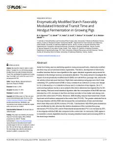
Figure 4. Phase contrast microscopy (10×) micrographs of two-dimensional LoVo cell monolayers (A−D) and SEM micrographs of LoVo MCTs (E−G). A and E, no gluten (control group); B and F, uG; C, 750 µg/mL AcG heat-treated at 90 °C for 0.75 h; and D and G, 750 µg/mL AcG heat-treated at 90 °C for 3 h.
inhibited cell growth in a dose-dependent fashion: At concentrations of 125, 250, 500, and 750 µg/mL, the inhibition was 7, 24, 38, and 53%, respectively (Figure 3), in comparison to the untreated cells. Heat-treated AcGs at the dose of 125, 250, and 500 µg/mL do not inhibit cell growth; only at the high concentration of 750 µg/mL was the inhibition 25% for AcG heated at 90 °C for 0.75 h and 21% for AcG heated at 90 °C for 3 h, in comparison to untreated cells. In Figure 4B), at phase contrast microscopy, we observed at the highest concentration of gluten (750 µg/mL) severe cell destruction with morphological abnormalities, characterized by changes in cell shape and size, and a large number of floating cells. Phase contrast microscopy revealed that cells exposed to heat-treated AcG (750 µg/mL) closely resembled the untreated controls (Figure 4, cf. C and D with A), with only a partial growth inhibition but the absence of cell destruction and morphological abnormalities. MCTSs, after 7 days of culture, were treated at the highest dose (750 µg/mL) of uG and heat-treated AcG (90° for 3 h). SEM revealed that MCTSs treated for 5 days with heat-treated AcG (Figure 4G) closely resemble the untreated spheroids (Figure 4E); they maintained a spherical/ovoidal shape with smooth boundaries and regular surfaces. MCTSs treated with
uG (Figure 4F) assumed a disklike shape, lost their threedimensional structure, and collapsed. These data confirmed the absence of important cytotoxic effects of the tested acid heatingtreated gluten proteins, as compared to the uG, both in twoand in three-dimensional cultures. Previously, it was shown that the LoVo cell line revealed to be a suitable model for studying celiac-related gliadin properties, because it was responsive to gliadins but not to other food proteins (30, 36). Therefore, it is another indication that acid-heating treatment of gluten, by removing or modifying the structure responsible for gluten cytotoxicity, decreases its celiac-triggering potential, irrespective of specific symptom typologies. TGase Reaction: Immunochemical Properties. The competitive ELISA used in this study was shown to be a reliable method to evaluate chemical modifications of gluten proteins related to celiac IgA-AGA immunospecificity. The IgA-AGA immunoreactivity to gluten proteins, as well as the gluten cytotoxicity, was decreased by the acid-heating treatment presumably by inducing a chemical deamidation of Gln residues. To further deepen the role of deamidation/modification of Gln residues on celiac IgA-AGA immunoreactivity to gluten proteins, a microbial TGase was used as a tool to modify
J. Agric. Food Chem., Vol. 55, No. 6, 2007
Celiac-Related Properties of Gluten Proteins Table 3. Celiac IgA-AGA Immunoreactivity to Gliadins Submitted to TGase Treatment sample
TGase
gliadins
− +
triptic-digested gliadins
− +
cysteamine − + − +
immunoreactivity (%) 100 185 ± 55 24 ± 5 100 130 ± 27 32 ± 8
enzymatically gluten proteins, acting directly on Gln residues according to the TGase specificity. Given the insolubility of gluten and considering that mostly gliadins contain epitopes recognized by celiac AGAs, gliadin samples were used for the enzyme treatments. TGase treatments on gliadins were performed either in the presence of cysteamine as the acylic acceptor, to favor the incorporation of thioethanoamide moieties on gliadin polypeptide by Gln residue transamidation, or in the absence of added acylic acceptors, to favor Gln residue deamidation. The low amount of peptide-bound lysine (1 mol %) in gliadins excludes a significant formation of intermolecular isopeptide bonds during the TGase treatments of gliadins. Ammonia released after TGase treatment of gliadins in the absence of added acylic acceptor was roughly the same, regardless if gliadin samples were or not predigested with trypsin (0.18 ( 0.03 vs 0.13 ( 0.02 µmol/mg). Transamidation performed by TGase treatment of gliadins in the presence of cysteamine led to thiol groups incorporation of 0.04 ( 0.01 µmol/mg. Considering the Gln residue content in gliadins (40 mol %) (13), it could be supposed that about 2% of Gln residues was deamidated by TGase treatment and less than 1% was transamidated by TGase treatment in the presence of cysteamine. The IgA-AGA immunoreactivity (Table 3) to gliadins after the TGase treatment was slightly increased, about 1.3-fold (up to 1.8-fold), with respect to that of untreated gliadins. When cysteamine was included in the TGase treatment of gliadins, the AGA immunoreactivity to the treated samples (Table 3) decreased at least three-fold (up to four-fold) with respect to that of the untreated gliadins. On the whole, the reported results suggest that the chemical deamidation lowers the IgA-AGA immunoreactivity whereas it is increased when deamidation is carried out by TGase. The different behavior could be explained by the different specificity of the chemical and TGase-mediated deamidations. The specificity of the chemical deamidation depends on the accessibility of the amide groups, whereas the specificity of the TGase action is directed mainly toward some Gln residues inserted in a particular sequence context (12, 37). TGase deamidation of gliadins seems to be involved in the CD-related humoral immunoresponse (38) leading to the generation of T cell and B cell epitopes (39, 40). Although targeted Gln residues in gliadins of the CD-involved human TGase and the microbial TGase could be different, here, we show that the microbial TGase deamidation of gliadins generates molecular structures recognized by IgA-AGA better than those of the nondeamidated gliadins. To deepen this effect, we tried to modify the specific target of TGase action by transamidation performed in the presence of cysteamine. The incorporation of cysteamine moieties, forming N-(2-mercaptoethyl) L-glutamine residues by TGase-mediated transamidation, decreased the IgA-AGA immunoreactivity. It may be due to changes in the TGase-targeted structure of gluten with respect to not only its native structure
2487
but also the TGase target structure modified by TGase-mediated deamidation (i.e., in the absence of cysteamine). LITERATURE CITED (1) Schofield, D. J.; Booth, M. R. Wheat proteins and their technological significance. DeV. Food Protein 1983, 2, 1-65. (2) Day, L.; Augustin, M. A.; Batey, I. L.; Wrigley, C. W. Wheatgluten uses and industry needs. Trends Food Sci. Technol. 2006, 17, 82-90. (3) Kinsella, J. E. Functional properties of proteins in foods: a survey. Crit. ReV. Food Sci. Nutr. 1976, 7, 219-280. (4) Matsudomi, N.; Kato, A.; Kobayashi, K. Conformation and surface properties of deamidated gluten. Agric. Biol. Chem. 1982, 246, 1583-1586. (5) Lens, J. P.; Mulder, W. J.; Kolster, P. Modification of wheat gluten for nonfood applications. Cereal Foods World 1999, 44, 5-9. (6) Finley, J. W. Deamidated gluten: a potential fortifier for fruit juice. J. Food Sci. 1975, 40, 1283-1285. (7) Wu, C. H.; Nakai, S.; Powrie, W. D. Preparation and properties of acid solubilized gluten. J. Agric. Food Chem. 1976, 24, 504510. (8) Hamada, J. S. Modification of food proteins by enzymatic methods. In Biochemistry of Food Proteins; Hudson, B. J. F., Ed.; Elsevier Science Publisher: London, 1992; pp 247-270. (9) Hamada, J. S. Deamidation of food proteins to improve functionality. Crit. ReV. Food Sci. Nutr. 1994, 34, 283-292. (10) Howell, N.; Bristow, E.; Copeland, E.; Friedli, G. L. Interaction of deamidated soluble wheat protein with sodium alginate. Food Hydrocolloids 1998, 12, 317-324. (11) Larre´, C.; Denery-Papini, S.; Popineau, Y.; Deshayes, G.; Desserme, C.; Lefebvre, J. Biochemical analysis and rheological properties of gluten modified by transglutaminase. Cereal Chem. 2000, 77, 32-38. (12) Piersma, S. R.; Pijpekamp, V. A.; Wijngaards, G.; Gruppen, H.; Boumans, H. Quantitation and localisation of (in vitro) transglutaminase-catalysed glutamine hydrolation using mass spectrometry. Enzyme Microb. Technol. 2002, 30, 266-272. (13) Wieser, H. Relation between gliadin structure and coeliac toxicity. Acta Paediatr. Suppl. 1996, 412, 3-9. (14) Murray, J. A. The widening spectrum of celiac disease. Am. J. Clin. Nutr. 1999, 69, 354-65. (15) Hill, I. D.; Bhatnagar, S.; Cameron, D. J.; De Rosa, S.; Ma¨ki, M.; Russell, G. J.; Troncone, R. Celiac disease: Working group report of the first world congress of pediatric gastroenterology, hepatology, and nutrition. J. Pediatr. Gastroenterol. Nutr. 2002, 35, S78-88. (16) Jones, R. B.; Robins, G. G.; Howdle, P. D. Advances in celiac disease. Curr. Opin. Gastroenterol. 2006, 22, 117-123. (17) Maiuri, L.; Ciacci, C.; Ricciardelli, I.; Vacca, L.; Raia, V.; Auricchio, S.; Picard, J.; Osman, M.; Quaratino, S.; Londei, M. Association between innate response to gliadin and activation of pathogenic T cells in coeliac disease. Lancet 2003, 362, 3037. (18) Shewry, P. R.; Tatham, A. S. The prolamin storage proteins of cereal seeds: Structure and evolution. Biochem. J. 1990, 267, 1-12. (19) Dieterich, W.; Ehnis, T.; Bauer, M.; Donner, P.; Volta, U.; Riecken, E. O.; Schuppan, D. Identification of tissue transglutaminase as the autoantigen of celiac disease. Nat. Med. 1997, 3, 797-801. (20) Schuppan, D.; Hahn, E. G. Biomedicine: Gluten and the gut lessons for immune regulation. Science 2002, 297, 2218-2220. (21) Shan, L.; Molberg, O.; Parrot, I.; Hausch, F.; Filiz, F.; Gray, G. M.; Sollid, L. M.; Khosla, C. Structural basis for gluten intolerance in celiac sprue. Science 2002, 297, 2275-2279.
2488
J. Agric. Food Chem., Vol. 55, No. 6, 2007
(22) Di Cagno, R.; De Angelis, M.; Auricchio, S.; Greco, L.; Clarke, C.; De Vincenzi, M.; Giovannini, C.; D’Archivio, M.; Landolfo, F.; Parrilli, G.; Minervini, F.; Arendt, E.; Gobbetti, M. Sourdough bread made from wheat and nontoxic flours and started with selected lactobacilli is tolerated in celiac sprue patients. Appl. EnViron. Microbiol. 2004, 70, 1088-1096. (23) Di Cagno, R.; De Angelis, M.; Alfonsi, G.; De Vincenzi, M.; Silano, M.; Vincentini, O.; Gobbetti, M. Pasta made from durum wheat semolina fermented with selected lactobacilli as a tool for a potential decrease of the gluten intolerance. J. Agric. Food Chem. 2005, 53, 4393-4402. (24) Berti, C.; Dolfini, E.; Forlani, F. Effects on celiac activity of gluten proteins modified by chemical deamidation. Pol. J. Food Nutr. Sci. 2002, 11, 135-137. (25) American Association of Cereal Chemists (AACC). Method 3810. In ApproVed Methods of the AACC, 8th ed.; American Association of Cereal Chemists: St. Paul, Minnesota, 1983. (26) Capelli, L.; Forlani, F.; Perini, F.; Guerrieri, N.; Cerletti, P.; Righetti, P. G. Wheat cultivar discrimination by capillary electrophoresis of gliadins in isoelectric buffers. Electrophoresis 1998, 19, 311-318. (27) Mamone, G.; Ferranti, P.; Melck, D.; Tafuro, F.; Longobardo, L.; Chianese, L.; Addeo, F. Susceptibility to transglutaminase of gliadin peptides predicted by a mass spectrometry-based assay. FEBS Lett. 2004, 562, 177-182. (28) Ellman, G. L. Tissue sulfhydryl groups. Arch. Biochem. Biophys. 1959, 82, 70-77. (29) Laemmli, U. K. Cleavage of structural proteins during the assembly of the head of bacteriophage T4. Nature 1970, 227, 680-685. (30) Dolfini, E.; Elli, L.; Dasdia, T.; Bufardeci, B.; Colleoni, M. P.; Costa, B.; Floriani, I.; Falini, M. L.; Guerrieri, N.; Forlani, F.; Bardella, M. T. In vitro cytotoxic effect of bread wheat gliadin on the LoVo human adenocarcinoma cell line. Toxicol. In Vitro 2002, 16, 331-337. (31) Berti, C.; Trovato, C.; Bardella, M. T.; Forlani, F. IgA antigliadin antibody immunoreactivity to food proteins. Food Agric. Immunol. 2003, 15, 217-223. (32) Walker-Smith, J. A.; Guandalini, S.; Schmitz, J.; Shmerling, D. H.; Visakorpi, J. K. Revised criteria for diagnosis of celiac disease. Arch. Dis. Child. 1990, 65, 909-911.
Berti et al. (33) Battaglia, M.; Pozzi, D.; Grimaldi, S.; Parasassi, T. Hoechst 33258 staining for detecting mycoplasma contamination in cell cultures: A method for reducing fluorescence photobleaching. Biotech. Histochem. 1994, 69, 152-156. (34) Dolfini, E.; Roncoroni, L.; Elli, L.; Fumagalli, C.; Colombo, R.; Ramponi, S.; Forlani, F.; Bardella, M. T. Cytoskeleton reorganization and ultrastructural damage induced by gliadin in a threedimensional in vitro model. World J. Gastroenterol. 2005, 11, 7597-7601. (35) Takeda, K.; Matsumura, Y.; Shimizu, M. Emulsifying and surface properties of wheat gluten under acidic conditions. J. Food Sci. 2001, 66, 393-399. (36) Dolfini, E.; Elli, L.; Ferrero, S.; Braidotti, P.; Roncoroni, L.; Dasdia, T.; Falini, M. L.; Forlani, F.; Bardella, M. T. Bread wheat gliadin cytotoxicity: A new three-dimensional cell model. Scand. J. Clin. Lab. InVest. 2003, 63, 135-141. (37) Piper, J. L.; Gray, G. M.; Khosla, C. High selectivity of human tissue transglutaminase for immunoactive gliadin peptides: implications for celiac sprue. Biochemistry 2002, 41, 386-393. (38) Caputo, I.; Amato, A. D.; Troncone, R.; Auricchio, S.; Esposito, C. Transglutaminase 2 in celiac disease: Minireview article. Amino Acids 2004, 26, 381-386. (39) Osman, A. A.; Gu¨nnel, T.; Dietl, A.; Uhlig, H. H.; Amin, M.; Fleckenstein, B.; Richter, T.; Mothes, T. B cell epitopes of gliadin. Clin. Exp. Immunol. 2000, 121, 248-254. (40) Andersson, R. P.; Degano, P.; Godkin, A. J.; Jewel, D. P.; Hill, A. V. S. In ViVo antigen challenge in celiac disease identifies a single transglutaminase-modified peptide as the dominant Agliadin T-cell epitope. Nat. Med. 2000, 6, 337-342. Received for review September 13, 2006. Revised manuscript received December 15, 2006. Accepted January 10, 2007. The study was supported by “Associazione Italiana CeliachiasRegione Lombardia ONLUS” (Milano, Italy) and by “Compagnia di San Paolo” (Torino, Italy).
JF062623N