min each and fixed in 10% (vol/vol) formalin overnight at 4 C before ... alized by fluorescence microscopy on a Carl Zeiss axioskop (Jena, Ger- many), and the ..... Liu J-P, Backer J, Perkins AS, Robertson EJ, Efstratiadis A 1993 Mice carrying.
0013-7227/02/$15.00/0 Printed in U.S.A.
Endocrinology 143(4):1530 –1537 Copyright © 2002 by The Endocrine Society
Increased Islet Cell Proliferation, Decreased Apoptosis, and Greater Vascularization Leading to -Cell Hyperplasia in Mutant Mice Lacking Insulin ´ ,* C. CURRIE, T. CHRONES, D. BUCCHINI, J. JAMI, R. L. JOSHI, B. DUVILLIE
AND
D. J. HILL
Medical Research Council Group in Fetal and Neonatal Health and Development (B.D., C.C., T.C., D.J.H.), Lawson Health Research Institute, London, Ontario, Canada N6A4V2; Departments of Physiology, Medicine, and Paediatrics (D.J.H.), University of Western Ontario, London, Ontario, Canada N6A 5A5; and U257 Institut National de la Sante´ et de la Recherche Me´dicale (D.B., J.J., R.L.J.), 75014 Paris, France The targeted disruption of the two nonallelic insulin genes in mouse was reported previously to result in intrauterine growth retardation, severe diabetes immediately after suckling, and death within 48 h of birth. We have further used these animals to investigate the morphology and cell biology of the endocrine pancreas in late gestation and at birth when insulin is absent throughout development. Pancreatic -cells were identified by detecting the activity of the LacZ gene inserted at the Ins2 locus. A significant increase in the mean area of the islets was found at embryonic d 18.5 (E18.5) and in the newborn in Ins1ⴚ/ⴚ, Ins2ⴚ/ⴚ animals compared with Ins1ⴚ/ⴚ, Ins2ⴙ/ⴚ and wild-type controls, whereas the blood glucose levels were unaltered. The individual size of the -cells in the insulin-deficient fetuses was similar to controls, suggesting that the relative increase in islet size was due to an increase in cell number. Immunohistochemistry for proliferating cell nuclear antigen within the pancreatic ductal epithelium showed no differences in labeling index between insulin-deficient and control mice, and no change in the number of -cells associated with ducts, but the relative size distribution of the islets was altered so that fewer islets under 5,000 m2 and more islets greater than 10,000 m2 were present in Ins1ⴚ/ⴚ, Ins2ⴚ/ⴚ animals. This suggests that the greater mean islet size seen in insulin-deficient animals represented an en-
I
NSULIN IS A major anabolic hormone for the growth and development of the mammalian embryo and fetus, and its production by the pancreatic -cells within the islets of Langerhans must be closely entrained to the demands of the growing organism. Insulin availability can be controlled not only by varying the release from each -cell, but by plasticity in the number and size of developing islets. The -cell mass is thought to be controlled by a balance between islet cell neogenesis, proliferation, and remodeling by apoptosis. The formation of new -cells can take place by replication of already differentiated -cells or neogenesis from putative islet stem cells within the pancreatic ductal epithelium, both being used in early life (1). Postnatally, the rate of -cell replication decreases and is generally very low in the adult (⬍3%) (2). We have shown that the optimal development of -cell mass depends on the actions of locally expressed Abbreviations: DAB, Diaminobenzidine; E18.5, embryonic d 18.5; IR, insulin receptor; IRS, insulin receptor substrate; PCNA, proliferating cell nuclear antigen; VEGF, vascular endothelial growth factor.
largement of formed islets and was not associated with an increase in islet neogenesis. The proportional contribution of ␣- and -cells to the islets was not altered. This was supported by an increase in the number of cells containing immunoreactive proliferating cell nuclear antigen in both islet ␣- and -cells at E18.5 in insulin-deficient mice, and a significantly lower incidence of apoptotic cells, as determined by molecular histochemistry using the terminal deoxynucleotidyl transferase-mediated deoxy-UTP nick end labeling reaction. The density of blood vessels within sections of whole pancreas, or within islets, was determined by immunohistochemistry for the endothelial cell marker CD31 and was found to be increased 2-fold in insulin-deficient mice compared with controls at E18.5. However, no changes were found in the steady-state expression of mRNAs encoding vascular endothelial growth factor, its receptor Flk-1, IGF-I or -II, the IGF-I and insulin receptors, or insulin receptor substrates-1 or -2 in pancreata from Ins1ⴚ/ⴚ, Ins2ⴚ/ⴚ mice compared with Ins1ⴚ/ⴚ, Ins2ⴙ/ⴚ controls. Thus, we conclude that the relative hyperplasia of the islets in late gestation in the insulin-deficient mice was due to an increased islet cell proliferation coupled with a reduced apoptosis, which may be related to an increased vascularization of the pancreas. (Endocrinology 143: 1530 –1537, 2002)
growth factors, such as IGF-II, and an appropriate maternal diet (3, 4). To date, little is known about the autocrine or paracrine effects of insulin itself on the development of the -cells and on islet form and mass. Because insulin is able to induce the phosphorylation of the insulin receptor and its primary substrates, insulin receptor substrate-1 (IRS-1) and IRS-2 in rat pancreatic islets, this suggests that a functional insulinsignaling pathway is present in islets and that insulin could act as an autocrine or paracrine factor (5). Leibiger et al. (6) also reported that exocytosis of insulin from the -cell can promote insulin gene transcription via the insulin receptor and PI3K, and other kinases suggesting an autoregulation of insulin release. There are two nonallelic insulin genes in the mouse, Ins1 and Ins2, that encode two very similar proteins (7). As their expression in the mouse embryo was detected as soon as E9.5 in the primary pancreatic bud, it has been suggested that insulin is an early marker of pancreatic development (8). Maternal insulin is considered not to cross the placental barrier to the fetus in biologically meaningful
1530
Duvillie´ et al. • Pancreatic Islet Hyperplasia in Insulin-Deficient Mice
amounts (9). We showed previously that disruption of the two insulin genes in the mouse by homologous recombination resulted in no embryonic lethality, but severe intrauterine growth retardation (10). These animals developed a severe diabetes immediately after suckling and they died within 48 h of birth with ketoacidosis (10). A pancreas was present with exocrine and islets, the latter containing all endocrine cell types including -cells, which were detected by using a -galactosidase marker derived from a LacZ gene inserted at the Ins2 locus. Inactivation of either the Ins1 or Ins2 locus individually resulted in a compensatory increase in -cell mass compared with wild-type animals at 2– 4 months of age (11). We have now used this model further to identify the effects of a complete lack of endogenous insulin on the morphometry and cell biology of the developing endocrine pancreas. Materials and Methods Animals The generation of the insulin-deficient mutant mice was described in Duvillie´ et al. (10). Briefly, Ins1⫺/⫺, Ins2⫹/⫺ mice generated onto a C57Bl/6 background were intercrossed and the resulting offspring were killed at E17.5, E18.5, and at birth, and Ins1⫺/⫺, Ins2⫺/⫺ animals identified by Southern blot analysis of tail-derived DNA. Five wild-type C57Bl/6, Ins1⫺/⫺, Ins2⫹/⫺, or Ins1⫺/⫺, Ins2⫺/⫺ animals were used within each analysis and for each age studied. The Ins1⫺/⫺, Ins2⫹/⫺ fetuses from the same litters as the Ins1⫺/⫺, Ins2⫺/⫺ mice were used as controls to avoid genetic background effects. Mice were maintained in strict compliance with the Animal Care Committee of the University of Western Ontario and in accordance with guidelines of the Canadian Council for Animal Care. Fetuses were obtained following caesarian section and newborn animals were killed by decapitation immediately after birth. Animals were weighed before organ dissection and individual wet organ weights were recorded for the pancreas, heart, lung, thymus, brain, liver, and tibia. The pancreata were either fixed for immunocytochemistry, or snap-frozen in liquid nitrogen for later RNA preparation. For blood glucose measurements, 50 l of blood was collected after decapitation of each fetus or newborn and the glycemia quantified using a glucometer (Roche Molecular Biochemicals, Mannheim, Germany).
X-gal staining Pancreata from Ins1⫺/⫺, Ins2⫺/⫺ fetuses at E18.5 and Ins1⫺/⫺, Ins2⫹/⫺ controls were rinsed in 0.1 m phosphate buffer, pH 7.3 [23 mm sodium phosphate monobasic, 77 mm sodium phosphate dibasic (Sigma, St. Louis, MO)] at room temperature for 10 min, and fixed for 15 min in 0.1 m phosphate buffer (pH 7.3) containing 0.2% (vol/vol) glutaraldehyde, 5 mm EGTA, and 2 mm MgCl2. They were subsequently washed three times for 5 min each in a wash buffer [2 mm MgCl2, 0.01% (wt/vol) deoxycholate, 0.01% (vol/vol) Nonidet-P40, in 0.1 m sodium phosphate, pH 7.3] and incubated for 24 h in wash buffer containing 5 mm K3Fe(CN)6 and 1 mg/ml 5-Br-4-Cl-3-indolyl-galactoside (X-gal), before photomicroscopy.
RT-PCR analysis Total RNA was extracted from the dissected pancreata from Ins1⫺/⫺, Ins2⫺/⫺ fetuses at E18.5 and Ins1⫺/⫺, Ins2⫹/⫺ controls as described (12). The preparation was treated with RNase-free DNase and checked for the absence of DNA contamination. RT-PCR was performed on 1 g of RNA/50 l reaction volume using an Access PCR kit (Promega Corp., distributed by Fisher Scientific, Nepean, Ontario, Canada) as recommended by the suppliers. Thirty cycles of DNA amplification were used for each reaction, which produced radioactive hybridization signal intensities within the linear ranges. Experimentation with 20 cycles did not yield a hybridization signal, while 40 cycles gave a stronger, but maximal signal intensity. Signal intensities were quantified
Endocrinology, April 2002, 143(4):1530 –1537 1531
using a laser densitometer. The reaction was also carried out in the absence of reverse transcriptase to ensure that the amplified material had derived from RNA. Negative controls were done for each reaction by performing an RT-PCR on 9 l of sterile water. An amplification for -actin with the primers 5⬘ CGTGGGCCGCCCTAGGCACCA 3⬘/5⬘ TTGGCCTTAGGGTTCAGGGGGG 3⬘ to check that all reactions were done with the same quantity of RNA.
Transcripts were detected with the primer pairs 5⬘ GGACCAGAGACCCTTT GCGGGG 3⬘5⬘ GGCTGCTTTTGTAGGCTTCAGTGG 3⬘ for IGF I, 5⬘ CGCCCCAGCGAG ACTCTGTGC 3⬘/5⬘ GCCCACGGGGTATCTGGGGAA 3⬘ for IGF II, 5⬘ GAGACGGCTT CTCTGCAGTA 3⬘/5⬘ GGCAGAGAGGGAAGGCAGAG 3⬘ for IGF I receptor, 5⬘ GCGAA GATCCCTTGAAGAGGTGGG 3⬘/5⬘ GCCCCGCTCCAGGGCAAAATGCTTCCG 3⬘ for the insulin receptor (IR), 5⬘ CAGCAGCAGCAACAGCAGCAGCA 3⬘/5⬘ TTGACGAGG ACAACCTATCTGCAT 3⬘ for IRS-1, and 5⬘ ATACACTCTCATGAGGGCCA 3⬘/5⬘ TCCGTTTACTGGGAAGGTCC 3⬘ for IRS-2. The RT-PCR products were assessed by electrophoresis on a 2% agarose gel. For vascular endothelial growth factor (VEGF), the primer pair 5⬘ CCTTGGC TTGTCACATCTGCAAG 3⬘/5⬘ CAGATCATGCGGATCAAACCTCACCAA 3⬘ was used and for Flk 1: 5⬘ CACCAAAGAGAGGAACG 3⬘/5⬘ ACAGGCAGAAACCAGTAG 3⬘. Primers for VEGF and Flk-1 were kindly provided by Dr. Guo Fong, Lawson Health Research Institute (London, Ontario, Canada).
Immunocytochemistry Following excision, the pancreata were washed twice in PBS for 10 min each and fixed in 10% (vol/vol) formalin overnight at 4 C before embedding in paraffin. Histological sections of pancreas (5 m) were cut from paraffin blocks with a rotary microtome and mounted on glass microscope slides (Superfrost plus, Fisher Scientific). Sections of whole pancreata were stained using the following specific primary antibodies: guinea pig anti-insulin (1:15) (provided by Dr. T. MacDonald, Department of Medicine, University of Western Ontario, Ontario, Canada), rabbit antiporcine glucagon (1:50, C-terminal 04A antiserum, kindly provided by Dr. R. Ungar, Dallas, TX), mouse antiendothelial cell CD31 (1:50, DAKO Corp., Santa Barbara, CA), mouse antiproliferating cell nuclear antigen (PCNA) (1:750, Sigma), mouse anticyclin D1 (1:750, Zymed Laboratories, Inc., South San Francisco, CA), rabbit anti-Nek2 (1:750, Zymed Laboratories, Inc.), and rabbit antipancreatic and duodenal homeobox factor 1 (Pdx-1) (a gift from Dr. C. V. Wright, Vanderbilt Medical Center, Nashville, TN). The secondary antibodies were either biotinylated antimouse (1:100), antiguinea pig (1:500), or antirabbit IgGs (1:30) (Vector Laboratories, Ltd., Burlington, Ontario, Canada). Visualization of the ligands was achieved using immunoperoxidase staining. All antisera were diluted in 0.01 m PBS (pH 7.5) containing 1% (wt/vol) BSA and 0.02% (wt/vol) sodium azide (100 l per slide). Peptide immunoreactivity was visualized by incubation with fresh 1.89 mm diaminobenzidine (DAB) tetrahydrochloride (Fast DAB tablets, Sigma) for 2 min. Tissue sections were counterstained with Carrazi’s hematoxylin, dehydrated in ascending series of alcohols (50%, 70%, 90%, 100%, vol/ vol), cleared in xylene and mounted under glass coverslip with Permount (Fisher Scientific). The presence of Pdx-1 was detected by immunofluorescence. Each method has been described by us in detail previously (3, 4). Dual staining for PCNA and insulin, was performed by first undertaking immunohistochemistry for PCNA as described above using DAB as the chromogen. Before counterstaining and dehydration, the sections were then subjected to immunohistochemistry for insulin as described above, using alkaline phosphatase (blue) (alkaline phosphatase substrate kit III, Vector) as the chromogen. After incubation with antisera against insulin, antiguinea pig or antiporcine alkaline phosphatase conjugate (Sigma) was applied to the sections for 1 h, followed by incubation with alkaline phosphatase substrate for 20 min before washing and counter-staining with Mayer’s hemalum. Sections were mounted under glass coverslips with an aqueous mounting solution (Aquamount, Polysciences, Warrington, PA). To demonstrate that pancreatic ductal tissue and acinar tissue could be appropriately identified for morphometric analysis, immunohistochemistry was performed with mouse antihuman cytokeratin 20 (1:50)
1532
Endocrinology, April 2002, 143(4):1530 –1537
(DAKO Corp.), or rabbit antihuman a amylase (1:2000) (Sigma), respectively. For the visualization of cytokeratin, tissues were first incubated with Bacto-Trypsin (0.015% wt/vol in Trizma buffer, pH 7.6) (Difco Laboratories, Detroit, MI) for 45 min at 37 C.
Duvillie´ et al. • Pancreatic Islet Hyperplasia in Insulin-Deficient Mice
TABLE 1. Mean body weight (⫾SEM) (g) for Ins1⫺/⫺, Ins2⫺/⫺ fetal mice at E18.5 compared with Ins1⫺/⫺, Ins2⫹/⫺ control fetuses, mean weight of the organs (mg) and the percent contribution of organ weights to total body weight
Apoptosis Body weight Pancreas
Apoptotic nuclei were detected with a Promega Corp. kit (distributed by Fisher Scientific) under the conditions provided by the supplier. Briefly, the apoptotic detection system measured the fragmented DNA of apoptotic cells by catalytically incorporating fluorescein-12-UTP at the 3⬘ DNA ends using the enzyme terminal deoxynucleotidyl transferase, which forms a polymeric tail using the principle of the terminal deoxynucleotidyl transferase-mediated deoxy-UTP nick end labeling assay. The fluorescein-12-deoxy-UTP-labeled DNA could then be visualized by fluorescence microscopy on a Carl Zeiss axioskop (Jena, Germany), and the apoptotic nuclei were counted using automated imaging software as described below.
Liver
Morphometric and statistical analysis
Tibia
Morphometric analysis was performed using a Carl Zeiss transmitted-light microscope at a magnification of ⫻250 or ⫻400. Analyses were performed using the Northern Eclipse version 2.0 morphometric analysis software (Empix Imaging Co., Mississauga, Ontario, Canada). The area of the pancreatic islets, the size of the -cells, and the number of insulin-, glucagon-, PCNA-, cyclin G1-, Nek2-, Cd31-, Pdx-1-positive cells and apoptotic nuclei were each calculated for five tissue sections from each pancreas representing predominantly the head regions for each age. Individual cell area and total areas of immunoreactive cells within islets were circled for image analysis and selected by gray-level threshold. Pancreatic -cell mass was calculated following immunohistochemistry for insulin or the visualization of X-gal on sections obtained throughout pancreata of known weight. -Cell mass was estimated by calculating the mean percentage area of tissue containing cells immunoreactive for insulin per sectional area of pancreas (five sections per organ). This was then expressed as milligrams -cell mass based on the total pancreatic wet weight for individual animals. Differences between mean values for variables within individual experiments were compared statistically by two-way ANOVA, followed by a Scheffe´ ’s test. For RT-PCR analysis, three separate RNA preparations, each from a separate animal, were used for each target gene product.
n ⫽ 5 animals,
Results Growth retardation and metabolism of the insulin-deficient fetuses
As the insulin-deficient mice developed a severe diabetes immediately after birth, analyses were performed during late fetal development and at birth. The Ins1⫺/⫺, Ins2⫹/⫺ adult mice were intercrossed and Ins1⫺/⫺, Ins2⫺/⫺ fetuses were obtained at E17.5 and E18.5 at the ratio of 1:4, as expected for the segregation of two independent genes. The mean level of blood glucose was 3.13 ⫾ 0.58 mm (n ⫽ 7) in the Ins1⫺/⫺, Ins2⫺/⫺ fetuses at E18.5, vs. 3.20 ⫾ 0.67 mm (n ⫽ 19) in the Ins1⫺/⫺ Ins2⫹/⫺ controls, demonstrating that fetal glycemia was not altered by the absence of insulin. The Ins1⫺/⫺, Ins2⫺/⫺ fetuses were relatively growth retarded at E18.5 with a body weight 0.17 g (15%) less than that of heterozygous pups, but the relative proportions of the organ sizes remained unaltered (Table 1). At birth, the relative reduction in body weight was 22%. Morphometry of the pancreatic islets
The presence of -cells was detected by using a -galactosidase enzymatic reaction, which was possible because of the LacZ gene inserted at the Ins2 locus in the animals with
Lung Heart Thymus Brain
a
Ins1⫺/⫺, Ins2⫹/⫺
Ins1⫺/⫺, Ins2⫺/⫺
1.29 ⫾ 0.02 5.29 ⫾ 0.52 0.41 ⫾ 0.10% 9.8 ⫾ 3.61 0.76 ⫾ 0.28% 6.19 ⫾ 3.35 0.48 ⫾ 0.26% 4.77 ⫾ 1.54 0.37 ⫾ 0.12% 83.85 ⫾ 6.32 6.50 ⫾ 0.49% 46.44 ⫾ 5.03 3.60 ⫾ 0.39% 9.54 ⫾ 4.12 0.74 ⫾ 0.32%
1.12 ⫾ 0.05a 5.15 ⫾ 2.46 0.46 ⫾ 0.22% 10.75 ⫾ 3.47 0.96 ⫾ 0.31% 7.05 ⫾ 2.01 0.63 ⫾ 0.18% 5.71 ⫾ 2.80 0.51 ⫾ 0.25% 76.72 ⫾ 10.19 6.85 ⫾ 0.91% 39.12 ⫾ 5.04 3.40 ⫾ 0.45% 7.84 ⫾ 3.13 0.70 ⫾ 0.28%
P ⬍ 0.01 vs. Ins1⫺/⫺, Ins2⫹/⫺, F ⫽ 12.2.
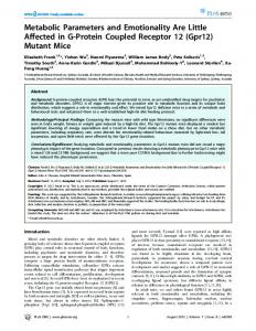
disrupted insulin genes (Fig. 1). In wild-type and Ins1⫺/⫺, Ins2⫹/⫺ animals, islets were visualized after immunostaining with an antiinsulin antibody (Fig. 1A). No significant differences were observed between the mean area (⫾sem) of the islets in wild-type C57Bl/6 islets and Ins1⫺/⫺, Ins2⫹/⫺ animals at E17.5, E18.5 or at birth (E18.5, wild-type, 4974 ⫾ 435 m2 vs. Ins1⫺/⫺, Ins2⫹/⫺, 6090 ⫾ 668 m2), suggesting that only one allele of the insulin gene is sufficient for a normal islet morphological development. The mean individual -cell volume was found to be 720 m3 and did not alter with genotype. A significant increase of the mean islet area was observed in the Ins1⫺/⫺, Ins2⫺/⫺ fetuses compared with the Ins1⫺/⫺, Ins2⫹/⫺ controls at E18.5 and at postnatal d 1 but not at E17.5 (Figs. 1B and 2). The total estimated -cell mass also increased progressively with the inactivation of insulin genes (E18.5, WT 182 ⫾ 15 g; Ins1⫺/⫺, Ins2⫹/⫺ 226 ⫾ 14 g; Ins1⫺/⫺, Ins2⫺/⫺ 348 ⫾ 18 g, P ⬍ 0.01 vs. WT). However, the mean area (⫾sem) of the individual X-gal-positive cells was similar between the Ins1⫺/⫺, Ins2⫺/⫺ and the Ins1⫺/⫺, Ins2⫹/⫺ fetuses (30.2 ⫾ 2.8 m2 vs. 31.2 ⫾ 2.9 m2, respectively). The relative increase in mean islet area in insulin-deficient animals was therefore likely to be due to an increase in the number of the -cells and not to an increase in individual -cell size. The presence of ␣-cells was determined by immunocytochemistry for glucagon (Fig. 1, C and D). The total estimated ␣-cell mass was also increased in insulin-deficient animals (E18.5, WT 38 ⫾ 8 g; Ins1⫺/⫺, Ins2⫹/⫺ 45 ⫾ 6 g; Ins1⫺/⫺, Ins2⫺/⫺ 68 ⫾ 11 g, P ⬍ 0.01 vs. WT). We also determined whether the number of islets differed between WT, Ins1⫺/⫺, Ins2⫺/⫺ and the Ins1⫺/⫺, Ins2⫹/⫺ fetuses. Islet number, defined by clusters of endocrine cells with an area greater than 300 m2, did not differ. However, the relative size distribution of arbitrarily defined small, medium, or large islets significantly differed between the Ins1⫺/⫺, Ins2⫺/⫺ and wild-type control mice, with the insulin-deficient animals showing a greater percentage of large islets at the expense of smaller islets (Table 2). To determine whether the neogenesis of islets from the pancreatic ductal epithelium was altered, -cells, which individu-
Duvillie´ et al. • Pancreatic Islet Hyperplasia in Insulin-Deficient Mice
Endocrinology, April 2002, 143(4):1530 –1537 1533
FIG. 1. Detection of insulin (A) by immunohistochemistry in pancreatic sections from C57Bl/6 wild-type fetuses, X-gal activity (B) in the pancreatic -cells of Ins1⫺/⫺, Ins2⫺/⫺ fetuses, and glucagon detected by immunohistochemistry in Ins1⫺/⫺, Ins2⫹/⫺ islets (C) and Ins1⫺/⫺, Ins2⫺/⫺ (D). Fetuses were collected at E18.5. Magnification bar, 10 m.
demonstrated that the rate of DNA synthesis within the pancreatic ductal cells was not significantly different in the Ins1⫺/⫺, Ins2⫺/⫺ animals (5.5%) compared with Ins1⫺/⫺, Ins2⫹/⫺ controls, or WT controls. Thus, the relative increase in mean islet size seen in the insulin-deficient animals does not appear to derive from an increased formation of new islets within the pancreatic ducts, but from a more rapid growth rate of the islets once they are formed. Islet cell proliferation and apoptosis
FIG. 2. Analysis of the size of the pancreatic islets of Ins1⫺/⫺, Ins2⫹/⫺ and Ins1⫺/⫺, Ins2⫺/⫺ fetuses at E17.5, E18.5 and postnatal d 1. Mean values SEM are shown (n ⫽ 5), P ⬍ 0.05 vs. Ins1⫺/⫺, Ins2⫹/⫺ animals. TABLE 2. Percent distribution (mean ⫾ SEM) of arbitrarily defined small (⬍5,000 m2), medium (5,000 –10,000 m2), or large (⬎10,000 m2) pancreatic islets in WT C57/B16, Ins1⫺/⫺, Ins2⫺/⫺ or Ins1⫺/⫺, Ins2⫹/⫺ fetuses at E18.5 Islet area (number studied)
WT C57/B16 (85) Ins1⫺/⫺, Ins2⫹/⫺ (111) Ins1⫺/⫺, Ins2⫺/⫺ (40) n ⫽ 5 animals,
a
⬍5,000 m2 5,000 –10,000 m2 ⬎10,000 m2
63 ⫾ 8 49 ⫾ 10 12 ⫾ 13a
30 ⫾ 9 33 ⫾ 8 55 ⫾ 17
P ⬍ 0.05 vs. WT, F ⫽ 4.3;
b
7⫾3 18 ⫾ 4 33 ⫾ 11b
P ⫽ 0.01, F ⫽ 4.9.
ally, or as clusters of up to three cells, contained immunoreactive insulin or X-gal activity, were counted and expressed as a percentage of ductal cells immunopositive for cytokeratin. In pancreatic ducts from WT animals, 5.9 ⫾ 0.8% cells were identified as -cells, whereas values were 4.4 ⫾ 1.5% for Ins1⫺/⫺, Ins2⫹/⫺ animals and 2.6 ⫾ 0.7% for Ins1⫺/⫺, Ins2⫺/⫺ mice. Immunohistochemistry for PCNA
Islet cell DNA synthesis was determined by immunohistochemistry for PCNA (Fig. 3A), and showed a significant increase in labeling index at E18.5 (P ⬍ 0.05) in insulindeficient fetuses, which was not present at E17.5 or postnatal d 1 (Fig. 4). To determine which islet cell populations contribute to a changed labeling index, codetection of insulin or X-gal and PCNA was performed on the same sections to identify -cells, and for glucagon and PCNA on sections from the same islet to identify ␣-cells (Fig. 3, B and C). Both - and ␣-cells contributed to an increased DNA synthesis in islets from Ins1⫺/⫺, Ins2⫺/⫺ mice at E18.5 (Table 3). The proportional area occupied by either ␣- or -cells was calculated, and was found to remain unchanged in Ins1⫺/⫺, Ins2⫺/⫺ mice with relative islet cell hyperplasia (Table 4). Because PCNA is not entirely specific to S phase of the cell cycle but is also expressed during G1, we further characterized cell cycle kinetics using immunhistochemistry for cyclin D1 (Fig. 3D), a G1 marker, and Nek2, a marker of G2. In pancreata from Ins1⫺/⫺, Ins2⫺/⫺ animals at E18.5, 2.7 ⫾ 0.6% (mean ⫾ sem) of the islet cells were in G1 phase vs. 2.0 ⫾ 0.3% in the Ins1⫺/⫺, Ins2⫹/⫺ controls, and 7.5 ⫾ 1.0% in G2 phase vs. 5.7 ⫾ 0.9% (n ⫽ 5) in Ins1⫺/⫺, Ins2⫹/⫺ animals. Thus, the percentage of islet cells found to be involved in cell cycle replication was increased in insulin-deficient animals, and this was likely to involve multiple endocrine cell types.
1534
Endocrinology, April 2002, 143(4):1530 –1537
Duvillie´ et al. • Pancreatic Islet Hyperplasia in Insulin-Deficient Mice
TABLE 3. Percentage (%) of cells containing immunoreactive insulin (or X-gal), or glucagon, which costained for PCNA in pancreatic islets from either Ins1⫺/⫺, Ins2⫺/⫺ or Ins1⫺/⫺, Ins2⫹/⫺ mice between E18.5 and birth E17.5
Ins1⫺/⫺, Ins2⫹/⫺ -cells ␣-cells Ins1⫺/⫺, Ins2⫺/⫺ -cells ␣-cells
E18.5
Newborn
4.8 ⫾ 0.4 3.4 ⫾ 0.3
8.7 ⫾ 0.7a 5.3 ⫾ 0.2b
7.3 ⫾ 0.5 5.1 ⫾ 0.4
4.9 ⫾ 0.5 3.2 ⫾ 0.4
11.4 ⫾ 1.2c 8.5 ⫾ 0.8d
6.8 ⫾ 1.0 5.4 ⫾ 0.6
Mean ⫾ SEM, n ⫽ 5, a P ⬍ 0.05 vs. E17.5, F ⫽ 4.1; b P ⬍ 0.001, F ⫽ 15.0; c P ⬍ 0.05, F ⫽ 3.9; d P ⬍ 0.001, F ⫽ 11.4. TABLE 4. Percentage area (%) of islets containing immunoreactive insulin (or X-gal), or glucagon in pancreatic islets from either Ins1⫺/⫺, Ins2⫺/⫺ or Ins1⫺/⫺, Ins2⫹/⫺ mice between E18.5 and birth (mean ⫾ SEM, n ⫽ 5)
Ins1⫺/⫺, Ins2⫹/⫺ -cells ␣-cells Ins1⫺/⫺, Ins2⫺/⫺ -cells ␣-cells FIG. 3. Detection by immunohistochemistry of PCNA (A) or glucagon (C) in the same islet, the colocalization of PCNA (brown) and X-gal (B) and cyclin D1 (D) in pancreatic islets of Ins1⫺/⫺, Ins2⫺/⫺ fetuses at E18.5. Arrows indicate examples of cells immunopositive for PCNA (A), glucagon (C), cyclin D1 (D), and the colocalization of X-gal and PCNA (B). Magnification bar, 10 m.
FIG. 4. Analysis of the percent of PCNA-positive cells by immunohistochemistry within the islets of Ins1⫺/⫺, Ins2⫹/⫺ and Ins1⫺/⫺, Ins2⫺/⫺ fetuses at E17.5, E18.5 and postnatal d 1. Mean values ⫾ SEM are shown (n ⫽ 5), P ⬍ 0.05 vs. Ins1⫺/⫺, Ins2⫹/⫺ animals.
The presence of islet cell apoptosis was measured by cell counting after terminal deoxynucleotidyl transferase-mediated deoxy-UTP nick end labeling staining (Fig. 5). The level of apoptosis was significantly decreased in the Ins1⫺/⫺, Ins2⫺/⫺ mice at E18.5 and at birth (Fig. 6), and this was seen in all regions of the islets. The mean percent apoptotic islet cells in WT animals at E18.5 (4.6 ⫾ 0.6%, n ⫽ 5) was similar to that in Ins1⫺/⫺, Ins2⫹/⫺ animals. Because the apoptotic cells were compacted with little cytoplasm, it was not possible to demonstrate a colocalization of either insulin, glu-
E17.5
E18.5
Newborn
72 ⫾ 5 21 ⫾ 3
75 ⫾ 6 18 ⫾ 3
74 ⫾ 5 19 ⫾ 4
75 ⫾ 3 19 ⫾ 4
77 ⫾ 4 16 ⫾ 3
77 ⫾ 4 16 ⫾ 3
cagon, or X-gal. This result suggests that a reduced programmed cell death also participated to the islet hyperplasia in insulin-deficient mice. Expression of Pdx-1 and vascularity
To investigate the expression of Pdx-1, a transcription factor normally required for both -cell differentiation and insulin gene expression, immunofluorescent localization were performed on sections of pancreas from insulin-deficient mice at E18.5 (Fig. 7). The percentage of Pdx-1-positive islet cells was not significantly different in the Ins1⫺/⫺, Ins2⫺/⫺ animals compared with the Ins1⫺/⫺, Ins2⫹/⫺ controls [65 ⫾ 7% vs. 77 ⫾ 8% respectively (mean ⫾ sem)]. The localization of X-gal in adjacent sections confirmed that the presence of Pdx-1 was associated with -cells, and demonstrated that the presence of insulin gene expression is not necessary for Pdx-1 expression. Pdx-1 was also associated with approximately 5% of the pancreatic ductal epithelial cells, but the number did not differ between Ins1⫺/⫺, Ins2⫺/⫺ and Ins1⫺/⫺, Ins2⫹/⫺ animals. Capillaries were identified within pancreatic sections after immunocytochemistry for the endothelial cell marker CD31 (Fig. 8). Capillary densities in the pancreata of Ins1⫺/⫺, Ins2⫺/⫺ fetuses were significantly higher than in Ins1⫺/⫺, Ins2⫹/⫺ controls at E18.5 (23.5 ⫾ 0.1 blood vessels/mm2 of pancreas vs. 10.2 ⫾ 2.8; n ⫽ 5, P ⬍ 0.05). Similarly, capillary density within islets was greater in Ins1⫺/⫺, Ins2⫺/⫺ mice (38.7 ⫾ 0.5/mm2) vs. Ins1⫺/⫺, Ins2⫹/⫺ animals (22.1 ⫾ 0.4/mm2); P ⬍ 0.05). Detection of the expression of IGF-II, VEGF, and Flk-1 mRNAs
Because we have previously shown that IGF-II had a major role in islet cell hyperplasia, and inhibited -cell apoptosis (3), we visualized the expression of steady-state mRNAs
Duvillie´ et al. • Pancreatic Islet Hyperplasia in Insulin-Deficient Mice
Endocrinology, April 2002, 143(4):1530 –1537 1535
FIG. 5. Histological staining with Carazzi’s hematoxylin (A, B) of islets, and detection of apoptotic cells in the same tissue sections using immunofluorescence (C, D) for Ins1⫺/⫺ Ins2⫹/⫺ (A, C) and Ins1⫺/⫺ Ins2⫺/⫺ mouse pancreas (B, D) at E18.5. Arrows indicate the same apoptotic cells in panels A and C, and in B and D. Magnification bar, 10 m.
FIG. 6. Determination of the percent of apoptotic nuclei within islets of Ins1⫺/⫺ Ins2⫹/⫺ fetuses and Ins1⫺/⫺, Ins2⫺/⫺ fetuses at E17.5, E18.5 and postnatal d 1. Mean values ⫾ SEM are shown (n ⫽ 5), P ⬍ 0.05 vs. Ins1⫺/⫺, Ins2⫹/⫺ animals.
encoding IGF-II in insulin-deficient mouse pancreas using RT-PCR. No significant change in IGF-II RNA abundance was found compared with the controls. Similarly, there were no apparent changes within intact pancreas in the levels of mRNA expression of IGF-I, the insulin or IGF-I receptor, IRS-1 or -2. To possibly explain the higher number of blood vessels in pancreata from insulin-deficient mice, the expression of mRNAs encoding VEGF and its receptor, Flk-1 were examined by RT-PCR in Ins1-/⫺, Ins2⫺/⫺, and Ins1⫺/⫺, Ins2⫹/⫺ fetuses. No significant differences was found by this semiquantitative method. Discussion
These data show that, in the complete absence of endogenous insulin, a relative pancreatic islet cell hyperplasia oc-
curs from d 18.5 of fetal life until birth, which is likely to result from both an increased islet cell proliferation and reduced apoptosis. Animals with an Ins1⫺/⫺, Ins2⫹/⫺ genotype demonstrated an intermediate increase in mean islet size and total -cell mass compared with wild-type mice. This suggests that insulin is normally a negative regulator of the size of the pancreatic islets in utero, although the actions may be indirect and include a modulation of vascularity within the pancreas. The changes in islet cell number seen in insulin deficiency are contrary to the reduced tissue growth seen an all other tissues examined from these animals. A tissue-specific knockout of the insulin receptor in pancreatic -cells created an insulin secretory defect associated with a modest decrease (20 – 40%) of islet size in mice at 4 months age (13). In that model, metabolic disorders may have been responsible for the -cell hypoplasia seen postnatally, and it is difficult to equate these findings to the fetal phenotype studied here, despite the similarity of impaired insulin presence or release. Because IGF-II is a potent stimulus to islet cell proliferation and prevents islet cell apoptosis in vivo (3), a logical hypothesis was that an altered local expression of IGF-II was associated with the islet cell hyperplasia of the insulin-deficient mice. No changes were seen by RT-PCR in the abundance of IGF-II mRNA expression within whole pancreas, or in the abundance of mRNAs for the IR or IGF-I receptor (IGF-IR) receptor, or their common intracellular substrates, IRS-1 and -2. However, any changes in expression specific to the islets may have been diluted by extraction of the whole tissue. The interactions of members of the insulin family are complex due to alternate usage of the IR or IGF-IR (14). IGF-1 and IGF-II usually exert their mitogenic actions via the IGF-IR (15). IGF-II also binds to the IGF-IIR, that is believed to act as a degradation pathway for IGF-II (16). However, there is also evidence that the effects of IGF-II may be mediated by
1536
Endocrinology, April 2002, 143(4):1530 –1537
Duvillie´ et al. • Pancreatic Islet Hyperplasia in Insulin-Deficient Mice
FIG. 7. Visualization of Pdx-1 immunoreactivity in the islets of Ins1⫺/⫺, Ins2⫹/⫺ (A) and Ins1⫺/⫺, Ins2⫺/⫺ (B) fetuses at E18.5. Arrows indicate examples of cells immunopositive for Pdx-1. Magnification bar, 10 m.
FIG. 8. Visualization of blood vessels using the immunohistological localization of the endothelial cell marker, CD31 in pancreatic sections of Ins1⫺/⫺, Ins2⫹/⫺ (A) and Ins1⫺/⫺, Ins2⫺/⫺ (B) fetuses at E18.5. Arrows indicate examples of CD31-positive endothelial cells. Magnification bar, 10 m.
the IR. Firstly, Igf-II⫺/⫺, Igf1r⫺/⫺ mice display a more severe dwarfism than IgfIr ⫺/⫺ mice, suggesting that IGF-II interacts with an additional receptor (17). Genetic analysis of different mutants suggest that this receptor could be the IR (18). Secondly, in IgfIr⫺/⫺ mouse fibroblasts transfected with human IR, IGF-II stimulates cell proliferation through the insulin receptor (19). Thirdly, there are two isoforms of the IR (IR-A and IR-B) in human and rodents, resulting from a different splicing of exon 11 (14, 20, 21). IR-A, but not IR-B, was found to bind IGF-II with an affinity close to that of insulin (14). These data suggest that IGF-II could, perhaps compensate for the absence of insulin in the insulin knockout mice by binding and activation of the IR or IGF-IR, resulting in hyperplasia of the pancreatic islets. We cannot exclude any altered regulation of IGF-II at a posttranscriptional level in these studies. Complete disruption of IRS-2 in mice carrying an heterozygous mutation for the IGF-IR (Irs-2-/⫺, Igf1r⫹/⫺ mice) resulted in a severe absence of -cells in 4-wk-old animals (22). This phenotype was more pronounced than the 50 – 60% reduction in -cells observed in islets of Irs2⫺/⫺ mice (22). The analysis of Igf1r⫹/⫺ and Igf1r⫹/⫺, Irs2⫹/⫺ mice revealed also a reduction of 30 –50% in the islet area of insulinpositive cells, which was less severe than that in Igf1r⫹/⫺, Irs2⫺/⫺ animals. These observations suggest that the IGF-IR and IRS-2 signaling pathway is critical for -cell development. Interestingly, mice carrying a null mutation of IRS-1 and heterozygous mutation for IRS-2 (Irs1⫺/⫺, Irs2⫹/⫺) displayed insulin resistance associated with normal islet morphology but a 2-fold increase of the -cell area at 4 wk or 4 months of age (22). These data suggest that IRS-1 is not
necessary for the maintenance of the -cell mass, but that IRS-2 is crucial for a compensatory effect of the insulin resistance, causing islet hyperplasia. In the insulin-deficient animals studied here, it is likely that any compensation by an IGF leading to increased islet size would use the IGF-IR/ IRS-2 pathway. Alternatively, islet cell hyperplasia may have been driven in the absence of insulin by an increased expression of other growth factors known to be mitogenic for islet cells, such as fibroblast growth factors of hepatocyte growth factor (23, 24). However, the actions of these factors involves an increase in the number of islets through increased endocrine cell neogenesis within the pancreatic ductal tissue. This would involve both an increased mitogenic activity in the ductal epithelium, and an increased incidence of cells expressing the lineage-determining transcription factor, Pdx-1, neither of which were seen in the insulin-deficient mice. This demonstrates that Pdx-1 presence is not dependent on the presence of insulin gene expression, although Pdx-1 also functions as a regulator of insulin gene expression. The vascularization of the pancreas, and particularly of the islets, was dramatically increased in the pancreas of the Ins1⫺/⫺, Ins2⫺/⫺ mice compared with Ins1⫺/⫺, Ins2⫹/⫺ controls. We hypothesize that the higher number of capillaries may influence cell proliferation and apoptosis within the islets, as shown to occur in tumors (25). Folkman et al. (26) found that transgenic mice expressing an oncogene within the -cells that recapitulated a progression from normality to hyperplasia, to neoplasia showed a pronounced angiogenic response. Vascularization can decrease apoptosis in pancreatic tumors and is a potential mechanism contributing to islet growth in the insulin-deficient mice. Dahri et al. (27) found a simultaneous reduction of cell proliferation, islet size, islet vascularization, and insulin content in rat fetuses at E21.5, where the pregnant mothers were subjected to a low protein diet, also supporting a direct relationship between the degree of vascularization in the pancreas and islet cell proliferation and apoptosis. Among factors that control the formation of the blood vessels are VEGF and its receptor Flk-1, but no modification of the expression of these factors was detected in the pancreas of the insulin-deficient mice. However, because of the low amounts of RNA extractable from the pancreas of insulin-deficient mice, and the low availability of animal numbers, the only practical analysis available was by RT-PCR, which is relatively insensitive as a method of quantification. The mechanism underlying increased blood vessel density therefore remains unknown. We conclude that the insulin-deficient mouse displayed a relative pancreatic islet cell hyperplasia in late fetal life due
Duvillie´ et al. • Pancreatic Islet Hyperplasia in Insulin-Deficient Mice
to increased cell proliferation and a reduced apoptosis, and that this was associated with increased vascularization. Thus, insulin may normally act as a negative regulator of -cell mass within the developing pancreas to maintain equilibrium between insulin production and insulin demand within the growing fetus and neonate.
Endocrinology, April 2002, 143(4):1530 –1537 1537
11. 12. 13.
Acknowledgments We are grateful to the Juvenile Diabetes Research Foundation International and the Canadian Institutes of Health Research for financial assistance.
14.
15.
Received September 7, 2001. Accepted December 20, 2001. Address all correspondence and requests for reprints to: Dr. Bertrand Duvillie´ , U457 Institut National de la Sante´ et de la Recherche Me´ dicale, Hoˆ pital R. Debre´ , 48 Bd Serrurier, 75019 Paris, France. B.D. held a scholarship from the Canadian Diabetes Association. * Present address: U457 Institut National de la Sante´ et de la Recherche Me´ dicale, Hoˆ pital R. Debre´ , 48 Bd Serrurier, 75019 Paris, France.
16. 17. 18.
References 1. Nielsen JH, Svensson C, Calsgaard ED, Moldrup A, Billestrup N 1999  Cell proliferation and growth factors. J Mol Med 77:62– 66 2. Finegood DT, Scaglia L, Bonner-Weir S 1995 Dynamics of -cells mass in the growing rat pancreas. Diabetes 44:249 –256 3. Petrik J, Pell JM, Arany E, McDonald TJ, Dean WL, Reik W, Hill DJ 1999 Overexpression of insulin-like growth factor-II in transgenic miceis associated with pancreatic islet cell hyperplasia. Endocrinology 140:2353–2363 4. Petrik J, Reusens B, Arany E, Remacle C, Coehlo C, Hoet JJ, Hill DJ 1999 A low protein diet alters the balance of islet cell replication and apoptosis in the fetal and neonatal rat and is associated with a reduced pancreatic expression of insulin-like growth factor II. Endocrinology 140:4861– 4873 5. Velloso LA, Carneiro EM, Crepaldi SC, Boschero AC, Saad MJ 1995 Glucoseand insulin-induced phosphorylation of the insulin receptor and its primary substrates IRS-1 and IRS-2 in rat pancreatic islets. FEBS Lett 377:353–357 6. Leibiger IB, Leibiger B, Moede T, Berggren PO 1998 Exocytosis of insulin promotes insulin gene transcription via the insulin receptor/PI3 kinase/p70 s6 kinase and CaM kinase pathways. Mol Cell 1:933–938 7. Wentworth BM, Schaefer IM, Villa-Komaroff L, Chirgwin JM 1986 Characterization of the two nonallelic genes encoding mouse preproinsulin. J Mol Evol 23:305–312 8. Deltour L, Leduque P, Blume N, Madsen O, Dubois P, Jami J, Bucchini D 1993 Differential expression of the two non-allelic proinsulin genes in the mouse. Proc Acad Sci USA 90:527–531 9. Widness JA, Goldman AS, Susa JB, Oh W, Schwartz R 1983 Impermeability of the rat placenta during organogenesis. Teratology 28:327–332 10. Duvillie´ B, Cordonnier N, Deltour L, Dandoy-Dron F, Itier J-M, Monthioux
19. 20. 21.
22. 23. 24.
25. 26. 27.
E, Jami J, Joshi RL, Jami J 1997 Phenotypic alterations in insulin-deficient mutant mice. Proc Natl Acad Sci USA 94:5137–5140 Leroux L, Desbois P, Lamotte L, Duvillie´ B, Cordonnier N, Jackerott M, Jami J, Bucchini D, Joshi RL 2001 Compensatory responses in mice carrying a null mutation of Ins1 or Ins2. Diabetes 50(Suppl 1):S150 –S153 Chirgwin JM, Przybila AE, MacDonald RJ, Rutter WJ 1979 Isolation of biologically active ribonucleic acid from sources enriched in ribonuclease. Biochemistry 18:5204 –5209 Kulkarni RN, Bruning JC, Winnay JN, Postic C, Magnuson MA, Kahn CR 1999 Tissue-specific knock-out of the insulin receptor in pancreatic  cells creates an insulin secretory defect similar to that in type-2 diabetes. Cell 96:329 –339 Frasca F, Pandini G, Scalia P, Sciacca L, Mineo R, Costantino A, Goldfine ID, Belfiore A, Vigneri R 1999 Insulin receptor isoform A, a newly recognized, high affinity insulin-like growth factor II in fetal and cancer cells. Mol Cell Biol 19:3278 –3288 Ullrich A, GrayA, Tam W, Yang-Fen T, Tsubokawa M, Collins C, Henzel W, Le Bon T, Kathuria S, Chen E, Jacobs S, Francke U, Ramachandran J, FujitaYamaguchi Y 1986 Insulin-like growth factor-I receptor primary structure: comparison with insulin receptor suggests structural determinants that define functional specificity. EMBO J 5:2503–2512 Werner H, LeRoith D 1996 The role of the insulin-like growth factor system in human cancer. Adv Cancer Res 68:183–223 Liu J-P, Backer J, Perkins AS, Robertson EJ, Efstratiadis A 1993 Mice carrying null mutations of the genes encoding insulin-like growth factor-I (Igf-1) and type 1 IGF receptor (Igf1r). Cell 75:59 –72 Louvi A, Accili D, Efstratiadis A 1997 Growth promoting interaction of IGF-II with the insulin receptor during mouse embryonic development. Dev Biol 189:33– 48 Morrione A, Valentinis B, Xu S, Yumet G, Louvi A, Efstratiadis A, Baserga R 1997 Insulin-like growth factor II stimulates cell proliferation through the insulin receptor. Proc Natl Acad Sci USA 94:3777–3782 Vidal H, Auboeuf D, Beylot M, Riou JP 1995 Regulation of insulin receptor mRNA splicing in rat tissues. Effect of fasting, aging and diabetes. Diabetes 44:1196 –1201 Sbraccia P, Giaccari A, Dı´Adamo M, Caiola S, Morviducci L, Zorretta D, Maroccia E, Buongiorno A, Tamburrano G 1998 Expression of the two insulin isoforms is not altered in the skeletal muscle and liver of diabetic rats. Metabolism 47:129 –132 Whiters DJ, Burks DJ, Towery HH, Altamuro SL, Flint CL, White MF 1999 Irs-2 coordinates Igf-1 receptor-mediated -cell development and peripheral insulin signalling. Nat Genet 23:32– 40 Oberg-Welsh C, Welsh M 1996 Effects of certain growth factors on in vitro maturation of rat fetal islet-like structures. Pancreas 12:334 –339 Le Bras S, Miralles F, Basmaciogullari A, Czernichow P, Scharfmann R 1998 Fibroblast growth factor 2 promotes pancreatic epithelial cell proliferation via functional fibroblast growth factor receptors during embryonic life. Diabetes 47:1236 –1242 Folkman J 1998 Editorial: Is tissue mass regulated by vascular endothelial cells? Prostate as a first evidence. Endocrinology 139:441– 456 Folkman J, Watson K, Ingber D, Hanahan D 1989 Induction of angiogenesis during the transition from hyperplasia to neoplasia. Nature 339:58 – 61 Dahri S, Snoeck A, Reusens-Billen B, Remacle C, Hoet JJ 1991 Islet function in offspring of mothers on low-protein diet during gestation. Diabetes 40 (Suppl 2):115–120