trotoluene (DNT) (12). Several of the same parameters responsible for organ specificity are involved in cell-specific carcino- genesis. Included among these are ...
Environmental Health Perspectives Vol. 49, pp. 155-163, 1983
Cell Specificity in DNA Binding and Repair of Chemical Carcinogens by James A. Swenberg,* Douglas E. Rickert,* Bonnie L. Baranyit and Jay 1. Goodmant Many animal models for organ specific neoplasia have been developed and used to study the pathogenesis of cancer. Morphologic studies have usually concentrated on the response of target cells, whereas biochemical investigations have usually employed whole organ homogenates. Since hepatocytes comprise nearly 90% of the liver's mass and 70-80% of its DNA, alterations in DNA replication, covalent binding and DNA repair of nonparenchymal cells are usually obscured when whole organ homogenates are used. By utilizing cell separation methods, we have been able to demonstrate differences between hepatocyte and nonparenchymal cell replication, DNA damage and repair following exposure to a variety of hepatocarcinogen. Differences in removal of simple 06alkylguanine and DNA replication correlate with cell specific carcinogenesis of simply alkylating agents. For several other procarcinogens, including 2-acetylaminofluorene and dinitroluene, cell specificity appears to reside primarily in the differential metabolic competence of hepatocytes and nonparenchymal cells. This results in greater covalent binding of the carcinogen to hepatocyte DNA, although the DNA adducts are removed at a similar rate in both cell types.
Introduction It has long been recognized that many chemical carcinogens are tissue- or organ-specific (1, 2). Principal mechanisms thought to be responsible for this tropism include the absorbtion and distribution of the compound, site(s) of biotransformation, DNA binding and repair, and the route of excretion. In addition to chemicals exhibiting organotropism, however, most compounds are also cell-specific. That is, the chemical selectively induces tumors in specific cell populations within the target organ. Several examples include the induction of gliomas in brains of rats by nitrosamides (3); pancreatic islet cell tumors by streptozotocin (4); liver angiosarcomas by 1,2-dimethylhydrazine (SDMH) (5, 6), dimethylnitrosamine (DMN) (7) and vinyl chloride (8); and hepatocellular carcinomas by diethylnitrosamine (7, 9), 2-acetylaminofluorene (2-AAF) (10, 11) and dinitrotoluene (DNT) (12). Several of the same parameters responsible for organ specificity are involved in cell-specific carcinogenesis. Included among these are differences in *Chemical Industry Institute of Toxicology, P.O. Box 12137, Research Triangle Park, NC 27709. tDepartment of Pharmacology and Toxicology, Center for Environmental Toxicology, Michigan State University, East Lansing, MI 48824.
metabolic competence for activating or inactivating carcinogens, selective effects on cell replication, differences in DNA repair, and cell specific enhancement or promotion. The ability to distinguish such differences between cell populations within the target organ requires that one be able to separate the different cell populations. Otherwise, differences present in smaller populations will be obscured by changes in the major cell type. For example, the liver is primarily composed of hepatocytes, bile duct cells, and sinusoidal lining cells composed of endothelial and Kupffer cells. As noted above, several chemical carcinogens induce neoplasms of only one of these cell types. If the chemical causes hepatocellular carcinomas, whole liver will probably provide a reasonable approximation, since 90% of the liver's mass is composed of hepatocytes (13). Conversely however, carcinogens that induce bile duct carcinomas or vascular tumors of the liver are probably not well approximated by whole liver, since bile duct and sinusoidal lining cells comprise only 10-20/ of the liver's cells. Thus, selective increases in DNA replication and covalent binding or impaired DNA repair could go undetected.
Methods for Liver Cell Separation Several methods are now available for separating hepatocytes and sinusoidal lining cells. High yields of sinusoidal lining cells can be obtained by pronase
156
S WENBERG ET AL.
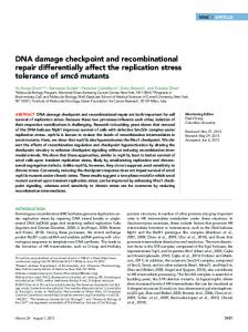
digestion of hepatocytes in a mixed liver cell suspension (14) following collagenase perfusion of the liver using the method of Berry and Friend (15). If both hepatocytes and sinusoidal cells are being investigated, either elutriation centrifugation (16, 17) or differential low speed centrifugation (18-20) can be used. The sinusoidal cells can be further separated into Kupffer and endothelial cells by elutriation centrifugation (16). These procedures can be performed aseptically if tissue culture studies are required. The techniques provide relatively pure populations of viable hepatocytes (Fig. la), sinusoidal lining cells (Fig. lb), endothelial cells (Fig. lc) and Kupffer cells (Fig. ld).
Factors Involved in Cell-Specific Carcinogenesis of Alkylating Agents Exposure of rats to 1,2-dimethylhydrazine (SDMH) results in colon tumors (5, 21) or liver
c
.j
(5, 6), depending on the dosing regimen. Nearly 100% of male Fischer-344 rats exposed to 30 ppm SDMH via the drinking water (-2 mg/kg/ day) developed angiosarcomas of the liver (6). Half of tumors
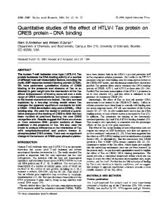
these animals had metastatic angiosarcoma in their lungs. Hepatocellular carcinomas also occurred, but in only 40% of the rats. Using this regimen, we have examined cell-specific DNA alkylation, repair and replication during continuous exposure for up to 4 weeks (6). Similar amounts of 7-methylguanine (7MG) were formed in both hepatocytes and sinusoidal lining cells, suggesting that differential alkylation is not the mechanism for cell specificity. The concentrations of 7MG rose from initial one day values of 320 and 470 pmole/mg DNA in sinusoidal lining cells and hepatocytes respectively, to 970 and 1020 pmole/mg DNA on day 3 (Fig. 2A). A slight decline to 750 pmole 7MG/mg DNA was noted in both cell populations after 4 weeks of exposure. This decrease in steady-state levels of 7MG could be associated with either decreased metabolic activation or
Sd
FIGURE 1. Photomicrographs of (a) hepatocytes, (b) sinusoidal lining cells, (c) endothelial cells and (d) Kupffer cells separated by centrifugal elutriation. H & E, x 285.
CELL SPECIFICITY IN DNA BINDING AND REPAIR
2000 -
marked difference in the removal of O6MG was also evident when one compared the O6MG/7MG ratios. 1500 lsThe sinusoidal lining cells showed an initial decrease, followed by a fairly constant ratio of 0.05. Hepatocytes exhibited an extremely rapid decrease in the O6MG/7MG ratios during the first 3 days and
oA. z< 0cj
8 a s~~~ "
°* iooo
0 8"*
I-0 XU Efi soo
0,,+
t°
*. 0
60
z
so50
So
I_
10
1S
25
30
B.
.-
. * 40 .\/
\s
X E 30 o~20 -'vs
=___
0
00 0
0.15
20
a
70 Z_
maintained
10
S
10
15 1S
2
2
0
20
25
30
-
o
C.
o.os
-
'b W *
*
OtO\8g_&_@ S 10 1S 0
5
\ ,
_@_20 _ 25_ _ __-,
a
ratio of 0.005 for the last 25 days of
4-week exposure (Fig. 2C).
.
: S
0
1,57
30,
DAYS EXPOSURE TO SDMH FIGURE 2. Normalized concentrations of (A) 7-methylguanine, (B) 0'-methylguanine and (C) the 06/N-7 alkylation ratios in sinusoidal lining cells (0) and hepatocytes (0) exposed to 30 ppm dimethylhydrazine in the drinking water for intervals up to 28 days. Each data point represents one animal, while the curve connects the means of three animals at each time point (6). From Cancer Research with permission.
increased removal of 7MG. Support for the former is available from previous studies in mice (22) which demonstrated alterations in DMN metabolism, resulting in lower formation of 7MG. 7MG-DNA-glycosylase activity has not been measured in liver cells during continuous oral exposure to SDMH; however, it was not increased in whole liver homogenates following repeated administration of DMN to rats (23). In marked contrast to 7MG, the concentration of the promutagenic DNA adduct, Oi-methylguanine (O6MG), was similar in hepatocytes and sinusoidal lining cells only at day 1 of exposure (6). Thereafter, the sinusoidal cells accumulated O6MG, while the hepatocytes efficiently removed it (Fig. 2B). The
a
We have recently measured the activity of the O6_ alkylguanine alkyl acceptor protein in both cell populations following continuous exposure of rats to SDMH (24). A 2- to 3-fold enhancement of this
alkyl
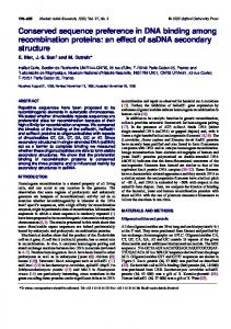
acceptor protein occurred in hepatocytes after 3 days exposure to SDMH. This high activity was maintained in hepatocytes throughout the 4-week exposure. In contrast, the sinusoidal lining cells showed an initial decline in alkyl acceptor protein activity, followed by a return to control levels. Thus, the difference in alkylation at the 06 position of guanine can be explained by cell-specific differences in DNA repair. Of equal importance, however, is the cell-specific mitogenic response to continuous SDMH exposure (25). The sinusoidal lining cells undergo a marked increase in de novo DNA synthesis, whereas a much smaller increase in de novo DNA synthesis occurs in hepatocytes (Fig. 3). One can approximate the probability of mutation due to mispairing of 'MG in replicating DNA templates for each cell population by multiplying the
concentration the amount of deatnovo theO6MG, and of total amount of DNA riskDNA synthesis (Fig. DPM 70
60
-
6
50
40
-
-
30
/
10 l 10 15 20 25 30 DAYS OF EXPOSURE FIGURE 3. De novo DNA synthesis in (0) sinusoidal lining cells and (0) hepatocytes from rats exposed to dimethylhydrazine in the drinking water for various times (25). From Cancer Research with permission. 0
5
S WENBERG ET AL.
158
4° 2.5 x 2.0
have low to moderate 06-alkylguanine alkyl acceptor protein activity (24). O-Alkylated pyrimidines, such as O2_ and 04-thymine, occur in greater proportion relative to 06-alkylguanine following exposure to ethylating agents (27). Since the 0-alkylpyrimidines are removed from liver DNA more slowly (28, 29), they may represent the primary lesions responsible for initiation in those cell types, such as hepatocytes, that have high 06-alkylguanine alkyl acceptor protein activity.
-
-
uJ
0
Z
1.5
-
z
1.0 o l~--
14 10 18 22 26 30 DAYS EXPOSURE TO SDMH FIGURE 4. Initiation Index for SDMH exposure of rats represents the probability of mutation due to O6MG. Data points represent the product of O6MG concentration (6) and de novo DNA synthesis (25) per mg DNA times the amount of DNA at risk per cell population for (0) sinusoidal lining cells and (0) hepatocytes.
2
6
4). It is readily apparent that the sinusoidal lining cells have a greater probability for mutation than do hepatocytes. These data are consistent with carcinogenesis studies at the same dosing regimens (6). Similar data have been obtained from mice exposed to 100 ppm DMN in their drinking water for up to 32 days (20). A progressive mitogenic response was evident in sinusoidal lining cells from 4 to 32 days of DMN exposure. Hepatocytes maintained a relatively constant elevated rate of de novo DNA synthesis from 4 to 32 days exposure. The sinusoidal lining cells had a progressive increase of O6MG from 2 to 32 days of DMN exposure, while hepatocytes maintained relatively constant low concentrations of O6MG. At 32 days, the sinusoidal lining cells had 12 times more OIMG per milligram DNA than hepatocytes. Thus, the probability of GC --
AT transitions due to
miscoding
of O6MG is
again
much greater in sinusoidal lining cells, the target cells for DMN carcinogenesis in the mouse (26). It must be emphasized that these data only reflect the contribution of O6MG to the initiation of carcinogenesis. The data must be fortified with the probability of mutation due to other forms of promutagenic DNA damage, since it is the sum of mutations due to all promutagenic lesions that is believed to comprise initiation. Cell specificity in carcinogenesis due to alkylating agents will depend on the ability of different cell populations to activate the carcinogen, their mitogenic response, their capacity to remove promutagenic DNA lesions and the nature of the alkylating agent. For instance, the major promutagenic lesion produced by SN1 methylating agents is O6MG (27). It probably represents the principal lesion responsible for initiation in those cell types, such as liver sinusoidal cells, that
Factors Involved in Cell-Specific Carcinogenesis of N-Substituted Aryl Compounds The amount of information available on cell-specific factors involved in N-substituted aryl carcinogenesis is more limited than that available for alkylating agents. Hepatocytes represent the principal target cell of most hepatocarcinogenic N-substituted aryl compounds (30). Tumors of bile duct epithelium also occur, whereas tumors of sinusoidal lining cells are rare.
2-Acetylaminofluorene 2-Acetylaminofluorene (AAF) is a model hepatocarcinogen, inducing a high incidence of hepatocellular carcinomas. Multiple pathways exist for activation of AAF to a reactive electrophile. The importance of N-hydroxylation of AAF as the first step in the hepatocarcinogenic process was confirmed when it was found that N-hydroxyacetylaminofluorene (N-OH-AAF) was more potent than the parent compound in producing tumors of the liver, mammary gland, small intestine and ear duct of the rat when given in an equimolar dose (10). In addition, N-OH- AAF produced tumors at the site of administration, while AAF was locally inactive (10). Esterification of the N-hydroxy group of N-OH-AAF as a result of sulfotransferase or N,O-acyltransferase activity, and subsequent formation of a reactive electrophile are steps in the metabolic pathway thought to be important in hepatic carcinogenesis following treatment of animals with AAF (10,31-34). We have utilized the cell separation methods referred to above to compare binding of carcinogen adducts to DNA of hepatocytes and sinusoidal lining cells following administration of equimolar doses of AAF or N-OH-AAF. An assessment of the binding of [ring-3H]-AAF (1.8 ,umole/100 g) to DNA of hepatocytes and sinusoidal lining cells following a single IP injection is presented in Figure 5. At the time of peak binding, 18 hours post-injection, the ratio of carcinogen adduct per milligram of hepatocyte DNA as compared
159
CELL SPECIFICITY IN DNA BINDING AND REPAIR
to sinusoidal lining cells was 2.7, indicating an initial quantitative selectivity of AAF for binding to DNA of hepatocytes (p < 0.05). After 3 days, 59% of the adducts remained in DNA of hepatocytes, and 50% of the adducts remained in DNA of the sinusoidal lining cells. The results of analogous studies with [ring-3H]-NOH-AAF are presented in Figure 6. Two hours after a single injection of [ring-3H]-N-OH-AAF (1.8 ,umole/100 g), the time of peak DNA binding, there was no significant difference in the amount of carcinogen bound per milligram of hepatocyte and sinusoidal lining cell DNA. Three days later, adducts had been removed from DNA of both cell populations to a similar extent, i.e., 54% of the adducts remained in DNA of hepatocytes, and 57% of the adducts remained in DNA of the sinusoidal lining cells. When these data are adjusted for the amount of 40
DNA at risk for the two cell populations, the results clearly show that the amount of carcinogen bound to the DNA of target cells, i.e., hepatocytes, is much greater (Table 1). This cell specificity is greatest for the procarcinogen, AAF, suggesting that hepatocytes have greater metabolic competence for N-hydroxylating AAF. Support for this hypothesis is provided by our results with N-OH-AAF, where similar amounts of covalent binding were present per milligram DNA. The total amount of N-OH-AAF bound to hepatocellular DNA versus sinusoidal lining cell DNA was considerably greater, however. Thus, cell specificity in N-OH-AAF binding to DNA would still favor the induction of hepatocellular tumors.
Previous studies of Tulp et al. reported greater binding of N-OH-AAF to hepatocyte nuclei than to stromal cell nuclei (35). Similar differential binding was present in DNA from these cells. The reason for the differing results is unknown; however, the studies may not be directly comparable since the studies of Tulp et al. utilized a different time point,
x
a~30 0D E 0
20
0
~0
10
~0
LE
90 18 Hours post-injection FIGURE 5. Binding of [ring-3H]AAF to DNA of hepatocytes (open bars) and sinusoidal lining cells (hatched bars). Following a single IP injection of [ring-3H]AAF (1.8 imole/100 g), the amount of carcinogen bound/mg DNA was determined at the time of peak binding (18 hr after injection) and 3 days later. Results are expressed as the mean value from six rats. The standard error is represented by a bar. At 18 hr after injection the amount of carcinogen bound to the DNA of sinusoidal lining cells was significantly different (p < 0.05) from that bound to hepatocytes, as determined by Student's t-test at p = 0.05.
2 72 Hours post- injection FIGURE 6. Binding of [ring-3H]N-OH-AAF to DNA of hepatocytes (open bars) and sinusoidal lining cells (hatched bars). Following a single IP injection of [ring-3H]N-OHAAF (1.8 ,Amole/100 g), the amount of carcinogen bound per mg DNA was determined at the time of peak binding (2 hr after injection), and 3 days later. Results are expressed as the mean value from six rats. Standard error is represented by a bar.
Table 1. Total binding of AAF and N-OH-AAF to liver cell DNA.
Carcinogen AAF
N-OH-AAF
Time, hr 18 90 2 72
DNA binding, pmole/total DNA Sinusoidal cell Hepatocyte 230 43 12 4
95±21
4±1
Hepatocyte/sinusoidal Cell ratio 19.2 23.8
141 ± 32 76 31
17 ± 3 10 3
8.3 7.6
160
SWENBERG ETAL.
a different cell separation procedure and a different DNA isolation method than we used. More recent studies from the same laboratories using AAF demonstrated similar amounts of the C-8 aminofluorene adduct in diploid stromal cells and tetraploid and octaploid hepatocytes, but much greater amounts of the C-8 acetylaminofluorene adduct in hepatocytes. Likewise, the total amount of covalent binding of AAF was greatest in hepatocytes, but removal of DNA adducts was similar in both cell populations
136).
Dinitrotoluene Dinitrotoluene (DNT) is a high volume commodity chemical used in the production of explosives and polyurethane foams. Recent bioassays demonstrated that DNT was carcinogenic for laboratory animals (12); however, the target organ differed with different conditions. 2-4DNT is a potent hepatocarcinogen in Fischer-344 rats (37), with initiating (38) and promoting (39) activity. It induces a 100% incidence of hepatocellular carcinomas in male rats within one year. Females also develop a high incidence of hepatocellular carcinomas; however, the latency period is somewhat longer. This sex difference is thought to be due to differences in the metabolism of DNT (40, 41). DNT is absorbed from the gastro-intestinal tract and transported to the liver, where it is metabolized to dinitrobenzyl alcohol and its glucuronide. In female rats, the glucuronide enters the general circulation and is primarily excreted in the uring, whereas in males it is excreted via the bile. The intestinal microflora hydrolyze the glucuronide and convert the aglycone to an as yet unidentified metabolite that is reabsorbed and transported to the liver. On this second pass through the liver, the unknown metabolite is covalently bound to hepatic macromolecules or is converted to a reactive species capable of covalent binding. We have initiated studies to characterize the extent and persistence of 2,6-DNt'covalent binding in hepatocytes and sinusoidal lining cells of male and female Fischer-344 rats in order to determine whether sex-dependent differences in DNA modification exist and whether covalent binding is greater in hepatocytes, the target cell for DNT carcinogenesis, than in the nontarget sinusoidal lining cell populations. Nine male and nine female Fischer-344 rats were dosed orally with 35 mg/kg 3H-2,6-DNT (250 ,Ci/rat). At 0.5, 4 or 7 days, the livers of three rats of each sex were perfused with collagenase and their hepatocytes and sinusoidal lining cells separated by differential centrifugation. DNA was isolated from hepatocytes of individual rats, whereas the sinusoidal lining cells from all three animals
were pooled prior to DNA isolation. The extent of covalent binding was determined by scintillation counting. DNA was measured using the diaminobenzoic acid method (42). Data expressed as DPM/mg DNA are shown in Figure 7, while total covalent binding per cell population is shown in Figure 8. DNA from male hepatocytes exhibited the greatest amount of covalent binding. DNA from female hepatocytes had slightly less initial binding. Covalently bound DNT was removed from hepatocyte DNA more rapidly during the first 4 days after exposure than during the subsequent 3 days. While the data on sinusoidal lining cells were limited due to pooling of samples, there was considerably less 2,6-DNT bound to their DNA and minimal evidence of DNA repair. For comparison, three germ-free male Fischer-344 rats were also examined for covalent binding to DNA 12 hours after exposure to 3H2,6-DNT. Hepatocellular DNA from the germ-free rats contained less than 10% of the amount of 2,6DNT bound to DNA of conventional animals (Figs. 7 and 8). When male rats were given weekly doses of 3H2,6-DNT (35 mg/kg, 250 MCi/rat) and killed 7 days later, covalently bound radioactivity accumulated only in hepatocellular DNA (Fig. 9). Thus, the extent of covalent binding of 3H-2,6-DNT exhibits a strong correlation in both cell and sex specificity. Additional investigations will be necessary to determine the nature of the covalently bound material. Preliminary data have demonstrated the presence of a single radioactive peak following HPLC separation of nucleosides. 400r
350p[ 300 4
z 250
-0
0
a
E
~~\