Effects Research Laboratory, U.S. Environmental Protection Agency, Research Triangle Park, North .... tent of washing particles remaining in airways into the al-.
Cellular and Biochemical Response of the Human Lung after Intrapulmonary Instillation of Ferric Oxide Particles John C. Lay, William D. Bennett, Andrew J. Ghio, Philip A. Bromberg, Daniel L. Costa, Chong S. Kim, Hillel S. Koren, and Robert B. Devlin Center for Environmental Medicine and Lung Biology, University of North Carolina; Clinical Research Branch, Human Studies Division, National Health and Environmental Effects Research Laboratory, U.S. Environmental Protection Agency, Chapel Hill; and Pulmonary Toxicology Branch, Experimental Toxicology Division, National Health and Environmental Effects Research Laboratory, U.S. Environmental Protection Agency, Research Triangle Park, North Carolina
Bronchoalveolar lavage (BAL) was used to sample lung cells and biochemical components in the lung air spaces at various times from 1 to 91 d after intrapulmonary instillation of 2.6 mm-diameter iron oxide particles in human subjects. The instillation of particles induced transient acute inflammation during the first day post instillation (PI), characterized by increased numbers of neutrophils and alveolar macrophages as well as increased amounts of protein, lactate dehydrogenase, and interleukin-8 in BAL fluids. This response was subclinical and was resolved within 4 d PI. A similar dose-dependent response was seen in rats 1 d after intratracheal instillation of the same particles. The particles contained small amounts of soluble iron (240 ng/mg) and possessed the capacity to catalyze oxidant generation in vitro. Our findings indicate that the acute inflammation after particle exposure may, at least partially, be the result of oxidant generation catalyzed by the presence of residual amounts of ferric ion, ferric hydroxides, or oxyhydroxides associated with the particles. These findings may have relevance to the acute health effects associated with increased levels of ambient particulate air pollutants. Lay, J. C., W. D. Bennett, A. J. Ghio, P. A. Bromberg, D. L. Costa, C. S. Kim, H. S. Koren, and R. B. Devlin. 1999. Cellular and biochemical response of the human lung after intrapulmonary instillation of ferric oxide particles. Am. J. Respir. Cell Mol. Biol. 20:631–642.
Epidemiologic studies have shown consistently that a modest rise in ambient air particle mass concentration, measured either as total suspended particulate mass or as the mass of particles < 10 mm mass median aerodynamic
(Received in original form March 4, 1998 and in revised form August 3, 1998) Disclaimer: Although the research described in this article has been supported by the United States Environmental Protection Agency through Cooperative Agreement CR824915 to the University of North Carolina Center for Environmental Medicine and Lung Biology, it has not been subjected to Agency review and therefore does not necessarily reflect the views of the Agency and no official endorsement should be inferred. Mention of trade names or commercial products does not constitute endorsement or recommendation for use. Address correspondence to: John C. Lay, D.V.M., Ph.D., University of N. Carolina, Ctr. for Environmental Medicine and Lung Biology, CB# 7310, US EPA Human Studies Facility, Chapel Hill, NC 27599-7310. Abbreviations: alveolar macrophage(s), AM(s); bronchoalveolar lavage, BAL; BAL fluid, BALF; ferric oxide, Fe2O3; ferric ion, Fe31; ferric chloride, FeCl3; Hanks’ balanced salt solution, HBSS; interleukin, IL; Limulus amebocyte lysate, LAL; lactate dehydrogenase, LDH; leukotriene, LT; superoxide anion, O22 ; prostaglandin E2, PGE2; post instillation, PI; reactive oxygen species, ROS; sterile physiologic saline solution, SPSS; thiobarbituric acid, TBA. Am. J. Respir. Cell Mol. Biol. Vol. 20, pp. 631–642, 1999 Internet address: www.atsjournals.org
diameter (PM10), is associated with exacerbation of respiratory disease (1–3) and excess human mortality (4–6). Increased morbidity associated with particulate air pollution is documented by increased numbers of hospital admissions for respiratory tract disease (3), increased absences of schoolchildren (7), and increased reporting of respiratory symptoms by persons keeping a daily diary of respiratory symptoms (1). These epidemiologic studies demonstrate a positive association between elevations in ambient particle concentrations and excess mortality (6, 8). At least one study (6) suggests that it is fine particles (< 2.5 mm), small enough to be deposited in the alveolar region, that are probably responsible for these adverse health effects. People most at risk of death are those with chronic cardiopulmonary disease (especially smokers) or lung cancer (6, 9). The mechanism(s) of lung injury after exposure to air pollution particles is not known. Injury has been postulated to be mediated by ultrafine particles (10), biologic agents (e.g., endotoxin) (11), acid aerosols (12), and polyaromatic hydrocarbons (13). Oxidant generation catalyzed by metals associated with particles could also mediate lung injury following exposure to particulate air pollutants. The in vitro generation of oxygen-derived free radicals by metals included in both emission source and ambient air pollu-
632
AMERICAN JOURNAL OF RESPIRATORY CELL AND MOLECULAR BIOLOGY VOL. 20 1999
tion particles has been documented (14–16). Iron is the metal found in highest concentration among particulate air pollutants studied, frequently in concentrations severalfold greater than all others (17, 18). We have previously reported our findings on the retention and clearance of particles in alveolar macrophages (AMs) following intrapulmonary instillation of relatively insoluble 2.6 mm–diameter ferric oxide (Fe2O3) particles in humans (19). Because we used bronchoalveolar lavage (BAL) to monitor particle retention in the alveoli and airways, we could also assess the lung’s response to the particles by quantifying changes in cell numbers and soluble mediators of inflammation in BAL fluid (BALF). This report examines cellular and biochemical changes associated with a transient pulmonary inflammatory response induced by the Fe2O3 particles. To assess the effect of dose (number of particles instilled per surface area of lung), we also examined BALF after intratracheal instillation of comparable doses of Fe 2O3 particles in rats. The Fe 2O3 particles used in this study were also compared with commercially available Fe 2O3 particles for their capacity to catalyze oxidant generation in vitro and to promote inflammation following intratracheal instillation in rats. Our findings indicate that intrapulmonary deposition of these particles induces transient mild acute inflammation that may, at least partially, be the result of oxidant generation catalyzed by the presence of residual amounts of ferric ion (Fe31) or ferric oxyhydroxides associated with the particles. These findings may have relevance to the acute health effects caused by exposure to ambient air particulate matter.
Materials and Methods Study Population The study population is the same as that reported previously (19), except that four additional subjects are included in the group studied at 1 d post instillation (PI), giving a total of 34 healthy, nonsmoking volunteers (27 male, 7 female), 19.6 to 35.5 yr of age (mean age 5 25.8 6 4.3 yr). Potential subjects were excluded from participation if they had a history of smoking, asthma, allergy, cardiac disease, chronic respiratory disease, recent acute respiratory illness, or extensive exposure to pollutants. All potential subjects underwent screening procedures, including completion of the Minnesota Multiphasic Personality Inventory and medical history form, physical examination, chest radiographs, pulmonary function tests, and routine hematologic and serum chemistry tests. Subjects were randomly assigned to groups. Subject groups were relatively matched with regard to age except that the mean age of the group lavaged 91 d PI (29.5 6 5.2 yr) was slightly greater than that of the other groups. Three of the female subjects were in the group lavaged at 1 d PI, two each were in the groups lavaged at 4 and 91 d PI, and there were no female subjects in the remaining groups (2 and 28 d PI). Subjects were informed of the purposes of the study, the procedures of the experiments, and the potential risk from participation, and each subject signed a statement of informed consent. This study was approved by the Committee on the Protection of
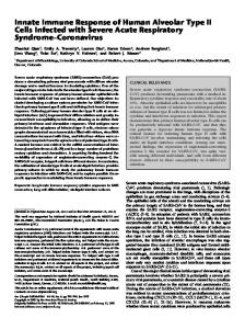
the Rights of Human Subjects of the University of North Carolina School of Medicine (Chapel Hill, NC). Study Design Subjects were randomly assigned to one of five groups of six subjects each (except that the group studied 1 d after exposure included 10 subjects). Each subject underwent two bronchoscopy procedures. During the first bronchoscopy, Fe2O3 microspheres suspended in nonpyrogenic, sterile physiologic saline solution (SPSS) (Baxter Healthcare Products, Deerfield, IL) were instilled into an identified subsegment of the lingula that could readily be wedged by advancing the bronchoscope. As a control, SPSS (without particles) was instilled into a segment (medial or lateral) of the right middle lobe. Subsequently, during the second bronchoscopy procedure, particles, cells, and soluble materials were recovered from the instillation sites by BAL at a specified time PI (1, 2, 4, 28, or 91 d). Each subject was thus lavaged only once, and none underwent serial lavages. Choice of Particles Our choice of Fe2O3 for studying retention and clearance of insoluble particles in the lavagable AM compartment was based on evidence that Fe2O3 is nontoxic, noncarcinogenic, and nonfibrogenic (20). Fe2O3 has been used previously by many investigators for studying mucociliary clearance in humans following inhalation of radiolabeled particles (21–25). It also has been used as a control particle for comparisons of the inhalation toxicity of different dust particles (26) and has been used extensively as a carrier particle for instillation studies (27–30) of the carcinogenicity of polyaromatic hydrocarbons. Control groups given Fe2O3 alone showed no changes in longevity and no evidence of carcinogenicity, chronic inflammation, or fibrosis (26, 29, 30). In one study of hamsters (30), intratracheal instillation of 50 mg Fe2O3 resulted in no long-term adverse health effects. Based on the amount of material per unit of alveolar surface area (mg Fe2O3/m2), this dose in hamsters is approximately 77 times the dose instilled in our human subjects. Particle Generation Particles were generated from colloidal Fe 2O3 made via the hydrolysis and hot dialysis of ferric chloride (FeCl 3) (31). Spherical Fe2O3 particles (nonradioactive, 2.6 mm count median diameter, sg 5 1.3) (Figure 1) were generated in multiple batches as previously described (19). Just before instillation, concentrated particles were suspended in 2 to 3 ml SPSS and placed in an ultrasonic bath for 30 min. Particles were examined and counted in a hemacytometer to assure the dispersion of clumps and to quantify particle numbers. Finally, 3 3 108 particles were suspended in 10 ml SPSS and transferred to a sterile syringe for instillation. Some portion of these particles is lost to the syringe and tubing during the instillation process (see below). Sterilization and Testing for Endotoxin Initial particle batches were sterilized by autoclaving at 1218C for 30 min. Later batches were sterilized by baking the particles at 2508C for 3.5 h, which also insured the de-
Lay, Bennett, Ghio, et al.: Lung Cellular and Biochemical Response to Instilled Particles
Figure 1. The particles used for instillation in human subjects were smooth and spherical with a count median diameter of 2.6 mm and geometric standard deviation (sg) of 1.3. The particles were generated from colloidal Fe2O3 made from the hydrolysis of FeCl3. Bar in the top margin of the photograph 5 10 mm.
struction of any endotoxin activity that might be present in the particle suspension. Batches of all the particle suspensions were tested for endotoxin activity using a gelationcapillary method (Endotect; ICN Biomedical, Costa Mesa, CA) or using a semiquantitative method (performed by UNC Tissue Culture Facility), both of which are based on the Limulus amebocyte lysate (LAL) assay. The capillary LAL method detects endotoxin concentrations as low as 0.06 to 0.10 ng/ml and provides only a positive or negative indication of the presence of endotoxin. The semiquantitative LAL method is equally sensitive and provides an indication of the actual concentration of endotoxin present. All particle suspensions tested by the capillary method were negative. Particle suspensions tested by the semiquantitative LAL method were < 0.06 endotoxin units/ml (1 EU 5 0.1 ng endotoxin). Particle Instillation and BAL Bronchoscopy and BAL were performed as previously described (32). Before bronchoscopy, all subjects were premedicated intravenously with 0.6 mg atropine. The posterior pharynx was anesthetized by gargling with a saline solution containing 4% lidocaine, and the nasal passage was anesthetized with a lubricating jelly containing 2% lidocaine. The larynx, trachea, and bronchi were anesthetized with topical 2% lidocaine instilled through a fiberoptic bronchoscope (Olympus BF, type 1T20D; Olympus, Lake Success, NY) to control coughing. To instill the particles into the distal airways and alveoli, the bronchoscope was passed to an identified subsegmental bronchus of the lingula but was not wedged. A sterile Teflon catheter was passed through the biopsy channel and then extended 4 to 5 cm beyond the tip of the bronchoscope into a subsegment of the lingula. Subjects
633
were instructed to take deep, slow, regular breaths. A total of 10 ml SPSS containing 3 3 108 Fe2O3 microspheres was slowly instilled through the catheter coincident with inspirations to maximize aspiration of particles into the alveolar region. This was followed by an additional 10 ml SPSS from a different syringe (for a total of 20 ml) with the intent of washing particles remaining in airways into the alveoli. A total of 20 ml SPSS (without particles) was instilled, as described, into the medial segment of the right middle lung lobe to serve as a control. To assess the number of particles that were lost to the syringe and catheter during the instillation process, simulated instillations were performed in vitro by injecting particle suspensions through the catheter into a glass vial and counting the particles deposited in the vial. On the basis of these simulations, almost one-third of the particles (31.4 6 3.8%) were lost to the syringe and catheter, so the actual number of particles instilled into the lung is estimated to be about 2.1 3 108 particles (< 5 mg Fe2O3 particles). Segmental BAL was performed at a specific interval PI in the same lingular subsegment in which Fe2O3 (or saline) was previously instilled. The lavage of each segment comprised six washes using a total of 270 ml SPSS per segment. The first washing was done with only 20 ml SPSS and was considered to be enriched with materials from the bronchial airways (33). BALF from the first wash (bronchial fraction) was kept separate from five subsequent washings of 50 ml each. The control segment was lavaged in a similar manner. Cell Preparation BALF and cells were processed as previously described (32). BALF was centrifuged at 250 3 g for 10 min. The cell pellets from the five 50-ml washes (alveolar fraction) were combined. Supernatants from the first two 50-ml washes were combined for analysis and the supernatants from the remaining washes were discarded. Some of the fluid was used immediately for biochemical assays, and the remainder was frozen at 2708C for additional assays to be performed later. Cells from both the bronchial and alveolar fractions were washed twice with RPMI 1640 (Sigma Chemical Co., St. Louis, MO) and used immediately for phagocytic assays and superoxide anion (O22 ) assays. Total cell counts were obtained by light microscopy using a hemacytometer. Cell viability was determined by exclusion of Trypan blue dye. Differential cell counts were obtained using slides prepared in a cytocentrifuge (Cytospin 3; Shandon, Inc., Pittsburgh, PA) at 500 rpm for 3 min. Slides were stained with a modified Wright’s stain (Leukostat Stain; Fisher Scientific, Fairlawn, NJ), and at least 300 cells were counted and evaluated. Biochemical Assays Lactate dehydrogenase (LDH) was measured using a kit purchased from Sigma Chemical Co. The kit was modified to allow 0.1 ml BALF to be assayed using a more concentrated substrate solution in a microtiter plate reader. LDH activity was calculated as milliunits of LDH per milliliter. O22 produced by lavaged cells (from alveolar wash only) was quantified by measuring the kinetics of ferricytochrome-C reduction as described previously (32, 34). Briefly,
634
AMERICAN JOURNAL OF RESPIRATORY CELL AND MOLECULAR BIOLOGY VOL. 20 1999
cells (105 viable AMs) were incubated in Hanks’ balanced salt solution (HBSS) in 96-well polypropylene microtiter plates with and without stimulation by phorbol 12-myristate 13-acetate (PMA; Sigma) in the presence of 80 mg of ferricytochrome-C at 378C for 40 min. Optical density (550 nm) was read at intervals during the incubation, and O22 activity was calculated as nanomoles O22 per 100,000 cells per hour. At 1 d PI, and to a lesser degree at 2 d PI, the BAL cell mixture from particle-instilled lung segments contained contaminating neutrophils in addition to the previously stated number of AMs. Biochemical assays were performed on BALF as described previously (35). Total protein was assayed colorimetrically using a COBAS FARA II integrated centrifugal analyzer (Roche Diagnostic Systems, Branchburg, NJ) and commercially available reagents (Coomassie blue; Sigma). Interleukin (IL)-6 and IL-8 were quantified using commercially available enzyme-linked immunosorbent assay (ELISA) kits (Quantikine immunoassay kits; R&D Systems, Minneapolis, MN). Fibronectin and a1-antitrypsin levels were quantified with competitive ELISA assays using antibodies and antigens purchased from Calbiochem, Inc. (La Jolla, CA). Leukotriene C4 (LTC4), LTD4, and LTE4 were assayed by radioimmunoassay (RIA) using a commercially available kit from Amersham (tritiated tracer, cross-reactivity: 100% with LTC4 and LTD4, 41% with LTE4; Arlington Heights, IL). Prostaglandin E2 (PGE2) was assayed by RIA (125I tracer, 100% cross-reactivity with PGE2, 3.7% cross-reactivity with PGE1) using a commercially available kit from New England Nuclear Research Products (Boston, MA). Both of these arachidonic acid metabolites were measured in lavage fluids after first being extracted and concentrated (53 for bronchial wash, 103 for alveolar wash) on SepPack C18 cartridges (Waters Associates, Milford, MA) (36). Phagocytic activity of lavaged AMs (from alveolar fraction only) was assayed by measuring uptake of Candida albicans organisms (37). C. albicans (1 3 106), labeled with fluorescein isothiocyanate (35), were incubated (1:1 ratio) with 1 3 106 AM in suspension in 1 ml RPMI 1640 tissue culture medium (Sigma) at 378C for 60 min, with intermittent shaking. Phagocytosis was stopped with the addition of 3.5 ml ice-cold HBSS. The suspension was centrifuged at 250 3 g for 10 min, and the cell pellet was suspended in 1 ml cold HBSS. Fluorescence associated with the cells and C. albicans was measured using an Epics Profile II Flow Cytometer (Coulter Corp., Hialeah, FL). AM were differentiated from neutrophils and other cells on the basis of their respective flow-cytometric properties. The percentage of AMs containing one or more fluorescent C. albicans organism (phagocytic index) was determined as the percentage of AM with associated fluorescence following quenching of extracellular fluorescence by addition of Trypan blue dye to the cell suspension. The average number of C. albicans organisms per phagocytic AM was calculated by dividing the average fluorescence per phagocytic AM by the average fluorescence associated with C. albicans organisms. Particle Instillation and BAL in Rats Two different ancillary studies were performed in rats to assess the capacity of the particles to induce inflammation
in the lungs of rats. In the first study, Fe2O3 particles were instilled intratracheally to assess the effect of particle dose on induction of inflammation when the doses (Fe 2O3/m2 lung surface) were adjusted to compare to that used in humans. In the second study, the Fe2O3 particles used for instillation in humans were compared with two commercial Fe2O3 particles for their capacity to induce inflammation in rat lung. Study 1. Fe2O3 particles were instilled intratracheally in a volume of 0.5 ml SPSS in halothane-anesthetized Fischer 344 male rats (38) (250 g, 90 d old) assigned to four groups of five rats each (low-, medium-, and high-dosage levels and saline control group). The medium dose of particles instilled in the rats (77 3 10 6 particles) was calculated to approximate the dose instilled in the human subjects (1.88 3 10 8 particles/m2), on the basis of estimated alveolar surface area as indicated in Table 1. The estimation of alveolar surface area was based on published allometric comparisons of mammalian species (39), assuming that the volume (and surface area) of the lingular subsegment was 1/64 that of the total human lung. At 24 h after instillation of particles, the rats were anesthetized with halothane and killed by exsanguination via the abdominal aorta. The lungs were lavaged with SPSS (29 ml/kg body weight) via an intratracheal cannula fixed in place with a ligature using a syringe attached to the cannula. BALF and cells were then stored on ice until analyzed. Total cell counts were performed immediately using an automated cell counter (Coulter). The BALF and cells were then centrifuged at 250 3 g for 10 min and the supernatant was decanted from the cell pellet. Total protein and LDH activity in the supernatant were measured using the COBAS FARA II integrated centrifugal analyzer with commercially available reagents. Cytological slides were prepared, and differential cell counts were performed as previously described. Study 2. The Fe2O3 particles used for instillation in humans were compared with two commercial Fe2O3 products (Sigma; and Alfa Chemical Co., Ward Hill, MA) having particle sizes of 0.2 mm diameter and 1 to 5 mm diameter, respectively. Sixteen male Fischer 344 rats were divided into four groups of four each. Rats in three of the groups had 1 mg of particles from each of the three sources instilled intratracheally in 0.5 ml sterile saline as described previously. The control group received only sterile saline without particles. At 24 h after instillation, the rats were killed and their lungs lavaged, and total protein concentraTABLE 1
Dosage of intratracheally instilled Fe2O3 particles in rats
Group
High Medium Low Control
Total Particles (10 6 )
231 77 7.7 0
Particles/m2 Alveolar Surface Area* (10 8 )
Total Mass of Particles (mg)
mg/m2 of Alveolar Surface Area
5.63 1.88 0.188 0
4.83 1.61 0.16 0
11.78 3.93 0.39 0
*Alveolar surface area estimated to be 0.41 m2 for a 250-g rat based on Ref. 39.
Lay, Bennett, Ghio, et al.: Lung Cellular and Biochemical Response to Instilled Particles
TABLE 2
Cell counts in BALF of individual subjects at 1 d PI: alveolar fraction Neutrophils
Macrophages
Saline (10 6 cells)
Particles (10 6 cells)
Saline (10 6 cells)
Particles (10 6 cells)
1 2 3 4 5 6 7 8 9 10
0.30 0.22 1.16 0.14 0.47 0.44 1.06 0.30 0.56 0.48
0.49 237.00 1.71 252.00 56.60 13.10 5.78 31.70 3.51 2.26
43.10 31.40 26.90 20.60 16.10 23.90 14.80 10.20 13.80 18.00
35.40 173.00 39.00 174.00 103.00 55.70 15.60 26.30 20.10 16.30
Mean Standard error
0.51 0.11
60.40 31.20
21.90 3.10
77.80 22.90
Subject
tion and percentage of neutrophils in the lavagate were quantified as described previously. Assessment of Solubility and Oxidant-Generating Capacity of Particles Soluble iron associated with the various Fe 2O3 particles was assessed using inductively coupled plasma emission
635
spectroscopy to measure iron extracted in distilled, deionized water as previously described (14). Oxidant generation catalyzed by particles used in this study was compared with commercially available ferric oxides using thiobarbituric acid (TBA)–reactive products of deoxyribose. The pentose sugar 2-deoxy- D-ribose reacts with oxidants to yield a mixture of products. On heating with TBA at a low pH, these products form a pink chromophore that can be measured by its absorbance at 532 nm. This chromophore is indistinguishable from a TBA– malondialdehyde adduct. The reaction mixture containing 1.0 mM deoxyribose, 1.0 mM H2O2, 1.0 mM ascorbate, and 200 mg of either (1) the Fe2O3 particles used for instillation, (2) iron (III) oxide from Sigma, or (3) iron (III) oxide from Alfa was incubated in saline at 378C for 60 min with agitation and then centrifuged at 1,200 3 g for 10 min. One milliliter each of 1.0% (wt/vol) TBA and 2.8% (wt/ vol) trichloroacetic acid were added to 1.0 ml of supernatant, heated at 1008C for 10 min, and cooled in ice, and the chromophore concentration was determined in triplicate specimens by its absorbance at 532 nm. Statistical Evaluations All values are expressed as means 6 standard error. Differences between particle-instilled versus control segments and bronchial versus alveolar fractions for various parameters were analyzed using Student’s t test for paired samples (40). Comparisons of values for the various pa-
TABLE 3
Total and differential cell counts: alveolar fraction* Lymphocytes (%)
Polymorphonuclear Leukocytes (Neutrophils) (%)
Eosinophils (%)
Epithelial Cells (%)
91.83 (1.16)
5.20 (0.80)
2.50 (0.54)
0.30 (0.10)
0.23 (0.14)
2.93 (0.81)
94.20 (1.28)
4.80 (1.20)
0.80 (0.39)
0.27 (0.12)
0.00 (0.00)
4
2.76 (0.51)
93.11 (1.46)
5.94 (1.33)
0.72 (0.29)
0.00 (0.00)
0.33 (0.21)
28
2.22 (0.17)
91.80 (2.34)
6.20 (2.34)
1.53 (1.04)
0.07 (0.07)
0.47 (0.33)
91
1.62 (0.21)
92.06 (1.77)
4.83 (1.69)
0.83 (0.22)
0.28 (0.10)
0.78 (0.59)
13.20† (5.22)
67.68† (6.69)
4.80 (0.82)
27.08† (6.69)
0.30 (0.09)
0.13 (0.07)
2
2.98 (0.78)
85.94 (3.62)
6.06 (0.71)
6.89 (3.52)
0.89 (0.32)
0.33 (0.17)
4
3.15 (0.48)
91.50 (0.51)
7.00 (0.63)
0.89 (0.27)
0.33 (0.15)
0.39 (0.22)
28
3.16 (0.34)
93.33 (1.39)
4.39 (1.31)
2.22 (1.13)
0.00 (0.00)
0.11 (0.11)
91
2.29 (0.41)
93.22 (2.62)
5.89 (2.40)
0.72 (0.23)
0.22 (0.22)
0.11 (0.07)
Total Cells (3 107 )
AMs (%)
2.37 (0.32)
2
Days PI
Saline 1
Particles 1
* Numbers in parentheses represent standard error. † Significantly different from saline control values (P , 0.05).
636
AMERICAN JOURNAL OF RESPIRATORY CELL AND MOLECULAR BIOLOGY VOL. 20 1999
rameters at different times after instillation were assessed by analysis of variance (ANOVA) and Scheffe’s post hoc test. A P value of 0.05 was chosen as the level of significance. Comparisons of results from the rat studies also were performed using either ANOVA or t tests, whichever was appropriate for a particular comparison.
Results Cell Counts in BALF Intrapulmonary instillation of Fe2O3 particles induced a transient inflammatory response that was apparent in most subjects (8 of 10) lavaged 1 d PI. Total and differential cell counts for alveolar and bronchial fractions are listed in Tables 3 and 4, respectively. An influx of cells into the particle-instilled segment was apparent at 1 d PI and comprised markedly increased numbers of both AMs and neutrophils (Figure 2). The mean number of AMs returned to control levels by 2 d PI, and neutrophils were no
Figure 2. The total number of cells recovered by BAL from the alveolar region of the lingular (particle-instilled) segment was markedly elevated at 1 d PI relative to the saline-instilled control segment because of the influx of both neutrophils (left) and macrophages (right) into the lavagable air spaces. The total numbers of both cell types returned to near control levels by 2 d PI. In the bronchial fraction, only neutrophils were elevated at 1 d PI. *Significant increase relative to saline control (P , 0.05). TABLE 4
Total and differential cell counts: bronchial fraction*
Days PI
Saline 1 2
Lymphocytes (%)
Polymorphonuclear Leukocytes (Neutrophils) (%)
Eosinophils (%)
Epithelial Cells (%)
46.80 (4.90)
3.30 (0.71)
32.53 (5.34)
1.07 (0.32)
16.17 (4.11)
73.87 (8.55)
4.93 (0.84)
14.33 (6.80)
1.60 (0.96)
5.27 (3.39)
Total Cells (3 10 5)
AMs (%)
7.81 (1.75) 20.70 (13.9)
4
10.30 (2.91)
63.50 (4.25)
5.72 (0.81)
11.83 (2.12)
0.39 (0.16)
18.56 (3.23)
28
11.92 (7.28)
54.07 (2.93)
2.07 (1.11)
5.67 (2.03)
0.33 (0.26)
37.80 (5.28)
91
5.53 (6.98)
57.94 (8.91)
3.33 (0.86)
7.44 (3.69)
0.17 (0.07)
31.61 (8.50)
21.30 (7.48)
32.71 (3.22)
3.50 (0.57)
47.13† (5.44)
1.17 (0.50)
15.25 (3.73)
2
9.82 (2.80)
69.92 (10.02)
4.13 (0.81)
15.00 (5.50)
0.74 (0.13)
9.96 (4.25)
4
14.30 (4.77)
55.17 (3.48)
4.72 (1.00)
20.06 (4.89)
0.89 (0.82)
18.89 (3.51)
28
9.49 (1.11)
68.83 (7.47)
2.78 (1.06)
12.39 (3.53)
0.22 (0.11)
15.50 (6.51)
91
8.23 (1.32)
49.56 (8.64)
1.78 (0.57)
8.72 (2.93)
0.28 (0.13)
39.44 (6.07)
Particles 1
* Numbers in parentheses represent standard error. † Significantly different from saline control values (P , 0.05).
Lay, Bennett, Ghio, et al.: Lung Cellular and Biochemical Response to Instilled Particles
longer present at 4 d PI (Figure 2). A great deal of variation was observed at 1 d PI in the total number of neutrophils recovered in BALF (Figure 2 and Table 2). The percentage of neutrophils in alveolar BALF was elevated in the lingular segment at 1 d PI (Table 3) and was still marginally elevated at 2 d PI, but was no longer elevated above control values at 4 d PI and later. The percentage of neutrophils in the bronchial fraction of the lingular lavagate also was elevated at 1 d PI but not at later times (Table 4). As expected, the percentages of neutrophils present in the bronchial fractions of both particle-instilled and control segments were higher than in the alveolar fraction throughout the 91-d study period. Neither cell viability nor volume of BALF recovered from particle-instilled segments was different from that of control segments at any of the sampling times. Superoxide Generation by Lavaged Cells O22 generation by BALF cells from the alveolar fraction was examined to help assess activation of AM by the instilled particles. The proportional change in O22 generation by BAL cells following PMA stimulation appeared to be slightly greater for cells from the particle-instilled segment relative to the control at 1 to 4 d PI. This may at least partially result from the presence of neutrophils in BALF at 1
637
and 2 d PI. These differences, however, were not statistically significant. Phagocytosis Assays Phagocytosis of C. albicans by AM from the alveolar fraction was the other functional assay used to assess activation of AMs by the instilled particles. In nearly all instances, for both particle-instilled and control segments, a significantly greater phagocytic index and significantly greater number of organisms per AM were measured for opsonized than for unopsonized C. albicans. No significant differences were detected between AMs from particleinstilled versus control segments for either phagocytic index or the number of organisms phagocytized per AM. These results suggest no apparent activation of AMs following uptake of Fe2O3 particles. Biochemical Parameters in BALF The results of biochemical assays of BALF from alveolar and bronchial fractions are tabulated in Tables 5 and 6, respectively. In the alveolar fraction, total protein, LDH, and IL-8 were significantly elevated (P , 0.05) in BALF from the particle-instilled segment relative to controls at 1 d PI. Mean levels of IL-6 and fibronectin appeared elevated at 1 d PI; however, these differences were not statistically significant because of large within-group variance
TABLE 5
Biochemical parameters in BALF: alveolar fraction* Total Protein (mg/ml) Days PI
Particles †
2
LDH (mU/ml) Saline
Particles
IL-6 (pg/ml) Saline
Particles
160.66 (41.18)
†
6.48 (1.38)
2.70 (0.84)
55.69 (30.81)
166.55 (28.15)
173.36 (60.30)
5.31 (1.74)
2.07 (0.45)
4
128.57 (17.61)
114.05 (26.93)
2.68 (0.66)
28
246.7 (24.0)
201.1 (21.1)
91
279.1† (48.0)
176.7 (30.7)
1
522.30 (285.8)
Particles
Saline
4.87 (0.50)
54.44 (8.59)
28.18 (5.89)
5.53 (1.05)
4.68 (0.70)
29.13 (8.19)
13.72 (3.47)
1.98 (0.56)
4.76 (0.50)
3.93 (0.50)
34.18 (10.50)
25.48 (2.82)
2.39 (0.42)
2.56 (0.39)
3.65 (0.52)
4.15 (0.64)
26.57 (6.10)
37.33 (14.81)
5.22 (0.87)
3.00 (0.81)
3.49 (0.36)
3.13 (0.0)
47.40 (10.15)
17.23 (2.67)
a-1 Antitrypsin (mg/ml)
LTC4/D4/E4 (pg/ml) Saline
Saline
†
PGE2 (pg/0.1 ml) Particles
IL-8 (pg/ml)
Particles
Fibronectin (ng/ml)
Saline
Particles
Saline
Particles
Saline
1
5.91 (2.03)
4.09 (1.38)
13.4 (3.7)
11.4 (3.1)
7.32 (1.47)
7.13 (1.90)
147.82 (67.95)
40.47 (18.49)
2
7.61 (3.88)
4.39 (2.76)
8.1 (4.1)
8.3 (2.8)
17.13 (6.10)
14.11 (3.56)
61.67 (30.08)
35.14 (16.09)
4
8.90 (2.19)
7.73 (2.46)
15.3 (6.5)
9.7 (3.1)
12.19 (5.32)
11.51 (3.32)
15.41 (8.90)
10.35 (3.89)
28
0.13 (0.02)
0.20 (0.06)
11.2 (1.6)
20.1 (6.8)
4.51 (1.93)
3.35 (1.63)
59.52 (17.83)
44.53 (14.59)
91
0.11 (0.05)
0.04 (0.01)
16.3 (3.8)
18.9 (2.8)
2.88 (1.08)
3.65 (1.15)
89.90 (18.88)
78.83 (19.22)
* Numbers in parentheses represent standard error. † Represents statistically significant elevation over saline control (paired Student’s t test, P , 0.05).
638
AMERICAN JOURNAL OF RESPIRATORY CELL AND MOLECULAR BIOLOGY VOL. 20 1999
TABLE 6
Biochemical parameters in BALF: bronchial fraction* LDH (mU/ml)
Total Protein (mg/ml) Days PI
Particles
Saline
Particles
1
88.17 (21.29)
67.97 (19.76)
8.64 (1.92)
2
50.10 (7.15)
30.70 (6.40)
4
56.18 (26.58)
28 91
IL-6 (pg/ml) Particles
Saline
Particles
Saline
7.06 (1.88)
9.8 (4.55)
4.48 (0.87)
301.22 (173.14)
147.40 (42.81)
8.88 (6.02)
2.74 (0.90)
6.10 (1.90)
3.45 (0.35)
123.00 (67.22)
42.75 (15.75)
56.42 (12.02)
2.68 (0.66)
7.91 (2.75)
5.08 (1.48)
5.78 (1.50)
114.20 (76.22)
80.8 (24.80)
122.13 (15.82)
89.67 (26.29)
5.65 (1.59)
4.52 (1.32)
4.51 (0.75)
3.65 (0.52)
74.57 (27.06)
42.05 (9.04)
121.07 (23.73)
95.78 (15.59)
12.54 (1.55)
4.52 (1.32)
3.31 (0.18)
3.13 (0.0)
150.07 (36.52)
62.13 (10.67)
a-1 Antitrypsin (mg/ml)
LTC4/D4/E4 (pg/ml)
PGE2 (pg/0.1 ml) Particles
Saline
IL-8 (pg/ml)
Saline
Particles
Saline
Particles
Saline
Fibronectin (ng/ml) Particles
Saline
1
17.35 (3.73)
10.19 (2.01)
16.3 (15.0)
1.25 (0.0)
5.91 (2.40)
17.93 (7.56)
4.78 (1.71)
5.74 (3.37)
2
25.05 (11.86)
7.87 (0.03)
1.25 (0.0)
22.9 (21.7)
7.9 (2.78)
10.70 (7.00)
4.78 (2.95)
0.73 (0.18)
4
10.88 (3.89)
9.67 (2.50)
9.7 (8.4)
16.2 (14.9)
4.60 (2.19)
11.52 (3.30)
1.58 (0.87)
1.99 (1.09)
28
0.17 (0.05)
0.17 (0.05)
21.2 (3.2)
38.0 (9.0)
0.84 (0.23)
0.78 (0.49)
19.43 (3.03)
10.93 (3.86)
91
0.22 (0.14)
0.16 (0.14)
18.7 (2.9)
47.1 (7.0)
0.76 (0.10)
2.52 (2.12)
28.05 (3.81)
25.65 (5.69)
*Numbers in parentheses represent standard error.
(especially in the particle-instilled segments). Mean levels of fibronectin, IL-8, and LDH also appeared elevated (although not significantly) at 2 d PI. a1-antitrypsin was slightly elevated in both particle-instilled and control segments at 2 and 4 d PI relative to that at 1 d PI. PGE 2 appeared elevated in both lingular and control segments at 1 to 4 d PI, relative to 28 and 91 d PI. In the bronchial fraction, no significant differences were found between particle-instilled and control segments for any of the measured parameters, although trends toward increased values in particle-instilled segments were noted for some parameters. The average IL-8 values tended to be higher in the bronchial BALF of particle-instilled segments than in controls at all time points, and IL-6 and PGE2 appeared to be slightly elevated in particle-instilled segments at 1 and 2 d PI. Dose-response study in rats. The total number of cells recovered in BALF from particle-instilled rat lungs at 1 d PI was markedly elevated in the two highest particle-dose groups but was not changed in the low-dose group compared with control animals. This increase in cell numbers was due almost exclusively to the influx of neutrophils into the lung spaces (Figure 3). The cellular influx was accompanied by dose-dependent elevations of total protein and LDH in BALF from rats in the two highest particle-dose groups (Figure 4). Comparison with commercial Fe2O3 (rats). In comparison with saline alone and the two commercial Fe2O3 prod-
ucts, the particles used for instillation in the human subjects induced significant elevations (P , 0.05) of neutrophil numbers and total protein concentration in the BALF at 1 d following intratracheal instillation in rats (Figure 5). Solubility and Oxidant-Generating Capacity of Fe2O3 Particles Water extraction of Fe2O3 particles generated for use in this study demonstrated soluble iron at a concentration of 0.24 6 0.003 mg/mg of Fe2O3 sample. This translates to 0.036% of the total iron content of the particles assuming no water of hydration associated with the Fe2O3. Soluble iron was not detectable in the commercial Fe2O3 products. The Fe2O3 particles used for instillation in the human subjects produced significantly greater TBA absorbance at 532 nm (Figure 6) in an in vitro system, indicating significantly greater oxidant-generating capacity than that of saline or the commercial Fe 2O3 samples examined. This demonstrates that some portion of the iron was available for electron transfer and therefore capable of participation in Fenton-like reactions that could lead to lipid peroxidation and generation of an inflammatory response.
Discussion Our original and primary purpose for instilling particles in the lungs of humans was to examine the retention, clear-
Lay, Bennett, Ghio, et al.: Lung Cellular and Biochemical Response to Instilled Particles
Figure 3. Relative to saline controls, the high- and medium-dose levels of the Fe2O3 particles used for instillation in human subjects induced a marked increase in total cell numbers in the lungs of rats 1 d after intratracheal instillation. The increase in lavagable cell numbers was due primarily to an influx of neutrophils. *Significant increase relative to saline control (P , 0.05).
ance, distribution, and redistribution of inert and insoluble particles within the lavagable AM compartment as a function of time. We have reported on the inhomogeneity of particle distribution and disproportionate clearance of particle-containing cells from the AM compartment following intrapulmonary instillation of these particles (19). We chose Fe2O3 particles for these studies because they are insoluble in aqueous solution at neutral pH (41) and are nontoxic, noncarcinogenic, and nonfibrogenic (20). To accomplish our goals in examining retention and clearance of particles in lavaged AMs, it was necessary to instill a relatively high concentration of particles into a localized region of the lung. This allowed adequate numbers of particles to be recovered by lavage for counting and evaluation of their intracellular distribution. After making certain assumptions regarding the size of the lung segment to be in-
Figure 4. Total protein content and LDH activity in BALF from rats increased in a dose-dependent manner 1 d after intratracheal instillation of different amounts of the Fe 2O3 particles used for instillation in humans. Both parameters are significantly elevated in the high- and medium-dose groups but are unchanged in the low-dose group relative to saline controls. *Significant increase relative to saline control (P , 0.05).
639
Figure 5. Relative to saline alone and to commercial iron oxide products purchased from Alfa Chemical Co. (A) and Sigma Chemical Co. (B), the particles used for instillation in the human subjects (C) induced significantly elevated neutrophil numbers and total protein concentrations in BALF after intratracheal instillation in rats. *Significant increase relative to saline alone and to commercial iron oxides (P , 0.05).
stilled, we calculated that instillation of 3 3 108 particles into a lingular subsegment should result in a concentration of about 3.2 particles per AM within that region, based on published allometric data (39). This would be true if all of the particles reached the alveoli; it does not take into account that some particles are lost to the syringe and tubing and that many of the particles may be cleared quickly by mucociliary clearance. Thus, the actual number of particles reaching the alveoli is likely much lower. By our own observations, the vast majority of lavaged AMs contained
Figure 6. Fe2O3 particles used in this study (C) had significantly greater capacity for catalyzing the generation of ROS in vitro than did saline controls or two commercial iron (III) oxides purchased from Alfa Chemical Co. (A) and Sigma Chemical Co. (B). *Significant increase relative to saline control and to commercial iron oxides (P , 0.05).
640
AMERICAN JOURNAL OF RESPIRATORY CELL AND MOLECULAR BIOLOGY VOL. 20 1999
no particles, and of those that had taken up particles, most contained only one or two particles (19). Thus, it is probable that we underestimated the size of the lung compartment into which the particles were instilled. Based on these calculations and our own observations, it is unlikely that a condition of particle “overload” (42) was achieved. Intrapulmonary instillation of these Fe2O3 particles induced an influx of both neutrophils and AMs, and elevation of LDH, total protein, and IL-8 in BALF at 1 d PI. These changes were detectable only by BAL and were not accompanied by overt clinical symptoms (fever, pain, coughing, etc.). These changes indicate cell injury (LDH), altered vascular permeability (total protein), and elaboration of neutrophil chemoattractants (IL-8) by lung cells, and are consistent with acute inflammation. Although transient inflammation has been reported in a variety of species at 1 or 2 d after intratracheal instillation or inhalation of various types of particles, including polystyrene (43), aluminum oxide (44), titanium dioxide (44, 45), carbon particles (46), and Fe2O3 dust (47), we were nonetheless somewhat surprised to see the marked neutrophil response to these ostensibly inert particles at 1 d PI. In our study we did not observe the sustained elevation in the number of lavagable AMs that was described following instillation of carbon particles in mice (46). Functional differences between AMs from particleinstilled and control lung segments were negligible as assessed by differences in phagocytic capability and ability to generate O22 . Our inability to detect differences may stem from the fact that only a small percentage of the AMs actually phagocytized particles. The slightly lower phagocytic index measured in both groups at 1 d PI is interpreted to be the result of the pronounced influx of AMs, many of which were immature cells with monocyte-like morphology and limited capacity for phagocytosis. A likely mechanism underlying the acute inflammatory response to the particles used in this study is that of oxidant generation and subsequent cell injury (lipid peroxidation) by reactive oxygen species (ROS) (48). The particles used in this study were generated from colloidal Fe 2O3 made from the hydrolysis and hot dialysis of ferric chloride (FeCl3). This process results in the slow formation of Fe2O3 with some portion first forming the intermediate species ferric hydroxide (Fe[OH]3, unstable), ferrihydrite (Fe5HO8 · 4 H2O), or ferric oxyhydroxide (FeOOH) before transformation to Fe2O3 (31, 49). We have established that the particles instilled in the human subjects were capable of catalyzing the generation of ROS in vitro and that a small amount of soluble iron (240 ng soluble Fe/mg particles) was associated with the particles. This is approximately 0.036% of the total iron content of the particles. We interpret these findings to indicate incomplete conversion of some small portion of the soluble Fe31 ion to stable, insoluble Fe2O3 during the hydrolysis of FeCl 3. This soluble Fe31 (14), and possibly also residual intermediate iron oxide species (50, 51), may support electron transfer and participate in Fenton-type chemistry resulting in oxidant generation, lipid peroxidation, and inflammation. The studies in rats demonstrated a dose-dependent response to the particles generated from FeCl 3 and also showed that the inflammatory response was limited to the
laboratory-made particles. Fe2O3 particles bought commercially had no detectable soluble iron component, were much less capable of oxidant generation, and did not induce an inflammatory response when instilled into the lungs of rats. These ancillary studies support our interpretation that it was catalytically active iron that was responsible for the inflammation seen following instillation of the particles in the human volunteers. Although disparities in iron species and catalysis of oxidants are associated with the potential of the particles to induce an in vitro release of pertinent mediators and an in vivo inflammatory response, it is possible that size, shape, and surface charge of the particles also contribute to these differences. This interpretation of our results is consistent with the hypothesis advanced previously (14–16) that transition metals associated with airborne particles may underlie some of the acute effects associated with particulate air pollution. The inflammatory response following exposure to the iron-containing particle in our study is comparable to that seen following exposure to other metal chelates (52). This inflammatory response is assumed to result from an oxidant-sensitive activation of specific promoters by the iron-containing particle. This presumably would include nuclear factor-kB (53), whose activation results in increased release of several cytokines such as tumor necrosis factor and ILs (54, 55), which function as chemotactic agents for inflammatory cells. Tissue injury subsequently results from exposure to either (1) the free radicals catalyzed by the metal reacting with critical molecules in the lung environment, or (2) proteases and endogenous oxidants generated by the recruited inflammatory cells. Other possible mechanisms that may have contributed to the inflammatory response include release of neutrophil chemoattractants by AMs subsequent to the phagocytic stimulus (56), complement activation (57), or “accidental” (premature fusion of lysosome with phagosome) release of lysosomal contents (hydrolases, proteases, ROS, other products) following the sudden introduction of large numbers of particles and subsequent phagocytic activity. The lack of response to the instilled commercial iron oxides (no soluble iron) in rats in the present study suggests that these alternate mechanisms did not play a significant role in inducing inflammation. We instilled 5 mg of Fe2O3 into one subsegment of the lingula lobe of the lung, which we estimated to have a surface area of about 1.6 m2. However, because only 0.036% of the total iron content that was instilled was soluble, the volunteers received a dose of 0.78 mg of soluble iron per square meter of lung surface. A person breathing ambient air containing 100 mg/m3 of fine particles would deposit approximately 450 mg per 24 h, assuming a tidal volume of 0.5 liters, a ventilation rate of 15 breaths per min, and deposition fraction of about 40% (58). Ionizable iron makes up 1 to 2% of ambient PM material by weight and as much as 4 to 5% of specific emission sources such as oil fly ash (14). Thus, the 450 mg of particles retained in the lung in a 24-h period may contain as much as 4.5 to 22.5 mg soluble iron or 0.04 to 0.22 mg/m2 lung surface, assuming 102 m2 of alveolar surface area for the total lung (39). This suggests that the amount of soluble iron instilled into the volunteers was not too dissimilar to what might be inhaled in a
Lay, Bennett, Ghio, et al.: Lung Cellular and Biochemical Response to Instilled Particles
heavily polluted urban area during a 24- to 48-h period. Furthermore, patients with airways disease (e.g., chronic obstructive pulmonary disease) have enhanced deposition of particles in their airways (59–61), so that in terms of dose per surface area, the deposited dose is even more comparable to the amount instilled into the volunteers in this study. Having made these arguments, we also recognize that the route of exposure (intrapulmonary instillation) is not a normal mode of entry and that the dose of particles instilled is unlikely to occur except in the most polluted environment over a period of time. Thus, they may have limited relevance to actual ambient exposures. The mean percentage of neutrophils in BALF 24 h after instillation of particles in this study was considerably higher than that found 1 h (35) or 18 h (32) after a 2-h exposure to 0.4 ppm ozone (27% neutrophils for particles versus about 10% for ozone). This indicates that exposure to particles and associated soluble iron may have the potential for inducing an inflammatory response in the lung whose severity is similar to or greater than that induced by ozone. Additional studies are required to determine what dose of inhaled particles might be necessary to produce such a response. Ambient air particles with relatively high levels of soluble iron might reasonably be expected to produce a greater inflammatory response than those with a low level of soluble iron.
Conclusions This is the first study in humans to use bronchoscopy and intrapulmonary instillation to assess the lung’s response to an instilled burden of respirable-sized particles. We have demonstrated a transient acute pulmonary inflammatory response to Fe2O3 particles at 1 d after instillation, which is likely due to ROS induced by residual soluble Fe31 or catalytically active residual intermediate iron oxide species associated with the particles. These findings are consistent with the hypothesis that transition metals associated with airborne particles underlie some of the acute effects attributed to particulate air pollution. Acknowledgments: The authors acknowledge and thank Lisa Dailey, Jackie Quay, Joleen Soukup, Alex Chall, Debra Levin, Maryann Bassett, Jim Lehmann, and John McGee, and doctors Tim Gerrity, Brian Boehlecke, Mike Madden, Frank Biscardi, Mark Robbins, Greg Bottei, and Kirby Zeman for their efforts or advice in performing various aspects of this work.
References 1. Schwartz, J., D. Wypij, D. W. Dockery, J. Ware, S. Zeger, J. D. Spengler, and B. Ferris, Jr. 1991. Daily diaries of respiratory symptoms and air pollution: methodological issues and results. Environ. Health Perspect. 90:181– 187. 2. Chestnut, L. G., J. Schwartz, D. A. Savitz, and C. M. Burchfiel. 1991. Pulmonary function and ambient particulate matter: epidemiological evidence from NHANES 1. Arch. Environ. Health 46:135–144. 3. Pope, C. A., III. 1991. Respiratory hospital admissions associated with PM10 pollution in Utah, Salt Lake, and Cache Valleys. Arch. Environ. Health 46:90–97. 4. Schwartz, J., and A. Marcus. 1990. Mortality and air pollution in London: a time series analysis. Am. J. Epidemiol. 131:185–194. 5. Pope, C. A., III, J. Schwartz, and M. R. Ransom. 1992. Daily mortality and PM10 pollution in Utah Valley. Arch. Environ. Health 47:211–217. 6. Dockery, D. W., C. A. Pope, III, J. D. Spengler, J. H. Ware, M. E. Fay, B. G. Ferris, Jr., and F. E. Speizer. 1993. An association between air pollution and mortality in six U.S. cities. N. Engl. J. Med. 329:1753–1759. 7. Ransom, M. R., and C. A. Pope, III. 1992. Elementary school absences and PM10 pollution in Utah Valley. Environ. Res. 58:204–219. 8. Schwartz, J. 1991. Particulate air pollution and daily mortality in Detroit.
641
Environ. Res. 56:204–213. 9. Utell, M. J., and M. W. Frampton. 1995. Particles and mortality: a clinical perspective. Inhalation Toxicology 7:645–655. 10. Oberdörster, G., J. Ferin, and B. E. Lehnert. 1994. Correlation between particle size, in vivo particle persistence, and lung injury. Environ. Health Perspect. 102:173–179. 11. Anto, J. M., and J. Sunyer. 1990. Epidemiologic studies of asthma epidemics in Barcelona. Chest 90:185s–190s. 12. Schlesinger, R. B., and J. A. Graham. 1992. Health effects of atmospheric acid aerosols: a model problem in inhalation toxicology and air pollution risk assessment. Fundam. Appl. Toxicol. 18:17–24. 13. Diaz-Sanchez, D., A. R. Dotson, H. Takenaka, and A. Saxon. 1994. Diesel exhaust particles induce local IgE production in vivo and alter the pattern of IgE messenger RNA isoforms. J. Clin. Invest. 94:1417–1425. 14. Pritchard, R. J., A. J. Ghio, J. R. Lehman, D. W. Winsett, J. S. Tepper, P. Park, M. I. Gilmour, K. L. Dreher, and D. L. Costa. 1996. Oxidant generation and lung injury after particulate air pollutant exposure increases with the concentrations of associated metals. Inhalation Toxicology 8:457–477. 15. Ghio, A. J., J. Stonehuerner, R. J. Pritchard, C. A. Piantadosi, D. R. Quigley, K. L. Dreher, and D. L. Costa. 1996. Humic-like substances in air pollution particulates correlate with concentrations of transition metals and oxidant generation. Inhalation Toxicology 8:479–494. 16. Dreher, K. L., R. H. Jaskot, J. R. Lehmann, J. H. Richards, J. K. McGee, A. J. Ghio, and D. L. Costa. 1997. Soluble transition metals mediate residual oil fly ash induced acute lung injury. J. Toxicol. Environ. Health 50: 285–305. 17. Jacob, D. J., and E. W. Gottlieb. 1989. Chemistry of a polluted cloudy boundary layer. J. Geophys. Res. 94:12975–13002. 18. Munger, J. W., D. J. Jacob, J. M. Waldman, and M. R. Hoffmann. 1983. Fogwater chemistry in an urban atmosphere. J. Geophys. Res. 88:5109– 5121. 19. Lay, J. C., W. D. Bennett, C. S. Kim, R. B. Devlin, and P. A. Bromberg. 1998. Retention and intracellular distribution of instilled iron oxide particles in human alveolar macrophages. Am. J. Respir. Cell Mol. Biol. 18:687– 695. 20. Stokinger, H. E. 1984. A review of world literature finds iron oxides noncarcinogenic. Am. Ind. Hyg. Assoc. J. 45:127–133. 21. Wilkey, D. D., P. S. Lee, F. J. Hass, T. R. Gerrity, D. B. Yeates, and R. V. Lourenco. 1980. Mucociliary clearance of deposited particles from the human lung: intra- and inter-subject reproducibility, total lung clearance, and model comparisons. Arch. Environ. Health 35:294–303. 22. Ilowite, J. S., G. C. Smaldone, R. J. Perry, W. D. Bennett, and W. M. Foster. 1989. Relationship between tracheobronchial particle clearance rates and sites of initial deposition in man. Arch. Environ. Health 44:267–273. 23. Lourenco, R. V., M. F. Klimek, and C. J. Borowski. 1971. Deposition and clearance of 2m particles in the tracheobronchial tree of normal subjects— smokers and nonsmokers. J. Clin. Invest. 50:1411–1420. 24. Morrow, P. E., F. R. Gibb, and K. M. Gazioglu. 1967. A study of particulate clearance from the human lungs. Am. Rev. Respir. Dis. 96:1209–1221. 25. Bennett, W. D., W. F. Chapman, J. C. Lay, and T. R. Gerrity. 1993. Pulmonary clearance of inhaled particles 24 to 48 hours post deposition: effect of beta-adrenergic stimulation. J. Aerosol Med. 6:53–62. 26. Wright, J. L., N. Harrison, B. Wiggs, and A. Churg. 1989. Quartz but not iron oxide causes air-flow obstruction, emphysema, and small airways lesions in the rat. Am. Rev. Respir. Dis. 138:129–135. 27. Saffiotti, U., F. Cefis, and L. H. Kolb. 1968. A method for the experimental induction of bronchogenic carcinoma. Cancer Res. 28:104–124. 28. Stenbäck, F., J. Rowland, and A. Sellakumar. 1976. Carcinogenicity of benzo(a)pyrene and dusts in the hamster lung (instilled intratracheally with titanium oxide, aluminum oxide, carbon and ferric oxide). Oncology 33:29–34. 29. Schreiber, H., D. H. Martin, and N. Pazmino. 1975. Species differences in the effect of benzo(a)pyrene-ferric oxide on the respiratory tract of rats and hamsters. Cancer Res. 35:1654–1661. 30. Saffiotti, U., R. Montesano, A. R. Selladumar, F. Cefis, and D. G. Kaufman. 1972. Respiratory tract carcinogenesis in hamsters induced by different numbers of administrations of benzo(a)pyrene and ferric oxide. Cancer Res. 32:1073–1081. 31. Neidle, M., and J. Barab. 1917. Studies in dialysis: II. The hot dialysis of the chlorides of ferric iron, chromium, and aluminum, and the rapid preparation of their colloidal hydrous oxides. J. Am. Chem. Doc. 39:71–81. 32. Koren, H. S., R. B. Devlin, D. E. Graham, R. Mann, M. P. McGee, D. H. Horstman, W. J. Kozumbo, S. Becker, D. House, W. F. McDonnell, and P. A. Bromberg. 1989. Ozone-induced inflammation in the lower airways of human subjects. Am. Rev. Respir. Dis. 139:407–415. 33. Rennard, S. I., M. Ghafouri, A. B. Thompson, J. Linder, W. Vaughan, K. Jones, R. F. Ertl, K. Christensen, A. Prince, M. Stahl, and R. A. Robbins. 1990. Fractional processing of sequential bronchoalveolar lavage to separate bronchial and alveolar samples. Am. Rev. Respir. Dis. 141:208–217. 34. Pick, E., and Y. Keisari. 1981. Superoxide anion and hydrogen peroxide production by chemically elicited peritoneal macrophages—induction by multiple nonphagocytic stimuli. Cell Immunol. 59:301–318. 35. Devlin, R. B., W. F. McDonnell, S. Becker, M. C. Madden, M. P. McGee, R. Perez, G. Hatch, D. E. House, and H. S. Koren. 1996. Time-dependent
642
36. 37. 38. 39. 40. 41. 42. 43. 44.
45. 46. 47.
AMERICAN JOURNAL OF RESPIRATORY CELL AND MOLECULAR BIOLOGY VOL. 20 1999
changes of inflammatory mediators in the lungs of humans exposed to 0.4 ppm ozone for 2 hours: a comparison of mediators found in bronchoalveolar lavage fluid 1 and 18 hours after exposure. Toxicol. Appl. Pharmacol. 138:176–185. Matthay, M. A., W. L. Eschenbacher, and E. J. Goetzl. 1984. Elevated concentrations of leukotriene D4 in pulmonary edema of patients with adult respiratory distress syndrome. J. Clin. Immunol. 4:479–483. Bjerknes, R. 1984. Flow cytometry assay for combined measurement of phagocytosis and intracellular killing of Candida albicans. J. Immunol. Methods 72:229–241. Costa, D. L., J. R. Lehmann, W. M. Harold, and R. T. Drew. 1986. Transoral tracheal intubation of rodents using a fiberoptic laryngoscope. Lab. Anim. Sci. 36:256–261. Stone, K. C., R. R. Mercer, P. Gehr, B. Stockstill, and J. D. Crapo. 1992. Allometric relationships of cell numbers and size in the mammalian lung. Am. J. Respir. Cell Mol. Biol. 6:235–243. Snedecor, G. W., and W. G. Cochran 1967. Statistical Methods. Iowa State University Press, Ames, IA. Lide, David R., editor. 1996. Handbook of Chemistry and Physics. CRC Press, Boca Raton. Morrow, P. E. 1988. Possible mechanisms to explain dust overloading of the lungs. Fundam. Appl. Toxicol. 10:369–384. Lehnert, B. E., Y. E. Valdez, and S. H. Bomalaski. 1985. Lung and pleural “free-cell responses” to the intrapulmonary deposition of particles in the rat. J. Toxicol. Environ. Health 16:823–839. Lindenschmidt, R. C., K. E. Driscoll, M. Perkins, J. Higgins, J. K. Maurer, and K. A. Belfiore. 1990. The comparison of a fibrogenic and two nonfibrogenic dusts by bronchoalveolar lavage. Toxicol. Appl. Pharmacol. 102: 268–281. Oberdörster, G., J. Ferin, R. Gelein, S. C. Soderholm, and J. Finkelstein. 1992. Role of the alveolar macrophage in lung injury: studies with ultrafine particles. Environ. Health Perspect. 97:193–199. Bowden, D. H., and I. Y. R. Adamson. 1978. Adaptive responses of the pulmonary macrophagic system to carbon: I. Kinetic studies. Lab. Invest. 38:422–429. Kavet, R. I., J. D. Brain, and D. J. Levens. 1978. Characteristics of pulmo-
48. 49. 50. 51. 52. 53. 54. 55. 56. 57. 58. 59. 60. 61.
nary macrophages lavaged from hamsters exposed to iron oxide aerosols. Lab. Invest. 38:312–319. Winterbourn, C. C. 1995. Toxicity of iron and hydrogen peroxide: the Fenton reaction. Toxicol. Lett. 82/83:969–974. Schwertmann, U., and R. M. Cornell. 1991. Iron Oxides in the Laboratory. VCH, Weinheim, Germany. Wardman, P., and L. P. Candeias. 1996. Fenton chemistry: an introduction. Radiat. Res. 145:523–531. Goldstein, S., D. Meyerstein, and G. Czapski. 1993. The Fenton reagents. Free Radic. Biol. Med. 15:435–445. White, D. A., M. G. Krfis, and D. E. Stover. 1987. Bronchoalveolar lavage cell populations in bleomycin lung toxicity. Thorax 42:551–552. Hayashi, T., Y. Ueno, and T. Okamato. 1993. Oxidoreductive regulation of nuclear factor kB. J. Biol. Chem. 268:11380–11388. DeForge, L. E., A. M. Preston, E. Takeuchi, J. Kenney, L. A. Boxer, and D. G. Remick. 1993. Regulation of interleukin 8 gene expression by oxidant stress. J. Biol. Chem. 268:25568–25576. Simeonova, P. P., and M. I. Luster. 1995. Iron and reactive oxygen species in the asbestos-induced tumor necrosis factor-a response from alveolar macrophages. Am. J. Respir. Cell Mol. Biol. 12:676–683. Adamson, I. Y. R., and D. H. Bowden. 1982. Chemotactic and mitogenic components of the alveolar macrophage response to particles and neutrophil chemoattractant. Am. J. Pathol. 109:71–77. Warheit, D. B., L. H. Overby, G. George, and A. R. Brody. 1988. Pulmonary macrophages are attracted to inhaled particles through complement activation. Exp. Lung Res. 14:51–66. Stahlhofen, W., G. Rudolf, and A. C. James. 1989. Intercomparison of experimental regional aerosol deposition data. J. Aerosol Med. 2:285–308. Bennett, W. D., K. L. Zeman, C. Kim, and J. Mascarella. 1997. Enhanced deposition of fine particles in COPD patients spontaneously breathing at rest. Inhalation Toxicology 9:1–14. Isawa, T., K. Wasserman, and B. V. Taplin. 1970. Lung scintigraphy and pulmonary function studies in obstructive disease. Am. Rev. Respir. Dis. 102:161–172. Lin, M. S., and D. A. Goodwin. 1976. Pulmonary distribution of an inhaled radioaerosol in obstructive pulmonary disease. Radiology 118:645–651.