Schistosoma mansoni. ... infections with Schistosoma mansoni are reduced by more ...... a Schistosoma japonicum gene encoding a major immunogen.
INFECrION AND IMMUNITY, Jan. 1993, p. 146-154 0019-9567/93/010146-09$02.00/0 Copyright ©) 1993, American Society for Microbiology
Vol. 61, No. 1
Candidate Vaccine Antigens Identified by Antibodies from Mice Vaccinated with 15- or 50-Kilorad-Irradiated Cercariae of Schistosoma mansoni DANIA RICHTER AND DONALD A. HARN*
Department of Tropical Public Health, Harvard School of Public Health, Boston, Massachusetts 02115 Received 1 September 1992/Accepted 29 October 1992
In murine schistosomiasis, the highest levels of resistance to cercarial challenge are obtained by vaccination with radiation-attenuated cercariae. To identify candidate vaccine antigens relevant to the vaccine model, we examined parasite antigens recognized by antibodies from mice vaccinated with irradiated cercariae of Schistosoma mansoni. To optimize recognition of a wide spectrum of antigens, several factors that influence the level of protection in this model were varied; specifically, we examined the effect of (i) single versus multiple vaccinations with irradiated cercariae, (ii) the dose of irradiation (15 or 50 kilorads) administered to the cercariae, and (iii) the genetic background of mouse strains, high-responder (C57BL/6J) versus moderateresponder (CBA/J) mice. We found that the number of vaccinations did not alter antibody specificity but modified the relative antibody titers against particular antigens. The dose of irradiation used to attenuate the immunizing cercariae had a similar effect on antibody titers but in addition influenced antibody specificity. Only mice that had been vaccinated with moderately irradiated cercariae recognized cathepsin B (Sm3l) and Sm32. Interestingly, when vaccinated mice of the two strains, C57BL/6J and CBA/J, were compared, differences in antibody responses to particular antigens were observed. Both strains recognized the integral membrane protein Sm23, glutathione S-transferase, and cathepsin B, whereas Sm32 and paramyosin were recognized only by CBA/J mice, and heat shock protein 70 was recognized exclusively by C57BL16J mice. In this study, we conclusively identified six distinct antigens that are specifically recognized by the humoral immune response of vaccinated mice.
The helminth parasites Schistosoma spp. infect 5 to 10% of the world's population. Current strategies to reduce the impact of this disease on human health include the development of a defined antigen vaccine; several laboratories have selected an array of candidate vaccine antigens (1, 14, 27, 47, 57). However, none of the purified or recombinant antigens identified to date confers a level of protection to challenge infection in mice that is as high as that conferred by vaccination with radiation-attenuated cercariae (vaccine model). In such vaccinated mice, worm burdens of challenge infections with Schistosoma mansoni are reduced by more than 90% compared with those in naive mice (19, 48). Therefore, antigens recognized in the vaccine model may represent potent immunogens for a defined vaccine. The high levels of resistance obtained in mice after vaccination with irradiated cercariae require antibodies and T cells (25, 38, 52). Thus, elucidation of antigens relevant to both humoral and cellular responses may be critical for the development of an optimal vaccine. Although the antibody response is essential for resistance in the vaccine model (52), only serum of repeatedly vaccinated mice successfully transfers protection to naive recipients (11, 25, 38), suggesting a quantitative or qualitative modification of the antibody response with multiple boosts (9). Several factors affect the level of resistance in the vaccine model and may separately or in concert stimulate antibody responses to distinct antigens. Cercariae attenuated with moderate doses of irradiation (15 to 20 kilorads) stimulate higher levels of resistance than do highly irradiated cercariae (50 kilorads) (48, 54). The genetic background of vaccinated
*
mice also influences their resistance to challenge (24). C57BL/6J mice generally develop higher levels of resistance to challenge following vaccination with irradiated cercariae than do mice of any other inbred strain (21, 24, 36). Thus, we examined the patterns of schistosomal antigens recognized by antibodies obtained from high- or moderate-responder mouse strains after single or multiple vaccinations with cercariae irradiated with 15 kilorads or less protective 50kilorad-irradiated cercariae. MATERIALS AND METHODS
Abbreviations. The following abbreviations are used: C5715, C57BL/6J mice vaccinated with cercariae irradiated with 15 kilorads; C57-50, C57BL/6J mice vaccinated with cercariae irradiated with 50 kilorads; CBA-15, CBA/J mice vaccinated with cercariae irradiated with 15 kilorads; CBA-50, CBA/J mice vaccinated with cercariae irradiated with 50 kilorads; CERC, saline-soluble preparation of cercarial antigens; SCHLAP, saline-soluble preparation of schistosomular antigens; SWAP, saline-soluble preparation of adult worm antigens; DOC-SOM, deoxycholate extract of schistosomular antigens; HSP70, heat shock protein of 70 kDa; CP1+CP2, cysteine proteinases 1 and 2; GST, glutathione S-transferase; Sm23, integral membrane protein of 23 kDa; SDS-PAGE, sodium dodecyl sulfate-polyacrylamide gel electrophoresis; PBS, phosphate-buffered saline; PBS-T, PBS containing 0.3% Tween 20; and IgG, immunoglobulin G. Host animals and parasites. Female CBA/J and C57BL/6J mice were purchased from Bomholtgard (Ry, Denmark) and Jackson Laboratory (Bar Harbor, Maine). A Puerto Rican strain of S. mansoni was maintained in our laboratory by
Corresponding author. 146
VOL. 61, 1993
CANDIDATE VACCINE ANTIGENS OF SCHISTOSOMA MANSONI
passage through Biomphalana glabrata snails and Swiss Webster mice (Taconic Farms, Germantown, N.Y.). Parasites of various stages were also obtained from F. Lewis, Biomedical Research Institute, Rockville, Md. Exposure of mice to irradiated cercariae. At monthly intervals, groups of 6- to 8-week-old CBA/J and C57BL/6J mice were exposed to 500 irradiated cercariae of S. mansoni. Freshly harvested cercariae were diluted to 500 organisms per ml and immediately attenuated with either a 15- or 50-kilorad dose of irradiation emitted by a 'Co source. Mice were anesthetized with a mixture of ketamine HCl (0.08 mg/g) (Aveco, Fort Dodge, Iowa) and xylazine (0.005 mg/g) (Haver, Shawnee, Kans.) injected intramuscularly in the thigh and exposed to irradiated cercariae for 30 min via their shaved abdomen by the ring method (56). Antisera and monoclonal antibodies. Vaccinated mice were bled from the tail 26 days after each exposure to cercariae. In one experiment, mice were also bled as early as 9 days after each exposure. Sera from each experimental group of at least 10 mice were pooled. Control sera were obtained from strain- and age-matched naive mice. A monoclonal antibody against schistosomal paramyosin (clone 4B1) was a kind gift of A. Sher, National Institutes of Health, Bethesda, Md. A monoclonal antibody directed against the conserved ATPbinding domain of human HSP70 (clone SA5) was purchased from Affinity BioReagents (Neshanic Station, N.J.). Preparation of antigens. To optimize for the detection of parasite antigens, we examined both saline-soluble and detergent extracts of various parasite stages. CERC, SCHLAP, SWAP, and a saline-soluble preparation of egg antigens were produced from previously frozen cercariae, living mechanically transformed schistosomula (35), previously frozen adult worms, and freshly isolated eggs (15), respectively. With a glass homogenizer, parasites were intermittently homogenized in PBS containing 1 ,uM leupeptin (Boehringer Mannheim, Indianapolis, Ind.) for 1 h on ice. After centrifugation at 16,000 x g for 1 h at 4°C, supernatants were collected, aliquoted, and stored at -80°C. To enrich for membrane proteins, deoxycholate extracts of cercarial antigens or DOC-SOM were prepared from freshly harvested cercariae or living, mechanically transformed schistosomula, respectively, by resuspending washed parasites in a solution containing 4 mM deoxycholate (Sigma, St. Louis, Mo.), 28 mM Tris base, and 1 ,M leupeptin at pH 8.3. Detergent extraction was carried out for 1 h at 4°C on a nutator without mechanical disruption of the parasites. Thereafter, the suspension was centrifuged, and the supernatant was collected and stored as described above. Protein contents of all extractions were estimated by using the Micro-BCA colorimetric assay (Pierce, Rockford, Ill.). The large hydrophilic domain of the integral membrane protein Sm23 was expressed as maltose-binding fusion protein in our laboratory (49). Purified schistosomal paramyosin (31) was a kind gift of J. P. Laclette, Instituto de Investigaciones Biomedicas, Universidad Nacional Autonoma de Mexico, Mexico. Recombinant schistosomal cathepsin B and Sm32 as bacteriophage MS2-polymerase fusion proteins were a kind gift of M. Q. Klinkert, Istituto di Biologia Cellulare, Consiglio Nazionale delle Ricerche, Rome, Italy (29, 30). A mixture of two purified schistosomal cysteine proteinases (CP1+CP2) was a kind gift of C. L. Chappell, Verna and Marrs McLean Department of Biochemistry, Baylor College of Medicine, Waco, Tex. (3). Purification of HSP70 and GST. To isolate HSP70 from cercariae, ATP affinity chromatography was performed essentially according to the method of Welch and Feramisco
147
(59). Briefly, an ATP-agarose column linked by C8 (Signa) was preequilibrated with 20 mM Tris-acetate buffer, pH 7.5, containing 20 mM sodium chloride, 3 mM magnesium chloride, 15 mM 2-mercaptoethanol, and 0.1 mM EDTA (buffer D). CERC, in the presence or absence of phenylmethanesulfonyl fluoride, was applied to the column. Nonspecifically bound proteins were removed by sequential washes with buffer D supplemented with 0.5 M sodium chloride, with buffer D alone, and with 1 mM free GTP in buffer D. After the column was washed with buffer D alone, HSP70 was eluted with 3 mM ATP in buffer D. Fractions were analyzed by SDS-PAGE using discontinuous 12.5% polyacrylamide gels under reducing conditions followed by silver staining using the Phastsystem (Pharmacia, Piscataway, N.J.) according to the manufacturer's directions. All fractions that contained proteins of 70 kDa were pooled, aliquoted, and stored at -80°C. To isolate GST from cercariae, glutathione affinity chromatography (53) was performed with modifications. Briefly, a glutathione-agarose column was preequilibrated with 50 mM Tris buffer, pH 7. CERC was loaded onto the column and washed with Tris buffer, pH 7. Glutathionebinding proteins were eluted with 7 mM glutathione in 50 mM Tris buffer, pH 9.6. Purification was performed at 4°C. Fractions were analyzed as described above, and all fractions that contained proteins of 28 kDa were pooled, aliquoted, and stored at -80°C. SDS-PAGE and Western blot (immunoblot) analysis. SDSPAGE was performed according to the method of Laemmli (32) under reducing or nonreducing conditions. For reducing conditions, sample buffer contained a final concentration of 2.5% 2-mercaptoethanol. Approximately 20 ,ug of crude parasite extracts or 5 jig of purified antigens was loaded per lane, separated by SDS-PAGE, and electrophoretically transferred to a polyvinylidene fluoride membrane (Millipore, Bedford, Mass.) as described by Towbin et al. (58). After transfer, the membrane was quenched in PBS containing 0.3% Tween 20 (PBS-T) for 1 to 1.5 h at room temperature. Individual membrane strips were incubated in antisera or monoclonal antibody diluted in PBS-T for 3 h at room temperature. Antisera obtained from mice were diluted 1:800, and those from rabbits were diluted 1:1,000. Monoclonal antibodies derived from ascites were diluted 1:1,000. Following incubation with primary antibody, membrane strips were washed in PBS-T and incubated in gold-conjugated goat anti-mouse IgG or goat anti-rabbit IgG antibody (Bio-Rad, Richmond, Calif.) that was diluted 1:800 in PBS containing 1% nonfat milk and 10 mM N-2-hydroxyethylpiperazine-N'-2-ethanesulfonic acid at pH 7.0 for 15 to 24 h at room temperature. The signals were enhanced photochemically with a gold enhancement kit (Bio-Rad) according to the manufacturer's directions. In one experiment, anti-mouse IgM antibody was used as the secondary antibody; strips were incubated in alkaline phosphatase-conjugated goat antimouse IgM antibody (Boehringer Mannheim) at a dilution of 1:1,000 in PBS-T for 1 h at room temperature. Following washes with PBS-T, signals were deveioped according to the manufacturer's instructions (Bio-Rad). To visualize entire protein patterns after transfer, strips were incubated in PBS-T, washed with MilliQ H20, and stained with colloidal gold (Diversified BioReagents, Newton Centre, Mass.). Two-dimensional Western blot analysis. Two-dimensional Western blots were performed using approximately 45 ,ug of DOC-SOM per gel. The first dimension was separated by isoelectric focusing as described by O'Farrell (44) using pH 3 to 10 ampholytes (Pharmacia). Second-dimension SDS-
148
RICHTER AND HARN
INFECT. IMMUN.
B
A
kD go
116-_
kD
97-
116 97 -
66-
-
IM-
Ws
_~ A
66-yJ
__c
4M'
-
-
45-
45 -
3 1
-
31 -
40,II~ ~ N.ml1
21
21 -
-
14 -
4-
14-
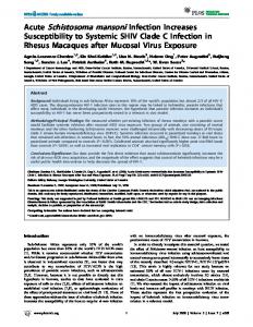
1234 5678 1 2 3 FIG. 1. Western blot analysis of SCHLAP probed with sera of mice vaccinated with irradiated cercariae. (A) Sera were obtained either 9 days after vaccination (lanes 1 to 4) or 26 days after vaccination (lanes 5 to 8) from C57BL/6J mice that were exposed once (lanes 1 and 5), twice (lanes 2 and 6), three times (lanes 3 and 7) or four times (lanes 4 and 8) to cercariae irradiated with 15 kilorads. (B) Sera were obtained from CBA/J mice vaccinated four times with cercariae irradiated with 15 kilorads (lane 1) or 50 kilorads (lane 2). Serum of naive mice is shown as a control (panel B, lane 3). (SDS-PAGE was run under nonreducing conditions.)
PAGE and immunoblotting above.
were
carried out
as
described
RESULTS
Antigens recognized by antibodies of C57BL/6J mice vaccinated with irradiated cercariae. All vaccination regimens were performed at least three times, and the pooled sera from each experiment were analyzed for differences in antigen recognition. No change in the specificity of sera was observed between experiments. Western blot analysis was performed to identify the molecular weight of schistosomal antigens recognized by antibodies of C57BL/6J mice that had been vaccinated with irradiated cercariae. In an initial study, we examined whether antigen recognition was affected by the time span between vaccination and bleeding or by the number of vaccinations. We collected sera 9 or 26 days after vaccination and tested them against SCHLAP. Serum obtained 9 days after the primary vaccination recognized antigens of 28 and 46 kDa, whereas serum collected 26 days after the same vaccination failed to recognize these antigens but bound weakly to antigens of 23 and 70 kDa (Fig. lA, lanes 1 and 5). Serum obtained at either time point after successive exposures to irradiated cercariae bound essentially identical antigens of 23, 28, 70, 116, and 200 kDa (Fig. lA, lanes 2 to 4 and 6 to 8) and resulted in stronger signals than the serum obtained after a single vaccination. The 46-kDa antigen was detected exclusively by sera that were obtained early after exposure to irradiated cercariae. A weak doublet at 97 kDa was only rarely and inconsistently observed. Sera obtained after the fourth vaccination appeared to bind strongest; thus, all subsequent experiments were performed using these sera. We concluded that following the primary vaccination, antibodies against particular antigens are generated at different rates and that the antigen pattern does not change after boosting.
1 2 3 4 5 6 7 8 9 101112 FIG. 2. Western blot analysis of CERC (lanes 1 to 3), SCHLAP (lanes 4 to 6), SWAP (lanes 7 to 9), and saline-soluble preparation of egg antigens (lanes 10 to 12) probed with sera of C57-15 mice (lanes 1, 4, 7, and 10) or C57-50 mice (lanes 2, 5, 8, and 11). Sera of naive mice are shown as controls (lanes 3, 6, 9, and 12). (SDS-PAGE was run under nonreducing conditions.)
We next compared the antigens recognized in various parasite stages by antibodies of C57BL/6J mice that had been repeatedly exposed to either moderately or highly irradiated cercariae. The two antigens that produced the strongest antibody responses, the 23- and 60- to 70-kDa antigens, were observed in all parasite stages examined (Fig. 2). The antibody titer of C57-15 mice generally seemed to be higher than that of C57-50 mice. Two weakly recognized antigens of 28 and 38 kDa were present in all stages, except for the schistosomular or adult stage, respectively. The recognition of a 14- and the 60- to 70-kDa antigens in the cercarial and schistosomular stages was restricted to sera of C57-15 mice. An antigen of 30 to 32 kDa recognized solely by serum of C57-15 mice was present predominantly in SWAP. Weaker bands of 100 to 200 kDa were found throughout all stages. Therefore, an effect of cercarial irradiation dose on antibody specificity in C57BL/6J mice was observed for the larva-associated 14- and 60- to 70-kDa antigens as well as for the SWAP-associated 30- to 32-kDa antigen. Specific antigens recognized by antibodies of CBA/J mice vaccinated with irradiated cercariae. To compare the antigens recognized by sera of CBA/J mice exposed to moderately or highly irradiated cercariae, SCHLAP was separated by nonreducing SDS-PAGE and probed with either serum. Sera of CBA-15 and CBA-50 mice bound to the same pattern of antigens of 23, 28, 46, 95, and 97 kDa (Fig. lB). However, sera of CBA-50 mice recognized the doublet of 95 to 97 kDa with stronger intensity than did the CBA-15 sera. In contrast, CBA-15 sera bound more intensely than CBA-50 sera to the 23-kDa antigen. Thus, sera of vaccinated CBA/J mice detect an antigen pattern partially different from that detected by sera of vaccinated C57BL/6J mice, and the intensity with which sera of vaccinated CBAIJ mice recognize particular antigens is dependent on the dose of irradiation used to attenuate the immunizing cercariae. Identification of the 23-kDa antigen as Sm23. To determine whether the 23-kDa antigen was identical to the previously described integral membrane protein Sm23 (16, 45, 49, 60), replicate lanes of a maltose-binding fusion protein of Sm23
CANDIDATE VACCINE ANTIGENS OF SCHISTOSOMA MANSONI
VOL. 61, 1993
kD :-.r-
kD
A
kD
11697-
9766-
149
B
11697-
66
--"
quam
4545-
453-r_
31-
3121 1 2 3 4 5 6 7 8 9 10 FIG. 3. Identification of Sm23 recognized by sera of C57BU6J and CBA/J mice vaccinated with irradiated cercariae. Western blot analysis of Sm23-maltose-binding fusion protein (lanes 1 to 6) and maltose-binding protein as a control (lanes 7 to 10) probed with sera of C57BL/6J (lanes 1, 2, and 7) or CBA/J mice (lanes 3, 4, and 8) vaccinated with cercariae irradiated with 15 kilorads (lanes 1, 3, 7, and 8) or 50 kilorads (lanes 2 and 4) or probed with rabbit anti-Sm23 serum (lanes 5 and 9). Sera of naive mice are shown as controls (lanes 6 and 10).
(49) were probed with sera of C57-15, C57-50, CBA-15, and CBA-50 mice or a rabbit serum that had been generated against purified native Sm23 (45). All five antisera bound to identical bands of 56 and 112 kDa (Fig. 3, lanes 1 to 5) but did not detect the control maltose-binding protein (Fig. 3, lanes 7 to 9). In addition, we compared the recognition of the 23-kDa antigen in DOC-SOM separated under reducing and nonreducing conditions. All antisera recognized the 23-kDa antigen separated under nonreducing conditions (Fig. 4A,
B
A
kD 200 - " 116-
-.
-
97-
66 4531
_as
21-
1 2 3 4 5 6 1 2 3 4 5 6 FIG. 4. Reduction-sensitive epitopes of Sm23. SDS-PAGE was run under nonreducing (A) or reducing (B) conditions. Western blot analysis of DOC-SOM probed with sera of C57BU6J (lanes 1 and 2) or CBA/J mice (lanes 4 and 5) vaccinated with cercariae irradiated with 15 kilorads (lanes 1 and 4) or 50 kilorads (lanes 2 and 5) or probed with rabbit anti-Sm23 serum (lanes 3). Sera of naive mice are shown as controls (lanes 6).
21-
211
2 3
1 2 3 4 5 6 78 FIG. 5. Identification of schistosomal HSP70 recognized by serum of C57BL6J mice vaccinated with irradiated cercariae. Western blot analysis of SWAP (A) and schistosomal HSP70 (B) purified in the absence (panel B, lanes 1 to 4) or presence (panel B, lanes 5 to 8) of phenylmethanesulfonyl fluoride, probed with serum of vaccinated C57BL/6J mice (panel A, lane 1; panel B, lanes 2 and 6) or anti-human-HSP70 monoclonal antibody (panel A, lane 2; panel B, lanes 3 and 7). Sera of naive mice are shown as controls (panel A, lane 3; panel B, lanes 4 and 8). Purified HSP70 was stained with colloidal gold for total protein (panel B, lanes 1 and 5). (SDS-PAGE was run under reducing conditions.)
lanes 1 to 5) but failed to do so if DOC-SOM was separated under reducing conditions (Fig. 4B, lanes 1 to 5). We concluded that reduction-sensitive epitopes on the cysteinerich integral membrane protein Sm23 (60) are recognized by both mouse strains vaccinated with cercariae irradiated with either dose. Identification of the 70-kDa antigen as HSP70. The major antigen recognized specifically by C57BL/6J mice had an approximate molecular mass of 70 kDa. To determine whether this antigen was schistosomal heat-shock protein (17, 18), we probed replicate lanes of SWAP with a monoclonal antibody against human HSP70 and C57-15 serum (Fig. 5A). A band with an identical molecular mass was recognized by the C57BLV6J serum and the monoclonal antibody. To substantiate further the identity of this antigen, we purified HSP70 from CERC by ATP affinity chromatography. The ATP-binding fractions contained two major proteins of 44 and 70 kDa that were recognized by the C57-15 serum and the monoclonal antibody against human HSP70 (Fig. 5B, lanes 1 to 3). The 44-kDa antigen elicited a stronger signal when probed with the monoclonal antibody than did the 70-kDa antigen. In contrast, C57-15 serum appeared to have stronger binding to the 70-kDa antigen than to the 44-kDa antigen. When HSP70 was purified in presence of phenylmethanesulfonyl fluoride, the yield of the 70-kDa protein was increased and that of the 44-kDa protein was reduced (Fig. SB, lanes 5 to 7). The 70-kDa antigen recognized by sera of vaccinated C57BL/6J mice appears to be schistosomal HSP70, which can be proteolyzed into an epitope-bearing 44-kDa fragment. Identification of the 30- to 32-kDa antigen as cathepsin B. The antigen of 30 to 32 kDa was recognized solely by C57-15 and CBA-15 sera and was found predominantly in SWAP. To determine whether this antigen was identical to enzymes
RICHTER AND HARN
150
A kD 116 9766-
B
INFECI'. IMMUN.
C
D
kD 11697-
''
45-
66lW
4531-
I
'0
i
21 1 2 3 4 5 6 1 2 3 4 5 6 1 2 34 5 6 1 2 345 6 FIG. 6. Identification of schistosomal cathepsin B (Sm3l) and Sm32 recognized by sera of C57-15 and CBA-15 mice vaccinated with irradiated cercariae. Western blot analysis of recombinant cathepsin B (A), Sm32 (B), MS2-polymerase control (C), and purified CP1+CP2 (D) probed with sera of C57-15 (lanes 2 and 3) or CBA-15 mice (lanes 4 and 5) vaccinated with cercariae irradiated with 15 kilorads (lanes 2 and 4) or 50 kilorads (lanes 3 and 5). Total protein was stained with colloidal gold (lanes 1). Sera of naive mice are shown as controls (lanes 6). (SDS-PAGE was run under reducing
conditions.)
associated with the parasite's digestion, i.e., cathepsin B (Sm3l) or Sm32 (29, 30), we probed recombinant MS2polymerase fusion proteins of these antigens with C57-15, C57-50, CBA-15, and CBA-50 sera. In addition, we tested purified CP1+CP2 (3). C57-15 and CBA-15 sera, but not C57-50 and CBA-50 sera, recognized recombinant cathepsin B and CP1+CP2 (Fig. 6A and D). Recombinant Sm32 was detected solely by CBA-15 serum (Fig. 6B). Therefore, cathepsin B and CP1+CP2 are recognized by mice of both strains when vaccinated with moderately irradiated cercariae, and the recognition of Sm32 is strain specific. Identification of the 28-kDa antigen as GST. To determine whether the 28-kDa antigen that was recognized early in the response and was weakly detected in all stages by sera of vaccinated C57BL/6J and CBA/J mice was identical to GST, glutathione affinity-purified GST was probed with sera of vaccinated C57BL/6J and CBA/J mice. No IgG binding could be demonstrated (data not shown). However, upon use of an anti-IgM antibody as the secondary antibody, antibodies of C57-15, C57-50, CBA-15, and CBA-50 were found to bind to GST (Fig. 7). Thus, GST-specific antibodies of vaccinated C57BL/6J and CBA/J mice are predominantly or exclusively of the IgM isotype. Identification of the 97-kDa antigen as paramyosin. A predominant antigen recognized by CBA-50 serum consisted of a doublet of 95 to 97 kDa. Because this molecular mass is similar to that described for schistosomal paramyosin (33), we probed a Western blot of DOC-SOM with CBA-50 serum as well as with a monoclonal antibody generated against schistosomal paramyosin (46). Both bound to identical bands of 95 to 97 kDa (Fig. 8A). In addition, two-dimensional Western blots of DOC-SOM probed with either the antiparamyosin monoclonal antibody or CBA-50 serum were superimposable with isomorph patterns of 92 to 97 kDa and a pI of about 5.5 (Fig. 8B). Furthermore, CBA-50 serum detected schistosomal paramyosin that was purified by col-
21 1412 3 4 5 FIG. 7. Identification of schistosomal GST recognized by IgM antibodies of C57BL/6J and CBA/J mice vaccinated with irradiated cercariae. Western blot analysis of purified schistosomal GST probed with sera of C57BL/6J (lanes 1 and 2) or CBA/J mice (lanes 3 and 4) vaccinated with cercariae irradiated with 15 kilorads (lanes 1 and 3) or 50 kilorads (lanes 2 and 4). Serum of naive mice is shown as a control (lane 5).
lagen affinity chromatography (Fig. 8C). Therefore, the high-molecular-weight antigen recognized strongly by the CBA-50 serum appears to be paramyosin. Analysis of different schistosomal antigen extracts showed that the apparent molecular weight of paramyosin differed depending on the method of antigen preparation. Paramyosin obtained from DOC-SOM resulted in antibody recognition of a doublet at 95 to 97 kDa, whereas the paramyosin bands in SCHLAP were shifted towards 75 to 80 and 95 kDa (Fig. 9, lanes 1 to 6). In addition, the intensity of paramyosin bands varied in different stages of the parasite; the paramyosin doublet in SWAP was much weaker than that found in CERC or SCHLAP, when identical amounts of protein were loaded per lane (Fig. 9, lanes 4 to 12). DISCUSSION Various defined antigens in their purified or recombinant forms have been used for vaccine studies in murine schistosomiasis (1, 14, 27, 47, 57). However, the administration of these nonliving parasite antigens has not yet achieved the high levels of protection that result from vaccination with radiation-attenuated living cercariae. The involvement of antibodies in the development of resistance in the vaccine model has been suggested by studies showing that serum of vaccinated mice can transfer protection to naive recipients (11, 25, 38). However, protection was observed only if the serum was obtained from repeatedly vaccinated mice (11, 25, 38). This suggests a quantitative or qualitative modification of the antibody response with multiple boosts (9). Another study compared humoral responses of different responder strains to surface-exposed proteins of schistosomula and found no differences (55). To avoid narrowing our study to a particular subset of parasite antigens, we examined proteins in saline-soluble as well as membrane-enriched
CANDIDATE VACCINE ANTIGENS OF SCHISTOSOMA MAIANSONI
VOL. 61, 1993
A
kD
116 97 -==
66
B
kD 11697-
pH_-o- 55
-
116 97 --
-v.
66-
-
C
kD
5.5
151
66 -
45-
45-
45 31
31
-
-
31
21 1 2 3
1
2
-
1 2 3
FIG. 8. Identification of schistosomal paramyosin recognized by serum of CBA-50 mice vaccinated with irradiated cercariae. Onedimensional Western blot analysis of DOC-SOM (A) and purified schistosomal paramyosin (C) and two-dimensional Western blot analysis of DOC-SOM (B) probed with serum of CBA-50 (lanes 1) or antiparamyosin monoclonal antibody (lanes 2). Sera of naive mice are shown as controls (lanes 3).
preparations (DOC extracts) of various parasite stages. As probes, we utilized antibodies obtained from two genetically distinct groups of mice with various levels of protection. Comparing sera of mice vaccinated once with irradiated cercariae with sera of repeatedly vaccinated mice, we noticed a general increase in antibody titer with boosts, confirming observations of an anamnestic response (47a). However, the specificity of antibodies was not influenced by boosting, in contrast to the results of Dalton and Strand (9). These differences may be due to the use of Western blotting in our study, whereas in their study metabolically labelled antigens were immunoprecipitated (9). Nevertheless, antibodies against particular antigens appeared to be generated at different rates after primary vaccination with irradiated cercariae. Specifically, responses to 28- and 46-kDa antigens were detected early and responses to 23-, 70-, 116-, and 200-kDa antigens were detected later, at a time when the IgG kD
116-
97-=_
_
66-
45-
31 1 2 3 4 5 6 7 8 9 1011 12 FIG. 9. Westem blot analysis of DOC-SOM (lanes 1 to 3), SCHLAP (lanes 4 to 6), CERC (lanes 7 to 9), and SWAP (lanes 10 to 12) probed with serum of CBA-50 (lanes 1, 4, 7, and 10) or antiparamyosin monoclonal antibody (lanes 2, 5, 8, and 11). Sera of naive mice are shown as controls (lanes 3, 6, 9, and 12).
responses generated by vaccination with irradiated cercariae may have been near their peak (7). With the methods employed in this study, the regimen of multiple exposures to irradiated cercariae appears to elevate the antibody titer but not to alter the antibody specificity. The degree of resistance against challenge infection varies in different mouse strains. The highest levels of protection have generally been observed in C57BL/6J mice (21, 24, 36). In this study, the specificity of antibodies from C57BL/6J mice was compared with that of antibodies from moderateresponder CBA/J mice. We identified three antigens that are differentially recognized by the humoral responses of these two mouse strains. CBA/J mice produced antibodies against Sm32 and paramyosin, whereas C57BV6J mice produced antibodies against HSP70. Simpson et al. observed no difference in antibody specificity when comparing the C57BL/6J strain with another moderate-responder strain, C3H/He (55). However, their study focused solely on immunoprecipitation of radiolabeled surface antigens of schistosomula and thus may have excluded certain antigens. Especially, HSP70 and paramyosin, which were detected in our study, are located in or below the tegument of schistosomes (39, 46, 51), and mechanically transformed schistosomula are not sufficiently developed to produce hemoglobinase. If the humoral immune response against the high-molecular-weight antigens contributes to the difference in protection between the two strains, the differential recognition of HSP70 and paramyosin might influence the level of resistance. HSP70 was the only antigen specifically recognized by the high-responder C57BL/6 mice. In other studies, humoral recognition of 70- to 75-kDa antigens has been associated with resistance (13, 26). In contrast to HSP70, paramyosin was recognized by the moderate-responder CBA/J mice. Therefore, antibodies specific for paramyosin might not positively correlate with the degree of protection. Similar observations regarding levels of antiparamyosin antibodies have been made in immunization studies with a nonliving vaccine (20, 22, 23). Further analysis of HSP70 showed differences in molecular weight and appearance in various preparations of different parasite stages. These observations may be explained by the ability of chymotrypsin to cleave HSP70 into a 60-kDa
152
RICHTER AND HARN
fragment and subsequently into a 44-kDa fragment (6). Cleavage of schistosomal HSP70 was reduced in the presence of a chymotrypsin inhibitor and not observed in schistosomal stages that produce less chymotryptic proteases (34) than cercariae. The presence of stage-specific proteases might similarly account for the various molecular weight bands of paramyosin in different parasite stages. In addition to the genetic background of the host, the dose of irradiation used to attenuate the immunizing cercariae is another factor that greatly influences the level of the host's resistance (19, 40, 48, 54). Our study showed that moderately irradiated cercariae provoke a specific antibody response against recombinant cathepsin B, Sm32, and purified CP1+CP2 that is not observed when highly irradiated cercariae were used for vaccination. However, using sera of mice vaccinated with moderately irradiated cercariae, Ruppel et al. failed to detect antibodies against 31- or 32-kDa antigens (50). Their inability to detect the Sm31/32 antigens with vaccine sera might be due to the use of different mouse strains or a less sensitive immunoblot technique. The purified CP1+CP2 mixture contains both cathepsin B and Sm32 (5). Whereas recombinant Sm32 was strain specifically recognized by CBA/J mice, recombinant cathepsin B and the purified CP1+CP2 mixture were detected by sera of both strains. These antigens are believed to be associated with the digestion of hemoglobin (3, 10, 12, 28). Hemoglobinase activity has been demonstrated in in vitro-cultured schistosomula as early as 8 days of culture (61), and specific antibody titers were observed in mice as early as 3 weeks after infection (4). Therefore, the stage-regulated expression of hemoglobin-digesting enzymes might explain why the antibody response against them is restricted to mice that were vaccinated with moderately irradiated cercariae which survive for a longer time in the host. The dose of irradiation applied to the cercariae influenced not only antibody specificity but also antibody titers against particular antigens. CBA-15 mice produced predominantly antibodies against low-molecular-mass antigens, 14, 23, 28, and 30 to 32 kDa, whereas CBA-50 mice generated a higher antibody titer against paramyosin. In contrast to that in CBAIJ mice, the titer increase in C57BL/6J mice associated with vaccination by moderately irradiated cercariae was directed against the entire antigen repertoire. Thus, it is likely that differences in time of survival and course of migration of cercariae attenuated with moderate or high doses of irradiation (37, 41) are manifest qualitatively as well as quantitatively in the humoral immune response. In this study, all four experimental groups generated anti-GST antibodies. However, the humoral responses of vaccinated mice to GST seemed to be restricted to antibodies of the IgM isotype, even when the sera were obtained a month after vaccination. Interestingly, mice of the P strain that are not protected by vaccination with irradiated cercariae fail to produce any IgM antibodies, suggesting a possible role of this isotype in the vaccine model (7). The integral membrane protein Sm23 was the antigen recognized most intensely independent of the host's genetic background and the irradiation dose of the immunizing cercariae. In other studies, sera of vaccinated CBA/Ca and C3HIHe mice detected a surface antigen of similar if not identical size (2, 55). Because the antibody response to this antigen seems not to be genetically restricted, the difference in protection between moderate- and high-responder mouse strains might depend not on the sole presence of antibodies against Sm23 but perhaps on quantitative or qualitative differences of the particular antibody response. Experiments
INFECT. IMMUN.
comparing titer and isotypes of anti-Sm23 antibodies, as well as of antibodies against the other antigens, generated by C57BL/6J and CBA/J mice vaccinated with moderately or highly irradiated cercariae are currently in progress. In conclusion, six distinct antigens recognized by the humoral responses of mice vaccinated with irradiated cercariae were identified. Their differential recognition by sera from vaccinated mice varies with the genetic background of the host as well as with the irradiation dose given to the immunizing cercariae. This confirms that vaccine studies should be performed in different mouse strains and that the ultimate vaccine against schistosomiasis will likely consist of a cocktail of immunogens to cover the diversity of immune responses within a population. Further, our study indicates that the site and duration of antigen presentation may be critical, because the irradiation dose directly influences the migratory route and/or the survival time of attenuated schistosomes (37, 41) and appears to indirectly affect the specific humoral immune response. In addition, the fact that these attenuated parasites deliver themselves to particular immune responsive sites in the host's body (8, 42, 43) and release antigens over an extended time (42) might explain the advantage of this vaccine model over conventional methods of antigen delivery. ACKNOWLEDGMENTS We thank S. R. Reynolds for providing the Sm23 fusion protein, J. J. Quinn for maintaining the life cycle, K. Palter for advice on the purification of HSP70, and J. A. Fuhrman and F.-R. Matuschka for helpful discussions. We appreciate the generous contributions of purified or recombinant antigens or monoclonal antibodies by C. L. Chappell, M. Q. Klinkert, J. P. Laclette, and A. Sher. This study was supported by grant AI24557 from the National Institutes of Health and grants from the Edna McConnel Clark Foundation, the UNDP/World Bank Program for Research and Training in Tropical Diseases, and the MacArthur Foundation. REFERENCES 1. Balloul, J.-M., J.-M. Grzych, R. J. Pierce, and A. Capron. 1987. A purified 28,000 dalton protein from Schistosoma mansoni adult worms protects rats and mice against experimental schistosomiasis. J. Immunol. 138:3448-3453. 2. Bickle, Q. D., M. Sacko, and D. A. A. Vignali. 1990. Induction of immunity against Schistosoma mansoni by drug (Roll-3128)-
3. 4.
5.
6. 7.
8.
9.
terminated infections: analysis of surface antigen recognition. Parasite Immunol. 12:569-586. Chappell, C. L., and M. H. Dresden. 1987. Purification of cysteine proteinases from adult Schistosoma mansoni. Arch. Biochem. Biophys. 256:560-568. Chappell, C. L., and M. H. Dresden. 1988. Antibody response to a purified parasite proteinase (SMw32) in Schistosoma mansoni infected mice. Am. J. Trop. Med. Hyg. 39:66-73. Chappell, C. L., M. H. Dresden, B. Gryseels, and A. M. Deelder. 1990. Antibody response to Schistosoma mansoni adult worm cysteine proteinases in infected individuals. Am. J. Trop. Med. Hyg. 42:335-341. Chappell, T. G., B. B. Konforti, S. L. Schmid, and J. E. Rothman. 1987. The ATPase core of a clathrin uncoating protein. J. Biol. Chem. 262:746-751. Corma-Oliveira, R., A. Sher, and S. L. James. 1984. Defective vaccine-induced immunity to Schistosoma mansoni in P strain mice. I. Analysis of antibody responses. J. Immunol. 133:15811586. Coulson, P. S., and A. P. Mountford. 1989. Fate of attenuated schistosomula administered to mice by different routes, relative to the immunity induced against Schistosoma mansoni. Parasitology 99:39-45. Dalton, J. P., and M. Strand. 1987. Schistosoma mansoni polypeptides immunogenic in mice vaccinated with radiation-
VOL. 61, 1993
CANDIDATE VACCINE ANTIGENS OF SCHISTOSOMA MANSONI
attenuated cercariae. J. Immunol. 139:2474-2481. 10. Davis, A. H., J. Nanduri, and D. C. Watson. 1987. Cloning and gene expression of Schistosoma mansoni protease. J. Biol. Chem. 262:12851-12855. 11. Delgado, V. S., and D. J. McLaren. 1990. Evidence for enhancement of IgGl subclass expression in mice polyvaccinated with radiation-attenuated cercariae of Schistosoma mansoni and the role of this isotype in serum-transferred immunity. Parasite Immunol. 12:15-32. 12. Dresden, M. H., and A. M. Deelder. 1979. Schistosoma mansoni: thiol proteinase properties of adult worm "hemoglobinase." Exp. Parasitol. 48:190-197. 13. Flanigan, T. P., C. H. King, R. R. Lett, J. Nanduri, and A. A. F. Mahmoud. 1989. Induction of resistance to Schistosoma mansoni infection in mice by purified parasite paramyosin. J. Clin. Invest. 83:1010-1014. 14. Harn, D. A. 1987. Immunization with schistosome membrane antigens. Acta Trop. 44(Suppl. 12):46-49. 15. Harn, D. A., M. Mitsuyama, and J. R David. 1984. Schistosoma mansoni: anti-egg monoclonal antibodies protect against cercarial challenge in vivo. J. Exp. Med. 159:1371-1387. 16. Harn, D. A., M. Mitsuyama, E. D. Huguenel, and J. R. David. 1985. Schistosoma mansoni: detection by monoclonal antibody of a 22,000-dalton surface membrane antigen which may be blocked by host molecules on lung stage parasites. J. Immunol.
29. 30.
31. 32. 33. 34.
35.
135:2115-2120. 17. Hedstrom, R., J. Culpepper, R. A. Harrison, N. Agabian, and G. Newport. 1987. A major immunogen in Schistosoma mansoni infections is homologous to the heat-shock protein Hsp7O. J. Exp. Med. 165:1430-1435. 18. Hedstrom, R., J. Culpepper, V. Schinski, N. Agabian, and G. Newport. 1988. Schistosome heat-shock proteins are immunologically distinct host-like antigens. Mol. Biochem. Parasitol. 29:275-282. 19. Hsu, S. Y. L., H. F. Hsui, and L. F. Burmeister. 1981. Schistosoma mansoni: vaccination of mice with highly X-irradiated cercariae. Exp. Parasitol. 52:91-104. 20. James, S. L. 1987. Induction of protective immunity against Schistosoma mansoni by a non-living vaccine. V. Effects of varying the immunization and infection schedule and site. Parasite Immunol. 9:531-541. 21. James, S. L., M. Labine, and A. Sher. 1981. Mechanisms of protective immunity against Schistosoma mansoni infection in mice vaccinated with irradiated cercariae. I. Analysis of antibody and T-lymphocyte responses in mouse strains developing differing levels of immunity. Cell. Immunol. 65:75-83. 22. James, S. L., and E. J. Pearce. 1988. The influence of adjuvant on induction of protective immunity by a non-living vaccine against schistosomiasis. J. Immunol. 140:2753-2759. 23. James, S. L., C. Salzman, and E. J. Pearce. 1988. Induction of protective immunity against Schistosoma mansoni by a nonliving vaccine. VI. Antigen recognition by non-responder mouse strains. Parasite Immunol. 10:71-83. 24. James, S. L., and A. Sher. 1983. Mechanisms of protective immunity against Schistosoma mansoni infection in mice vaccinated with irradiated cercariae. III. Identification of a mouse strain, P/N, that fails to respond to vaccination. Parasite Immunol. 5:567-575. 25. Jwo, J., and P. T. LoVerde. 1989. The ability of fractionated sera from animals vaccinated with irradiated cercariae of Schistosoma mansoni to transfer immunity to mice. J. Parasitol. 75:252-260. 26. Kely, E. A. B., and D. G. Colley. 1988. In vivo effects of monoclonal anti-L3T4 antibody on immune responsiveness of mice infected with Schistosoma mansoni. Reduction of irradiated cercariae-induced resistance. J. Immunol. 140:2737-2745. 27. King, C. H., R. R. Lett, J. Nanduri, S. El Ibiary, P. A. S. Peters, G. R. Olds, and A. A. F. Mahmoud. 1987. Isolation and characterization of a protective antigen for adjuvant-free immunization against Schistosoma mansoni. J. Immunol. 139:42184224. 28. Klinkert, M. Q., R. Felleisen, G. Link, A. Ruppel, and E. Beck 1989. Primary structures of Sm31/32 diagnostic proteins of
36. 37.
38.
39. 40.
41. 42.
43.
44. 45.
46.
47.
153
Schistosoma mansoni and their identification as proteases. Mol. Biochem. Parasitol. 33:113-122. Klinkert, M. Q., A. Ruppel, and E. Beck. 1987. Cloning of diagnostic 31/32 kilodalton antigens of Schistosoma mansoni. Mol. Biochem. Parasitol. 25:247-255. Klinkert, M. Q., A. Ruppel, R. Felleisen, G. Link, and E. Beck 1988. Expression of diagnostic 31/32 kilodalton proteins of Schistosoma mansoni as fusions with bacteriophage MS2 polymerase. Mol. Biochem. Parasitol. 27:233-240. Laclette, J. P., C. B. Shoemaker, D. Richter, L. Arcos, N. Pante, C. Cohen, D. Bing, and A. Nicholson-Weller. 1992. Paramyosin inhibits complement Cl. J. Immunol. 148:124-128. Laemmli, U. K. 1970. Cleavage of structural proteins during the assembly of the head of bacteriophage T4. Nature (London) 227:680-685. Lanar, D. E., E. J. Pearce, S. L. James, and A. Sher. 1986. Identification of paramyosin as schistosome antigen recognized by intradermally vaccinated mice. Science 234:593-596. Landsperger, W. J., M. A. Stirewalt, and M. H. Dresden. 1982. Purification and properties of a proteolytic enzyme from the cercariae of the human trematode parasite Schistosoma mansoni. Biochem. J. 201:137-144. Lazdins, J. K., M. J. Stein, J. R. David, and A. Sher. 1982. Schistosoma mansoni: rapid isolation and purification of schistosomula of different developmental stages by centrifugation on discontinuous density gradients of percoll. Exp. Parasitol. 53: 39-44. Lewis, F. A., and E. M. Wilson. 1982. Schistosoma mansoni: splenic lymphocyte responses of mice after initial exposure to highly irradiated cercariae. Exp. Parasitol. 54:21-32. Mangold, B. L., and D. A. Dean. 1984. The migration and survival of gamma-irradiated Schistosoma mansoni larvae and the duration of host-parasite contact in relation to the induction of resistance in mice. Parasitology 88:249-266. Mangold, B. L., and D. A. Dean. 1986. Passive transfer with serum and IgG antibodies of irradiated cercaria-induced resistance against Schistosoma mansoni in mice. J. Immunol. 136: 2644-2648. Matsumoto, Y., G. Perry, R. J. C. Levine, R. Blanton, A. A. F. Mahmoud, and M. Aikawa. 1988. Paramyosin and actin in schistosomal teguments. Nature (London) 333:76-78. Minard, P., D. A. Dean, R. H. Jacobson, W. E. Vannier, and K. D. Murrell. 1978. Immunization of mice with cobalt-60 irradiated Schistosoma mansoni cercariae. Am. J. Trop. Med. Hyg. 27:76-86. Minard, P., D. A. Dean, W. E. Vannier, and K. D. Murrell. 1978. Effect of immunization on migration of Schistosoma mansoni through lungs. Am. J. Trop. Med. Hyg. 27:87-93. Mountford, A. P., P. S. Coulson, and R. A. Wilson. 1988. Antigen localization and the induction of resistance in mice vaccinated with irradiated cercariae of Schistosoma mansoni. Parasitology 97:11-25. Mountford, A. P., and R. A. Wilson. 1990. Schistosoma mansoni: the effect of regional lymphadenectomy on the level of protection induced in mice by radiation-attenuated cercariae. Exp. Parasitol. 71:463-469. O'Farrell, P. H. 1975. High resolution two-dimensional gel electrophoresis of proteins. J. Biol. Chem. 250:4007-4021. Oligino, L. D., A. J. Percy, and D. A. Harn. 1988. Purification and immunochemical characterization of a 22 kilodalton surface antigen from Schistosoma mansoni. Mol. Biochem. Parasitol. 28:95-104. Pearce, E. J., S. L. James, J. Dalton, A. Barrall, C. Ramos, M. Strand, and A. Sher. 1986. Immunochemical characterization and purification of Sm-97, a Schistosoma mansoni antigen monospecifically recognized by antibodies from mice protectively immunized with a nonliving vaccine. J. Immunol. 137: 3593-3600. Pearce, E. J., S. L. James, S. Hieny, D. E. Lanar, and A. Sher. 1988. Induction of protective immunity against Schistosoma mansoni by vaccination with schistosome paramyosin (Sm97), a nonsurface parasite antigen. Proc. Natl. Acad. Sci. USA 85: 5678-5682.
154
RICHTER AND HARN
47a.Percy, A. J., and D. A. Harn. Unpublished data. 48. Reynolds, S. R., and D. A. Harn. 1992. Comparison of irradiated-cercariae schistosome vaccine models that use 15- and 50-kilorad doses: the 15-kilorad dose gives greater protection, smaller liver sizes, and higher gamma interferon levels after challenge. Infect. Immun. 60:90-94. 49. Reynolds, S. R., C. B. Shoemaker, and D. A. Harn. T and B cell epitope mapping of Sm23, an integral membrane protein of Schistosoma mansoni. J. Immunol., in press. 50. Ruppel, A., U. Rother, H. Vongerichten, R. Lucius, and H. J. Diesfeld. 1985. Schistosoma mansoni: immunoblot analysis of adult worm proteins. Exp. Parasitol. 60:195-206. 51. Scallon, B. J., B. J. Bogitsh, and C. E. Carter. 1987. Cloning of a Schistosoma japonicum gene encoding a major immunogen recognized by hyperinfected rabbits. Mol. Biochem. Parasitol. 24:237-245. 52. Sher, A., S. Hieny, S. L. James, and R. Asofsky. 1982. Mechanisms of protective immunity against Schistosoma mansoni infection in mice vaccinated with irradiated cercariae. II. Analysis of immunity in hosts deficient in T-lymphocytes, B-lymphocytes, or complement. J. Immunol. 128:1880-1884. 53. Simons, P. C., and D. L. VanderJagt. 1977. Purification of glutathione S-transferases from human liver by glutathioneaffinity chromatography. Anal. Biochem. 82:334-341. 54. Simpson, A. J. G., F. Hackett, T. Walker, R. De Rossi, and S. R. Smithers. 1985. Antibody response against schistosomulum surface antigens and protective immunity following immunization with highly irradiated cercariae of Schistosoma mansoni. Para-
INFECT. IMMUN. site Immunol. 7:133-152. 55. Simpson, A. J. G., S. L. James, and A. Sher. 1983. Identification of surface antigens of schistosomula of Schistosoma mansoni recognized by antibodies from mice immunized by chronic infection and by exposure to highly irradiated cercariae. Infect. Immun. 41:591-597. 56. Smithers, S. R., and R. J. Terry. 1965. The infection of laboratory hosts with cercariae of Schistosoma mansoni and the recovery of the adult worms. Parasitology 55:695-700. 57. Tarrab-Hazdai, R., F. Levi-Schaffer, V. Brenner, S. Horowitz, Z. Eshhar, and R. Arnon. 1985. Protective monoclonal antibody against Schistosoma mansoni: antigen isolation, characterization, and suitability for active immunization. J. Immunol. 135: 2772-2779. 58. Towbin, H., T. Staehelin, and J. Gordon. 1979. Electrophoretic transfer of proteins from polyacrylamide gels to nitrocellulose sheets: procedure and some applications. Proc. Natl. Acad. Sci. USA 76:4350-4354. 59. Welch, W. J., and J. R. Feramisco. 1985. Rapid purification of mammalian 70,000-dalton stress proteins: affinity of the proteins for nucleotides. Mol. Cell. Biol. 5:1229-1237. 60. Wright, M. D., K. J. Henkle, and G. F. Mitchell. 1990. An immunogenic Mr 23,000 integral membrane protein of Schistosoma mansoni worms that closely resembles a human tumorassociated antigen. J. Immunol. 144:3195-3200. 61. Zerda, K. S., M. H. Dresden, and C. L. Chappell. 1988. Schistosoma mansoni: expression and role of cysteine proteinases in developing schistosomula. Exp. Parasitol. 67:238-246.