(h) Filter unit (0.2 m M CN membrane, Nalgene, Catalog No. 450-0020). ...... Wash all labware with ELIMINase and rinse with dis- tilled or deionized dH 2O.
Chapter 8 DNA Barcoding in Mammals Natalia V. Ivanova, Elizabeth L. Clare, and Alex V. Borisenko Abstract DNA barcoding provides an operational framework for mammalian taxonomic identification and cryptic species discovery. Focused effort to build a reference library of genetic data has resulted in the assembly of over 35 K mammalian cytochrome c oxidase subunit I sequences and outlined the scope of mammalrelated barcoding projects. Based on the above experience, this chapter recounts three typical methodological pathways involved in mammalian barcoding: routine methods aimed at assembling the reference sequence library from high quality samples, express approaches used to attain cheap and fast taxonomic identifications for applied purposes, and forensic techniques employed when dealing with degraded material. Most of the methods described are applicable to a range of vertebrate taxa outside Mammalia. Key words: Mammalia, Molecular diagnostics, Molecular biodiversity, Molecular methods, DNA extraction, PCR, Primers, Sequencing, Cytochrome c oxidase subunit I
1. Introduction Mammals represent a minute fraction of biological diversity, with only ~5,500 species currently recognized. Despite their large size and charismatic nature, garnering much taxonomic scrutiny, it is projected that their global diversity is still severely underestimated with at least 7,000 mammalian species in existence (1). The present rate of species discovery in mammals is surprisingly high (~10% increase in 15 years (1, 2)) with more than half of newly recognized species classified as “cryptic” (2) and their discovery largely attributable to the use of non-morphological character complexes. The recently introduced Genetic Species Concept (3, 4) emphasizes genetic rather than reproductive isolation as key to mammalian species’ definition and utilizes genetic divergence to assess species boundaries. While this concept does not define species based on genetic divergence alone and is thus not analogous to molecular taxonomy, it calls for collecting large amounts of
W. John Kress and David L. Erickson (eds.), DNA Barcodes: Methods and Protocols, Methods in Molecular Biology, vol. 858, DOI 10.1007/978-1-61779-591-6_8, © Springer Science+Business Media, LLC 2012
153
154
N.V. Ivanova et al.
information on inter- and intraspecific genetic divergence as an important first step in the process of elaborating taxonomic hypotheses. This coincides with the DNA barcoding approach (5, 6), which offers an operational framework for species identification and discovery using short, standardized gene fragments. Although cytochrome b has been traditionally used for studying mammalian alpha-taxonomy (e.g., ref. 3), cytochrome c oxidase subunit 1 (COI) was the marker of choice for many groups outside Chordata, such as insects and certain marine invertebrates (7–9), and has been adopted as the standard barcoding marker for the animal kingdom (10). While COI evolves more slowly than cyt b (11), it performs equally well in mammalian diagnostics (12) and can yield complementary data for combined phylogenetic analyses (13). The utility of COI DNA barcoding in mammals has been demonstrated in a range of different studies, including bioinformatics (14–16), verification of field-made taxonomic assignments (17, 18), and assessment of genetic diversity patterns in regional (19, 20) and continental faunas (21). The standard animal DNA barcode region has been used in conjunction with other genes for taxonomic revision (13, 22–24) and new species’ description (25). As of 2010, more than 35 K mammalian sequences from over 1 K species have been assembled at the Biodiversity Institute of Ontario as part of an ongoing international effort to build the reference library of DNA barcodes. The sources from which our samples were obtained ranged from degraded archival specimens to high quality cryopreserved tissue. This experience has helped to outline the scope of mammal-related barcoding projects—from forensic cases to the creation of the reference DNA barcode library—and the array of methodological approaches that optimally suit each particular case. While one should keep in mind that mammalian DNA barcoding also intersects with important topics including the ethics of sampling vertebrate collections (26), approaches to conservation genetics (27), specimen examination (28), and the front-end logistics of the barcoding pipeline (29) for efficient high throughput molecular processing, these topics fall outside the scope of this paper and are discussed elsewhere. Here, we have attempted to summarize the methodological approaches employed for the molecular aspects of DNA barcoding in mammals. The molecular protocols are similar to those used in other animals (30), particularly vertebrates, and can be readily applied when dealing with sample sets which include multiple vertebrate groups. We describe three molecular pathways depending on application. 1. Routine barcoding—the assembly of the reference barcode library from high grade tissue samples. This approach frequently employs high throughput methodology in 96-well plate-based manual applications but is also applicable to robotic liquid handling protocols (31). The outcome is the generation
8
DNA Barcoding in Mammals
155
of high quality genomic DNA extracts suitable for long-term archival and bidirectional reads of the full-length barcode region of COI (657 bp). 2. Express barcoding—applied barcoding used in ecological surveys and rapid taxonomic assessments. Similar to routine methods, the approach generally utilizes high grade tissue sources and high throughput techniques, but can be scaled down for small numbers of samples. Express protocols use comparatively fewer reagents and require simpler equipment to be cost-effective, but the resulting DNA extracts are not of archival grade. Unidirectional short length (420 bp) sequences are often generated which are sufficient for reliable species-level identification in mammals (32). An offshoot of this approach is the design and use of microarrays (12, 14). 3. Forensic barcoding—applied barcoding aimed at generating DNA-based identifications when the DNA is degraded and contaminated with fungi or bacteria or when the samples are otherwise recalcitrant. This approach requires polymerase chain reaction (PCR) primers amplifying shorter fragments of DNA (33) and quality checks for cross-contamination. If the tissue is fresh, quick alkaline lysis can be used for DNA extraction; otherwise, we recommend following the DNA extraction protocol for routine barcoding (31). The protocols below are centered on high throughput approaches towards routine barcoding adopted for laboratories lacking robotic liquid handling equipment, but highlight the methodological deviations required for express and forensic barcoding. The utility of these protocols has been validated against a wide range of mammalian taxa representing all major extant orders.
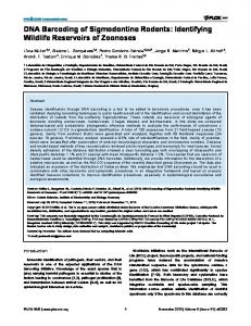
2. Materials 1. Tissue preservation (routine DNA barcoding): (a) Ethanol 96% (store in a flammable liquid cabinet). (b) ELIMINase (Decon Labs Inc.) for sterilizing instruments. (c) Cryotubes with O-ring caps. (d) Acid-free label paper (Rite-in-the-Rain or equivalent). (e) Fine smooth-tip forceps (Dumont or equivalent; see Note 1). 2. Tissue subsampling and lysis (routine DNA barcoding): (a) Ethanol 96% (store in a flammable liquid cabinet). (b) Hard-shell skirted microplate (Eppendorf twin-tec 96 PCR plate, Fisher Scientific; Fig. 1).
156
N.V. Ivanova et al.
Fig. 1. 96-Well microplate shown from below demonstrating the recommended amount of tissue to sample in each well.
(c) 12-Strip flat PCR caps (Thermo Scientific). (d) ELIMINase (Decon Labs Inc.). (e) 12 × 8 Cryotube holding rack for arranging tubes in plate format. (f) Fine forceps (Dumont or equivalent, see Note 1). (g) KimWipes (Kimberly-Clark, Inc.). (h) Glass jars: one 4 oz for ELIMINase and three 8 oz for tool rinsing. (i) Multichannel pipette 5–200 mL LTS or Liquidator (Rainin); see Note 2. (j) Proteinase K: Proteinase K (20 mg/ml) in 10 mM Tris– HCl pH 7.4, 50% glycerol v/v. (Add 20 ml of water and 0.5 ml of 1 M Tris–HCI pH 7.4, to a vial with 1 g of Proteinase K, close the lid, mix well by inverting, and do not shake. Pour into graduated cylinder, add water to 25 ml, then add 25 ml of glycerol, and mix well on magnetic stirrer. Do not filter.) (k) VLB buffer: 100 mM NaCl, 50 mM Tris–HCl pH 8.0, 10 mM EDTA pH 8.0, 0.5% SDS (20 ml 1 M NaCl, 10 ml 1 M Tris–HCl pH 8.0, 1 g sodium dodecyl sulfate (SDS), water to 200 ml). Store at room temperature for up to 6 months. (l) Lysis mix: Mix 5 ml of VLB and 0.5 ml of Proteinase K in sterile container.
8
DNA Barcoding in Mammals
157
3. DNA extraction [routine DNA barcoding: glass fiber (GF) method]; see Note 3: (a) ELIMINase (Decon Labs Inc.). (b) Ethanol 96% (store in a flammable liquid cabinet). (c) 8-Strip flat PCR caps (Thermo Scientific). (d) GF plate: AcroPrep 96 1 ml filter plate with 1.0 mm GF media (PALL, Catalog No. 5051). (e) Matrix Impact2 pipette, 15–1,250 ml with tips (Matrix Technologies). (f ) Square-well block (PROgene Deep-Well Storage Plate 2 ml, Ultident). (g) PALL collar (SBS Receiver Plate Collar, PALL) (see Note 4). (h) Filter unit (0.2 mM CN membrane, Nalgene, Catalog No. 450-0020). (i) Hard-shell skirted microplate; also used as collection plate for DNA eluates. (j) Aluminum Sealing Film (Axygene Scientific, VWR) (to seal DNA plate). (k) Clear Sealing Film (Axygene Scientific, VWR) (used to cover GF plates during centrifugation). (l) Refrigerated centrifuge with swinging deep-well plate bucket rotor (Allegra 25R, Beckman Coulter) (see Note 5). (m) 1 M Tris–HCI pH 8.0 [26.5 g Trizma base (SigmaAldrich)], 44.4 g Trizma HCl (Sigma-Aldrich, water to 500 ml) (see Note 6 for all stock solutions and buffers). (n) 1 M Tris–HCI pH 7.4 (9.7 g Trizma base, 66.1 g Trizma HCl, water to 500 ml). (o) 0.1 M Tris–HCI pH 6.4 (6.06 g Trizma base, water to 500 ml); dissolve in smaller volume than 500 ml, adjust pH with HCl to 6.4–6.5, and then add water to attain the final volume. (p) 1 M NaCl (29.22 g NaCl, water to 500 ml). (q) 1 N NaOH (20 g NaOH, water to 500 ml). (r) 0.5 M EDTA pH 8.0 (186.1 g EDTA, ~20.0 g NaOH, water to 500 ml). Vigorously mix on magnetic stirrer with heater. Disodium salt of EDTA does not dissolve until pH is adjusted to ~8.0 with NaOH. Briefly rinse NaOH granules with ddH2O in a separate glass before dissolving. Adjust pH to 8.0 with 1 N NaOH, before bringing to the final volume. (s) BB buffer: 6 M guanidinium thiocyanate (GuSCN), 20 mM EDTA pH 8.0, 10 mM Tris–HCl pH 6.4, 4%
158
N.V. Ivanova et al.
Triton X-100 (354.6 g GuSCN, 20 ml 0.5 M EDTA pH.8.0, 50 ml 0.1 M Tris–HCl pH 6.4, 20 ml Triton X-100, water to 500 ml). Store at room temperature in the dark for up to 2 months. Weigh dry components first, then add required volumes of stock solutions, followed by small volume of water; dissolve on a warm water bath while constantly stirring; add water to attain the final volume. Filter while solution is still warm. Although the filtering is optional, it helps to minimize crystallization. If crystallization occurs, heat to 56°C before use to redissolve any salts (caution: GuSCN is an irritant; see Note 7). (t) WB buffer: 60% Ethanol, 50 mM NaCl, 10 mM Tris– HCl pH 7.4, 0.5 mM EDTA pH 8.0 (600 ml ethanol 96%, 23.75 ml 1 M NaCl, 9.5 ml 1 M Tris–HCl pH 7.4, 0.950 ml 0.5 M EDTA pH 8.0, water to 950 ml; do not adjust pH); store at –20°C. (u) BM buffer: 250 ml of BB buffer, 250 ml ethanol 96% (store at room temperature in the dark for up to 1 month). (v) PWB buffer: 260 ml of BB buffer, 700 ml ethanol 96%, 40 ml water (store at room temperature in the dark for up to 1 month). 4. PCR (same for all pathways): (a) 10% Trehalose: 5 g of D-(+)-Trehalose dihydrate (SigmaAldrich, No. 90210-50g; see Note 8), molecular grade water to 50 ml. Store in 1–2 ml aliquots at –20°C. (b) 10× PCR Buffer, Minus Mg (Invitrogen); store at –20°C. (c) 50 mM Magnesium Chloride (Invitrogen); store at –20°C. (d) 10 mM Deoxynucleotide Solution Mix (Promega); store in 100 ml aliquots at −20°C. (e) 100 mM primer stock: Dissolve desiccated primer (IDT DNA, USA) in the amount of water indicated by the manufacturer to produce the final solution of 100 mM (i.e., add the number of nmol × 10 ml of water; see Note 9); store at –20°C. (f ) 10 mM primer stock: 20 ml of 100 mM stock, 180 ml of water; store at –20°C. (g) Platinum Taq DNA Polymerase (Invitrogen); store at –20°C. (h) Eppendorf Mastercycler ep gradient S Thermocycler (Eppendorf). (i) Heat sealer (Eppendorf) (used to apply heat sealing film on PCR plates prior to thermal cycling). (j) Aluminum Sealing Film (Axygene Scientific, VWR) (used to reseal DNA plate after use).
8
DNA Barcoding in Mammals
159
(k) Clear Sealing Film (Axygene Scientific, VWR) (used for temporary cover of PCR plates prior to use). (l) Heat sealing Film (GE Uniseal Al Heat Seal Film, Lab Planet) (used to seal PCR plate prior to thermal cycling). (m) Hard-shell skirted microplate (see Note 10). (n) Swinging bucket centrifuge with microplate rotor (Thermo Scientific). 5. PCR Product Check (same for all pathways): (a) Gel documentation system (AlphaImager, Alpha Innotech). (b) Pre-cast agarose gels (2% E-Gel 96, Invitrogen). (c) E-Base Integrated power supply (Invitrogen). 6. Cycle sequencing reaction (same for all pathways): (a) 10% Trehalose: 5 g of D-(+)-Trehalose dihydrate (SigmaAldrich, Catalog No. T9531-100g), molecular grade water to 50 ml. Store in 1–2 ml aliquots at –20°C. (b) 5× Sequencing Buffer (400 mM Tris–HCl pH 9.0 + 10 mM MgCl2). (c) 100 mM primer stock: Dissolve desiccated primer (IDT DNA, USA) in the amount of water indicated by the manufacturer to produce the final solution of 100 mM (i.e., add the number of nmol × 10 ml of water; see Note 9); store at –20°C. (d) 10 mM primer stock: 20 ml of 100 mM stock, 180 ml of water; store at –20°C. (e) BigDye Terminator v3.1 Cycle Sequencing Kit (Applied Biosystems). (f ) Microplate (any microplate can be used at this stage). (g) AirClean Systems Ductless PCR Workstation (Fisher Scientific). 7. Sequencing reaction cleanup (same for all pathways): (a) Sephadex G50 (Sigma). (b) Acroprep 96 Filter plate, 0.45 mM GHP (PALL Corporation Catalog No. 5030). (c) Pop-7 Polymer for 3730xl DNA Analyzers (Applied Biosystems). (d) 3730xl DNA Analyzer Capillary Array, 50 cm (Applied Biosystems). (e) 10× Running Buffer for 3730xl DNA Analyzers (Applied Biosystems). (f) Collection plate: MicroAmp 96-well reaction plate (Applied Biosystems).
160
N.V. Ivanova et al.
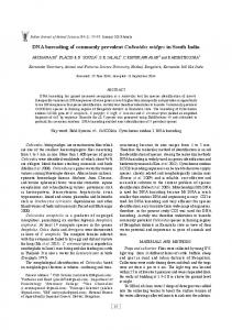
Fig. 2. FTA Elute blotting card with blood blots demonstrating the recommended amount of tissue to sample in each blotting circle.
(g) Hydroclave MC8 International).
Steam
Sterilizer
(Barnstead
(h) AirClean Systems Ductless PCR Workstation (Fisher Scientific). (i) 3730xl DNA Analyzer (Applied Biosystems). (j) 8-Channel Matrix multichannel pipette (Matrix Impact2 pipette, 15–1,250 ml, Matrix Technologies). 8. Tissue preservation (express DNA barcoding: FTA Elute blotting cards; Fig. 2): (a) Small cotton swabs, e.g., ear swabs for liquid tissue (see Note 11). (b) FTA Elute blotting cards (Whatman, GE Healthcare). 9. Tissue subsampling and lysis (express DNA barcoding: FTA Elute blotting cards): (a) Ethanol, 96%. (b) Harris Micropunch (1.2 mm, Sigma-Aldrich). (c) Harris self-healing cutting mat (Sigma Aldrich)—as support surface for blotting and punching. (d) Microplate (Eppendorf twin-tec PCR plate, Fisher Scientific). 10. DNA extraction (express DNA barcoding: FTA Elute blotting cards): (a) Swinging bucket centrifuge with microplate rotor (Thermo Scientific).
8
DNA Barcoding in Mammals
161
11. PCR (express DNA barcoding)—materials listed under item 4. 12. PCR product check (express DNA barcoding)—materials listed under item 5. 13. Cycle sequencing reaction (express DNA barcoding)—materials listed under item 6. 14. Sequencing reaction cleanup (express DNA barcoding)— materials listed under item 7. 15. Tissue preservation (forensic DNA barcoding): materials listed under item 1. 16. Tissue subsampling and alkaline lysis (forensic DNA barcoding)—materials listed under item 2. 17. DNA extraction (forensic DNA barcoding: alkaline lysis); see Note 3: (a) AL buffer: 0.1 N NaOH, 0.3 mM EDTA pH 13.0 (5 ml 1 N NaOH, 30 ml 0.5 M EDTA pH 8.0). Store in small aliquots at –20°C. (b) NT buffer: 0.1 M Tris–HCl pH 7.0 (6.06 g Trizma base, water to 500 ml). Adjust pH with HCl to 7.0; add water to the final volume. Store in small aliquots at –20°C. 18. PCR (forensic DNA barcoding)—materials listed under item 4. 19. PCR product check (forensic DNA barcoding)—materials listed under item 5. 20. Cycle sequencing reaction (forensic DNA barcoding)— materials listed under item 6. 21. Sequencing reaction cleanup (forensic DNA barcoding)— materials listed under item 7.
3. Methods 1. Tissue sampling and preservation (routine DNA barcoding): (a) Dissect and remove piece of skeletal muscle (see Note 12) with clean scissors/forceps. (b) Fill cryotube with 96% ethanol (see Note 13) and label the tube with individual specimen number (see Note 14) on the outside (ethanol-resistant ink or pencil) or on paper (use ethanol-resistant marker/pencil and acid-free paper). Ensure that at least a 10:1 ratio is maintained between fixative and tissue; e.g., the volume of tissue placed in a 2 ml cryotube should not exceed 5 × 5 × 5 mm. (c) Mince tissue in the tube thoroughly with scissors to allow fixative penetration; place label inside the tube (if applicable).
162
N.V. Ivanova et al.
(d) Between samples, remove visible tissue particles from tool tips, sterilize tool tips with detergent (e.g., ELIMINase) and rinse with several changes of water to remove detergent residue (see Note 15). (e) Replace fixative after 2–10 days of storage (see Note 16). (f) Store tissues at low temperatures (ideally, below 0°C) and away from light (see Note 17). 2. Subsampling and lysis (routine DNA barcoding; see Note 18): (a) Prefill a 96-well microplate with 30 ml (or one drop) of 96% ethanol per well. Cover plate with 12-cap strips. Do not use ethanol if the samples were previously fixed in another medium (see Note 16). (b) Assemble specimens and prepare a map, based on the alpha numeric grid of well and row location of sample locations in 96-well plate (see Note 19). (c) Using forceps, sample ca. 1 mm3 of tissue into each well of the plate. While working, keep only one row uncapped at a time to minimize the chance of error and crosscontamination. (d) Between samples, sterilize forceps by wiping with KimWipe, washing in ELIMINase and rinsing with three changes of distilled water. (e) Seal each row firmly with cap strips. (f) Prior to lysis, centrifuge plate for 1 min at 1,000 × g to remove ethanol and tissue from cap strips. (g) Remove cap strips and evaporate residual ethanol at 56°C (secure plate against workbench surface and remove caps slowly to prevent spraying of well contents). (h) Add 50 ml of Lysis Mix to each well and reseal with sterile 8-cap strips. (i) Incubate at 56°C for >12 h to allow digestion (see Notes 20 and 21). 3. DNA extraction—GF protocol for routine barcoding (see Note 22): This method utilizes a bind–wash–elute approach commonly used in commercial kits for DNA extraction, but delivers similar or even better results at a fraction of the cost. The DNA binds to a GF membrane in the presence of chaotropic salts, while contaminants are washed away using two different wash buffers. After two wash stages and drying, DNA is eluted from membrane using molecular grade water or low-salt buffer (see Note 23). (a) Centrifuge plates with lysed samples [from step 1(i)] at 1,500 × g for 1 min to remove any condensate from cap strips.
8
DNA Barcoding in Mammals
163
(b) Open cap strips (secure plate against workbench surface and remove caps slowly to prevent spraying of lysate). (c) Add 100 ml of BM buffer to each sample using multichannel pipette or Liquidator (see Note 2). (d) Mix lysate by gently pipetting; transfer lysate (about 150 ml) from microplate to corresponding wells of GF plate resting on square-well block. Seal GF plate with clear film. (e) Centrifuge at 5,000 × g for 5 min to bind DNA to GF membrane. (f ) Perform the first wash step: Add 180 ml of PWB buffer to each GF plate well. Seal with new cover and centrifuge at 5,000 × g for 2 min. (g) Perform the second wash step: Add 750 ml of WB buffer to each GF plate well. Seal with new clear film and centrifuge at 5,000 × g for 5 min. (h) Remove clear film and place GF plate over collection microplate. Incubate at 56°C for 30 min to evaporate residual ethanol. (i) Position PALL collar on collection microplate and place GF plate on top. Dispense 50–60 ml of ddH20 (prewarmed to 56°C) directly on each GF plate well membrane to elute DNA (see Note 23) and incubate at room temperature for 1 min. Seal GF plate with clear film. (j) Place plate assembly on clean square-well block to prevent collection plate cracking; centrifuge at 5,000 × g for 5 min to collect eluted DNA. (k) Remove GF plate and discard. (l) Cover DNA plate with strip cap or aluminum PCR foil. (m) Use 1–2 ml of DNA for PCR. DNA can be temporarily stored at 4 or at –20°C for long-term storage. (n) To reuse 2 ml square-well blocks, wash them with hot water and ELIMINase and rinse with deionized water. Dry and expose to UV light for >15 min prior to use. 4. PCR (routine DNA barcoding): M13-tailed primer cocktails are more effective than conventional degenerate primers, allowing barcode work on taxonomically diverse samples to be performed in a high throughput fashion. (a) Select primer cocktails, depending on application (Table 1); refer to volume proportions indicated in the column “Ratio in cocktail” of Table 1 (see also Note 24). (b) Prepare PCR reagent mix using volumes listed in Table 2.
FR1d_t1
FishR2_t1
VF2_t1
FishF2_t1
VR1i_t1
VR1_t1
VR1d_t1
LepR1_t1
VF1i_t1
VF1d_t1
VF1_t1
LepF1_t1
Primer name
Fish Cocktail [C_FishF1t1 + C_FishR1t1] TGTAAAACGACGGCCAGTCGACTAA TCATAAAGATATCGGCAC TGTAAAACGACGGCCAGTCAACCAA CCACAAAGACATTGGCAC CAGGAAACAGCTATGACACTTCAGG GTGACCGAAGAATCAGAA CAGGAAACAGCTATGACACCTCAGG GTGTCCGAARAAYCARAA
Mammal Cocktail (C_VF1LFt1 + C_VR1LRt1] TGTAAAACGACGGCCAGTATTCAAC CAATCATAAAGATATTGG TGTAAAACGACGGCCAGTTCTCAAC CAACCACAAAGACATTGG TGTAAAACGACGGCCAGTTCTCAAC CAACCACAARGAYATYGG TGTAAAACGACGGCCAGTTCTCAAC CAACCAIAAIGAIATIGG CAGGAAACAGCTATGACTAAACTTC TGGATGTCCAAAAAATCA CAGGAAACAGCTATGACTAGACTTC TGGGTGGCCRAARAAYCA CAGGAAACAGCTATGACTAGACTTC TGGGTGGCCAAAGAATCA CAGGAAACAGCTATGACTAGACTTC TGGGTGICCIAAIAAICA
Primer sequence
M13R
M13R
M13F
M13F
M13R
M13R
M13R
M13R
M13F
M13F
M13F
M13F
M13 seq primer
1
1
1
1
3
1
1
1
3
1
1
1
Ratio in cocktail
RB, EB
RB, EB
RB, EB
RB, EB
RB, EB
RB, EB
RB, EB
RB, EB
RB, EB
RB, EB
RB, EB
RB, EB
Application
Fish52
MammCOI
Thermocycler program
652
658
(46)
(46)
Product length (bp)a Reference
Table 1 Mammalian primer sets for various applications (RB routine barcoding and reference library, EB express barcoding and ecological surveys, FB forensic barcoding and museum samples)
164 N.V. Ivanova et al.
N/A N/A N/A N/A N/A N/A N/A N/A N/A N/A N/A N/A N/A N/A
Primers for forensic and express barcoding of bats TTCTCAACCAACCACAAAGACATTGG GCATGAGCTGTTACGATTACG CTGGGGCCCTATTAGGTGAT TATATCGGGTGCGCCAATTA GGGGGATTCGGTAATTGATT GGCTAGTGGTGGGTATACGG TACAGTTGAAGCTGGCGTTG AGCTCCAAGGATTGACGAAA TCTCTCTTCACCTAGCCGGA GGCAGTGATTAAAACGGATCA GCCCTCTCTCAATATCAAACAC TAGACTTCTGGGTGGCCAAAGAATCA
Sequencing primers TGTAAAACGACGGCCAGT CAGGAAACAGCTATGAC
VF1b BC1R BC2F BC2R BC3F BC3R BC4F BC4R BC5F BC5R BC6F VR1b
M13F (-21)c M13R (-27)c
RB, EB, FB FB, EB FB, EB FB, EB FB, EB FB, EB FB, EB FB, EB FB, EB FB, EB FB, EB RB, EB, FB
RB, EB RB, EB
FB, EB
FB, EB
RB, EB RB, EB
Application
Seq3.1
ExpressCOI
RatCOI
ExpressCOI
MammCOI54
COIfast
Thermocycler program
161
93
96
134
106
117
702
187
421
658
(50)
(31) This study This study This study This study This study This study This study This study This study This study (49)
(48)
This study
(12)
(47)
Product length (bp)a Reference
M13-tailed primer cocktails are more effective than conventional degenerate primers, allowing barcode work on taxonomically diverse samples to be performed in a high throughput fashion a Product lengths are given without primers b VF1/VR1 are universal vertebrate primers that can be used for some mammal species as a stand-alone primer pair; their tailed modification is used in the M13-tailed mammal cocktail c M13F/M13R are used for sequencing products generated with M13-tailed primers and their cocktails. In all other sequencing reactions, use the same primers as for the PCR reaction
N/A N/A N/A N/A N/A N/A N/A N/A N/A N/A N/A N/A
N/A N/A
N/A N/A
N/A
Rat primers [BatL5310 + R6036R] CCTACTCRGCCATTTTACCTATG ACTTCTGGGTGTCCAAAGAATCA
AquaF2
RonM_t1
BatL5310 R6036R
M13F
N/A N/A
Ratio in cocktail
N/A
Folmer degenerate [dgLCO-1490 + dgHCO-2198] GGTCAACAAATCATAAAGAYATYGG TAAACTTCAGGGTGACCAAARAAYCA
dgLCO-1490 dgHCO-2198
M13 seq primer
Forward primers for express and forensic barcoding TGTAAAACGACGGCCAGTGGMGCMC CMGATATRGCATTCCC ATCACRACCATCATYAAYATRAARCC
Primer sequence
Primer name
8 DNA Barcoding in Mammals 165
166
N.V. Ivanova et al.
Table 2 PCR reagent mix used in mammal DNA barcoding Each well (ml) regular PCR
96-Well plate (ml) regular PCR
Each well (ml) FTA elute disks
96-Well plate (ml) FTA elute disks
10% Trehalose
6.25
625.0
6.25
625.0
ddH2O
2.00
200
4.00
400
10× Buffer
1.25
125.0
1.25
125.0
50 mM MgCl2
0.625
62.5
0.625
62.5
10 mM primerA
0.125
12.5
0.125
12.5
10 mM primerB
0.125
12.5
0.125
12.5
10 mM dNTPs
0.0625
6.25
0.0625
6.25
Platinum Taq (5 U/ml)
0.060
6.0
0.060
6.0
Total
10.5
1/8 Aliquot
–
1,050 130
12.5 –
1,250 155
(c) Defrost reagents listed in Table 2 (except Taq polymerase) at room temperature; briefly vortex and spin down in a mini-centrifuge. Keep Taq polymerase at –20°C until immediately before use (see Notes 25 and 26); prior to use, spin the tube with Taq polymerase for 10–15 s in a mini-centrifuge. Do not mix Taq polymerase by vortexing or pipetting. (d) Add reagents to a 2 ml tube in volumes listed for “96-well plate” (Table 2). (e) Mix vigorously with vortex or pipette (vortexing traps liquid under the cap of tube necessitating a subsequent 15-s spin in a mini-centrifuge). (f ) Dispense 10.5 ml of PCR mix into well of the 96-well plate (the same pipette tip can be used for all transfers). (g) Add 1–2 ml of DNA template to each well (use new filter pipette tip for each sample). 5. PCR thermocycling (same for all pathways): (a) Place aluminum foil or heat-seal cover over the top of 96-well plate and centrifuge for 1 min at 1,000 × g. (b) Place the plate in a thermocycler block, close the lid and run thermocycling program appropriate for the primer cocktail employed (Table 1). Apply thermal cycling conditions from Table 3. (c) The resulting PCR plate can be stored for several weeks at 4°C or several months at –20°C until ready for sequencing.
8
DNA Barcoding in Mammals
167
Table 3 PCR programs and thermal cycling parameters for different primer sets used in mammal DNA barcoding PCR program
Primer combinations
Thermal cycling conditions
MammCOI
Mammal Cocktail (C_VF1LFt1 + C_VR1LRt1)
94°C for 2 min; 5 cycles (94°C for 30 s, 50°C for 40 s, and 72°C for 1 min); 35 additional cycles (94°C for 30 s, 55°C for 40 s, and 72°C for 1 min); final extension at 72°C for 10 min; then hold indefinitely at 10°C
Fish52
Fish Cocktail (C_FishF1t1 + C_FishR1t1)
94°C for 2 min; 40 cycles (94°C for 30 s, 52°C for 40 s, and 72°C for 1 min); final extension at 72°C for 10 min; then hold indefinitely at 10°C
MammCOI54
421 bp Fragment (RonM_t1 + C_VR1LRt1)
94°C for 2 min; 40 cycles (94°C for 30 s, 54°C for 40 s, and 72°C for 1 min); final extension at 72°C for 10 min; then hold indefinitely at 10°C
MammCOI54
419 bp Fragment (RonM_t1 + C_FishR1t1)
Same as above, but use 52°C for annealing
RatCOI
Rat primers (34) (BatL5310 + R6036R)
94°C for 2 min; 35 cycles (94°C for 30 s, 60°C for 30 s, and 72°C for 1 min); final extension at 72°C for 5 min; then hold indefinitely at 10°C
COIfast
Folmer degenerate primers (dgLCO-1490 + dgHCO-2198)
94°C for 2 min; 5 cycles (94°C for 30 s, 45°C for 40 s, and 72°C for 1 min); 35 additional cycles (94°C for 30 s, 51°C for 40 s, and 72°C for 1 min); final extension at 72°C for 10 min; then hold indefinitely at 10°C
ExpressCOI
~200 bp Fragment (AquaF2 + C_VR1LRt1) or (AquaF2 + C_FishR1t1)
94°C for 2 min; 40 cycles (94°C for 30 s, 51°C for 40 s, and 72°C for 30 s); final extension at 72°C for 10 min; then hold indefinitely at 10°C
6. PCR product check (E-gel, same for all pathways; see Note 27): (a) Preset program EG on Mother E-base; set the run time to 4 min. (b) Remove gel from package and detach plastic comb. Slide gel into electrode connections on Mother E-Base (caution: gel contains ethidium bromide; see Note 27). (c) Dispense 12 ml of ddH2O into each E-gel well. (d) Load 4 ml of PCR product into each corresponding e-gel well. (e) Begin electrophoresis by briefly pressing “pwr/prg” button. Red indicator light should change to green during the run. End of run is signaled by flashing red light and sound alarm; press and release the “pwr/prg” button when finished.
168
N.V. Ivanova et al.
Fig. 3. E-gel image of PCR products generated from an FTA Elute card.
(f ) Remove gel from base and capture a digital image (we use Alpha Imager documentation system; Fig. 3). PCR products can be visualized under any UV-emitting source. (g) If success rate is over 75%, all samples from the PCR plate should be sequenced. Success rates below 75% require hitpicking of amplified products and failure tracking (amplification of failed samples with alternative primer sets) of the remaining samples (see Note 28). Hit-picked PCR products obtained with different M13-tailed primers (e.g., mammal and fish cocktails) can be combined in the same plate for sequencing; ensure that the resulting position of rearranged samples is plotted in the new plate map. 7. Cycle sequencing (same for all pathways): (a) Defrost reagents (Table 4) at room temperature. BigDye is light sensitive and should not be exposed to light for more than a few minutes. Do not vortex undiluted BigDye stock; gently mix by pipetting instead. (b) Prepare “forward” and “reverse” sequencing mixes: Combine forward primer with reagents from Table 4 in a single 2 ml tube in proportions listed under “96-well plate”; repeat the procedure for reverse primer. Mark tubes accordingly. (c) Mix contents of each tube gently but thoroughly by pipetting or vortexing for about 5–10 s (vortexing traps liquid under the cap of tube necessitating a subsequent 15-s spin in a mini-centrifuge).
8
DNA Barcoding in Mammals
169
Table 4 Cycle sequencing reaction recipe used in mammal DNA barcoding Reagents
Each well (ml)
96-Well plate (ml)
Primer
1
104
2.5× SEQ Buffer
1.875
195
BigDye
0.25
26
ddH2O
0.875
91
10% Trehalose
5
520
Subtotal
9
936
DNA template
1–1.5
N/A
Total
10–10.5
N/A
1/8 Aliquot
115
N/A
(d) Assemble “forward” and “reverse” sequencing plates: Dispense 9.0 ml of forward-sequencing mix into each well of an empty 96-well plate; repeat the procedure for reverse mix. Use a new set of pipette tips to transfer forward- and reverse-sequencing mixes (tips can be reused between transfers within each plate). Mark plates accordingly. (e) Add 1–1.5 ml of nonpurified PCR product (see Notes 29 and 30) from the PCR plate [step 6(g)] to each well of the “forward” and “reverse” sequencing plates (use new pipette tip for each well). (f) Place aluminum foil or heat-seal cover over sequencing plates and centrifuge at 1,000 × g for 1 min to collect sequence cocktail and PCR template at the bottom of the wells. (g) Place each sequencing plate in a thermocycler block and start program Seq3.1: initial denaturation at 96°C for 2 min; followed by 30 cycles at 96°C for 30 s, annealing at 55°C for 15 s, and extension at 60°C for 4 min; followed by indefinite hold at 4°C. (h) Store processed sequencing plates for up to 1 week at 4°C in a dark enclosure to avoid degradation of light-sensitive sequencing products (see Note 31). 8. Sequencing cleanup (same for all pathways; see Note 32): (a) Measure dry Sephadex G50 with black column loader into 350 ml PALL filter plate. Prepare two plates to balance
170
N.V. Ivanova et al.
each other or prepare a single plate and appropriate counterbalance. (b) Add 300 ml molecular grade water to plate wells prefilled with Sephadex powder; leave mixture to hydrate overnight at 4°C or for 3–4 h at room temperature before use. (c) Join Sephadex plate with MicroAmp collection plate to catch water flow through and secure with two rubber bands. (d) Balance two sets of plates for centrifuging; use additional rubber bands as needed. (e) Centrifuge at 750 × g for 3 min to remove excess water and generate a packed Sephadex column. (f ) Add the entire sequencing reaction to the center of Sephadex columns by pipette. Do not insert the pipette tip into the column; dispense liquid onto the upper surface of the column. Damaging the column with a pipette tip or dispensing solution onto the side of the column may result in incomplete removal of unincorporated BigDye terminators which will adversely affect sequencing results. (g) Add 25 ml of 0.1 mM EDTA to each well of a new MicroAmp collection plate (see Note 33). (h) Elute clean sequencing reaction by attaching the collection plate containing EDTA to the bottom of Sephadex plate and securing with rubber bands. (i) Balance two sets of plates for centrifuging; use additional rubber bands as needed. (j) Centrifuge at 750 × g for 3 min and remove Sephadex plate. (k) Cover the top of collection plate with septum. (l) Place collection plate into black plate base of capillary sequencer and attach white plate retainer. (m) Stack assembled plates into ABI 3730xl capillary sequencer and import plate records using Plate Manager module of the Data Collection software (Applied Biosystems). (n) Begin sequencing run within Run Scheduler. 9. Tissue sampling and preservation (express DNA barcoding: FTA Elute blotting card; see Note 34): (a) Dissect and remove a 3–4-mm3 piece of skeletal muscle with clean scissors/forceps. If sampling from live animal (see Note 11), collect ca. 10–20 ml of blood on a cotton swab. (b) Dab muscle or cotton swab against blotting circle of the FTA Elute card. Do not oversample (see Note 35).
8
DNA Barcoding in Mammals
171
(c) If employing reusable tools, sterilize them between samples as per step 1(d); if using swabs, discard them after each sample. (d) Map the position of samples on the recording portion of the card (see Note 36). (e) Leave the card to air dry with the blotting portion opened (see Note 37). Do not expose to direct sunlight. (f) Store the card away from light at room temperature in a dry environment or in a sealed bag with desiccant (e.g., silica gel; see Note 38). 10. Tissue lysis (express DNA barcoding: FTA elute blotting card): (a) Wipe Harris mat with ethanol; open FTA Elute card and slide mat under blotting surface (filter paper portion). (b) Punch FTA elute card (filter portion only) using a 1.2 mm Harris Micropunch. (c) Place one punch into each well of 96-well microplate and create sample map as above [step 2(b)]. (d) Between sampling, clean micropunch by punching clean filter paper. 11. DNA extraction—(express DNA barcoding: FTA elute blotting card—muscle blot or blood): (a) Ensure that all supplies are ready. Protocol must be completed in timed consecutive steps (prolonged incubation in water results in DNA loss). (b) Add 100 ml of water to each well of 96-well microplate from step 10(c) using pipette or Liquidator; seal with aluminum film. Vortex plate for 10–15 s and centrifuge immediately for 1 min at 1,000 × g. (c) Aspirate and discard water from each well using pipette or Liquidator (make sure that disks remain in their wells after water is removed). Incubate open plate with disks at 56°C for 5 min to evaporate residual water. Do not cover the plate because disks easily stick to film or cap strips during movement. After drying, disks can be used directly in PCR reaction. 12. PCR (express DNA barcoding): (a) Select primer cocktails for express barcoding (Table 1); refer to volume proportions indicated in the column “Ratio in cocktail” (see also Note 24). (b) Prepare PCR reagent mix using volumes indicated for FTA Elute disks in Table 2. (c) Follow step 4(a–d) for routine barcoding.
172
N.V. Ivanova et al.
(d) Dispense 12.5 ml of PCR mix into each well of plate containing prewashed FTA disks. Change pipette tips after each well. 13. PCR thermocycling (express DNA barcoding)—procedures listed under step 5. 14. PCR product check (express DNA barcoding)—procedures listed under step 6; see also Note 27. 15. Cycle sequencing (express DNA barcoding)—procedures listed under step 7. 16. Sequencing cleanup (express DNA barcoding)—procedures listed under step 8; see also Note 32. 17. Tissue sampling and preservation (forensic DNA barcoding; if applicable)—procedures listed under step 1. 18. Tissue subsampling and lysis (forensic DNA barcoding: alkaline lysis protocol). (a) Follow subsampling step 2(a–g), if applicable; when subsampling for immediate lysis, prefill plate with AL buffer instead of ethanol in step 2(a). 19. DNA extraction (forensic DNA barcoding: alkaline lysis protocol): (a) Add 35 ml of AL buffer to ~0.5–1 mm3 of fresh skeletal muscle or hairs with follicles in tubes or 96-well microplate. (b) Incubate for 5 min in a thermocycler at 95°C or in freshly boiled water in a water bath. (c) Centrifuge for 1 min to remove condensate from caps or plate cover. (d) Add 65 ml of NT buffer and mix by pipetting. (e) Use 1–2 ml of crude lysate for PCR reaction (label PCR plate for reference using sticker or alcohol-resistant marker). Store at –20°C for up to 2 weeks (see Note 39). 20. PCR (forensic DNA barcoding): (a) Select primer cocktails for forensic barcoding (Table 1); refer to volume proportions indicated in the column “Ratio in cocktail” (see also Note 24). (b) Prepare PCR reagent mix using volumes indicated for regular PCR in Table 2. (c) Follow step 4(a–g) for routine barcoding. 21. PCR thermocycling (forensic DNA barcoding)—procedures listed under step 5. 22. PCR product check (forensic DNA barcoding)—procedures listed under step 6; see also Note 27.
8
DNA Barcoding in Mammals
173
23. Cycle sequencing (forensic DNA barcoding)—procedures listed under step 7. 24. Sequencing cleanup (forensic DNA barcoding)—procedures listed under step 8; see also Note 32.
4. Notes 1. Do not use forceps with cross-hatched or serrated tips due to the difficulty of removing tissue remains during sterilization. 2. There are multiple manufacturers of single- and multichannel pipettes; however, pipetting accuracy and design features may vary. The most important feature for a multichannel pipette is the uniformity of tip loading and unloading. Pipettes should be made from durable material; if O-rings are used to seal tips, frequently check their integrity and have spare O-rings available for replacement. In a high throughput facility, Liquidator 96 from Rainin can be utilized for convenient and fast liquid transfers; this device offers manual pipetting of 96 samples at a time. Although the recommended range volume of the Liquidator is 5–200 ml, it can still be used for volumes of 1.5–2 ml. 3. All chemicals used for tissue lysis and DNA extraction should be of molecular biology grade or equivalent—of the highest purity. Wash all labware with ELIMINase and rinse with distilled or deionized dH2O. Weigh reagents using a clean spatula. Use molecular grade doubly distilled (dd) H2O in all buffer formulations. Millipore MilliQ purified water (18 MW) can be used for lysis and DNA extraction buffers, while commercially manufactured molecular grade water is preferred for PCR and sequencing. Do not use DEPC-treated water, as it may inhibit PCR and cycle sequencing reactions. Store molecular grade water in small aliquots to prevent contamination. Filter buffers through a 0.2 mM Nalgene filter into a clean sterile bottle; prepare working aliquots of smaller volume (e.g., 100 ml). Store stock solutions and working aliquots at 4°C. 4. A PALL collar is used to secure a GF plate on top of the Microplate during the elution stage. 5. The centrifuge for DNA extraction should have deep buckets to handle filter plate assemblies and should deliver at least 5,000 × g. 6. To prepare stock solutions and buffers, dissolve salts and other components in a smaller volume than that of the final solution and then add water to attain the final volume. 7. GuSCN does not dissolve without heating (this is an endothermic reaction). Caution: All GuSCN solutions are irritants
174
N.V. Ivanova et al.
and toxic and should be disposed of with care. Use nitrile gloves and goggles at all times and protective mask while handling powder; avoid contact with acids to prevent release of cyanide gas. 8. Some trehalose brands contain traces of pig DNA which may contaminate reactions. 9. To minimize the risk of contamination, do not mix 100 mM stocks by pipetting. After adding water, incubate tubes for 15–30 min at room temperature, vortex for 30 s, and briefly spin tubes in a tabletop mini-centrifuge prior to opening. 10. Although non-skirted microplates can be used in PCR setup, we recommend hard-shell skirted microplates, such as Eppendorf twin-tec 96 PCR plate, to prevent spraying of PCR products while reopening cap strips. Skirted microplates are compatible with heat sealing film, which is more convenient to use in a high throughput setting. Alternatively, 8-cap strips, sealing mats, aluminum-sealing film, or PCR-grade clear film can be used to cover plates prior to thermal cycling. Prior to running actual samples, test the sealing technique by thermal cycling of a plate filled with water. Most clear sealing films do not provide a good seal. 11. Although express barcoding is instrumental in analyzing tissue taken from live mammals (17, 18), this chapter does not cover the tools or methods used in biopsy and live sampling of liquid tissue or the respective animal handling techniques; refer to specialized literature (e.g., refs. 34, 35) for details. 12. While there are numerous guidelines on mammal tissue preservation, many existing tissue collections and protocols focus on studies of chemical contaminants or other specialized tasks (e.g., allozyme analyses) and promote collecting tissue from internal organs, such as liver (e.g., ref. 36). Both liver and muscle have high mitochondria content (37); however, our experience corroborates other studies (38, 39) which indicate that internal organs (e.g., liver and kidney) are suboptimal sources of mitochondrial DNA. The yield of full-length mitochondrial PCR products is lower or complicated by pseudogenes (numts) Fig. 4 which impede or prevent accurate sequencing. These effects appear inconsistent between samples, presumably dependent on the time between euthanasia and tissue collection, type and quality of initial fixation, and subsequent storage conditions. Liver is characterized by the early (