Dec 30, 1988 - (CDR 62, cerebellar degeneration-related 62-kDa protein) and a minor antigen of 34 .... 2.5) and the active fractions were combined, neutralized, and stored at -70'C. .... in cerebral cortex, basal ganglia, midbrain, and spinal cord. A. I. 97 -. 68- .... In the case ofa similar disorder, subacute sensory neuropathy ...
Proc. Natl. Acad. Sci. USA Vol. 86, pp. 2873-2877, April 1989 Medical Sciences
Characterization of a cDNA encoding a 34-kDa Purkinje neuron protein recognized by sera from patients with paraneoplastic cerebellar degeneration (tumor antigens/remote effects of cancer/autoantibodies/autoimmunity)
HENRY M. FURNEAUX*t, EDWARD J. DROPCHO*t, DENISE BARBUT*t, YAO-TSENG CHEN§, MARC K. ROSENBLUM¶, LLOYD J. OLD§, AND JEROME B. POSNER*t *George C. Cotzias Laboratory of Neuro-Oncology, Samuel Freeman Laboratory, and IDepartment of Pathology, Memorial Sloan-Kettering Cancer Center, 1275 York Avenue, New York, NY 10021; and tDepaartment of Neurology, Cornell University Medical College, New York, NY 10021
Contributed by Lloyd J. Old, December 30, 1988
ABSTRACT Paraneoplastic cerebellar degeneration is a neurological disorder of unknown cause occurring in patients with an identified or occult cancer. An autoimmune etiology is likely since autoantibodies directed against the Purkinje cells of the cerebellum have been found in the serum and cerebrospinal fluid of some patients. Two Purkinje cell-specific antigens are recognized by these autoantibodies, a major antigen of 62 kDa (CDR 62, cerebellar degeneration-related 62-kDa protein) and a minor antigen of 34 kDa (CDR 34). Our previous studies have described the isolation and characterization of a human cerebellar cDNA that encodes an epitope recognized by sera from patients with paraneoplastic cerebellar degeneration. We have now established by two independent methods that this gene is uniquely expressed in Purk' 'e cells of the cerebellum and corresponds to the minor antigen CDR 34. This antigen is also expressed in tumor tissue from a patient with paraneoplastic cerebellar degeneration.
91% of its sequence consisted of 34 almost exact tandem repeats of 6 amino acids (Fig. 1). In this report we present evidence that the cloned cDNA sequence corresponds to the CDR 34 antigen and that this antigen is expressed in tumor tissue from PCD patients. A portion of this work was presented at the annual meeting of the American Neurological Association. 1
MATERIALS AND METHODS Human Tissue. Human cerebellum was obtained at autopsy. Purkinje neurons were purified from cerebellar tissue according to the method of Yanagihara and Hamburger (8). Preparation of Agtll CDR2 Fusion Protein. Lysogens of recombinant CDR2 phage were prepared in Escherichia coli Y1089 (9) at a multiplicity of infection of 5. Cultures were grown at 320C to an OD550 of 0.5, followed by induction of lytic growth at 440C for 30 min, addition of isopropyl f3-D-thiogalactoside to a concentration of 10 mM, and incubation at 370C for 2 hr. Bacterial pellets were lysed in cold Tris'HCl/EDTA/lysozyme buffer, sonicated, and centrifuged. The supernatant was retained and stored at -70'C. Western Blotting. The indicated amounts of protein extract were electrophoresed on 10o SDS/polyacrylamide gels (10) and then transferred to nitrocellulose as described by Towbin et al. (11). The nitrocellulose filters were blocked by incubation with 5% Blotto and then incubated with the appropriate amount of antibody for 2 hr at room temperature. After washing with TBST (0.1 M Tris, pH 8.0/0.2 M NaCl/0.2% Triton X-100/0.1% bovine serum albumin), the nitrocellulose filters were incubated with 125I-labeled protein A (0.1 ,Ci/ml; 1 Ci = 37 GBq) for 1 hr at room temperature, washed with TBST, and apposed to x-ray film. Affiity Purification of Rabbit Anti-Peptide IgG. Antisera recovered from rabbits immunized with a synthetic peptide corresponding to amino acid residues 149-165 of the cloned CDR 34 gene was the kind gift of R. Lerner (Scripps Institute). A peptide affinity matrix was prepared by coupling 100 ,ug of peptide to 1 ml of Affi-Gel 10 according to the manufacturer's instructions. The column was then treated with ethanolamine (0.1%) to block remaining reactive groups and washed extensively (10 column volumes) with phosphate-buffered saline (PBS)/0.1% Nonidet P-40 (NP-40). Rabbit serum (2 ml) was then applied to the column and
Paraneoplastic cerebellar degeneration (PCD) is a rare and disabling neurologic disorder characterized by the unexplained loss of cerebellar Purkinje neurons in some patients harboring small, often asymptomatic cancers of lung, breast, or ovary or Hodgkin disease (1). The etiology of this syndrome is unknown but recent evidence suggests that PCD results from autoimmune-mediated damage to the nervous system. High-titer autoantibodies that react specifically with cerebellar Purkinje neurons have been found in the serum and cerebrospinal fluid of several patients with a clinical diagnosis of PCD (2, 3). Electrophoretic transfer (Western) blot analysis of purified Purkinje neurons has shown that the autoantibodies recognize at least two proteins: a major antigen of 62 kDa (CDR 62, cerebellar degeneration-related 62-kDa protein) and a minor antigen of 34 kDa (CDR 34) (4). These autoantibodies are a marker for PCD associated with ovarian and breast cancer (5). We have not found these antibodies in patients with other cerebellar diseases, other paraneoplastic syndromes, or cancer without neurological symptoms or in normal subjects (5). There are reports of an anti-Purkinje cell autoantibody in three patients with ovarian cancer without PCD (6), but these sera were not subjected to Western blot analysis and the identity of these autoantibodies remains unknown. We have previously reported the isolation of a cDNA clone (pCDR2) from a cerebellar expression library that encodes an antigenic determinant recognized by serum from a PCD patient (7). Sequence analysis of a full-length cDNA clone (pCDR13) revealed an open reading frame of 223 amino acids. The most striking feature of this putative protein was that
Abbreviations: PCD, paraneoplastic cerebellar degeneration: CDR,
cerebellar degeneration-related.
tPresent address: Department of Neurology, University of Alabama, Birmingham, AL 35294.
IFurneaux, H. M., Dropcho, E. J., Barbut, D., Chen, Y.-T., Rosen-
The publication costs of this article were defrayed in part by page charge payment. This article must therefore be hereby marked "advertisement" in accordance with 18 U.S.C. §1734 solely to indicate this fact.
blum, M. K., Old, L. J. & Posner, J. B., 113th Annual Meeting of the American Neurological Association, October 2-5, 1988, Phila-
delphia.
2873
2874
Medical Sciences: Furneaux et al. p
NH2
I-h
l
p
p
50
Proc. Natl. Acad. Sci. USA 86 (1989)
p
p
L
p
p
100
J
pC DR2
150
200
COOH
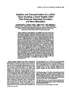
FIG. 1. Structure of the open reading frame encoded by the full-length cerebellar cDNA clone (pCDR13). Black triangles represent the 6-amino acid motif (Leu-Leu-Glu-Asp-Val-Asp) repeated throughout almost the entire coding region. The region present in the f3-galactosidase fusion protein (pCDR2) is shown enclosed by brackets.
unbound IgG was removed by extensive washing with PBS/0.1% NP-40 (10 column volumes). Specific IgG was then eluted with 0.1 M sodium citrate (pH 2.5) and the active fractions were combined, neutralized, and stored at -70'C. Immunobistochemistry. Six-micron frozen sections were cut on a cryostat, fixed for 10 min in acetone, and then washed with PBS. Sections were then treated at room temperature with 0.3% hydrogen peroxide diluted in PBS. After washing with PBS the sections were incubated with 10% goat serum (10 min at room temperature) to suppress any nonspecific binding. After excess serum was removed the sections were incubated (18 hr at 40C) with the indicated amounts of antibody diluted in PBS/1% bovine serum albumin. After washing with PBS the sections were incubated with either goat anti-human IgG or goat anti-rabbit IgG biotinylated secondary antibody. After washing with PBS the sections were incubated with avidin-biotin peroxidase complex (Vectastain ABC complex, Vector Laboratories) for 30 min. The sections were then washed with PBS and the substrate reactions were developed with 0.05% diaminobenzidine.
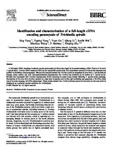
RESULTS Identification of the Cerebellar ODNA Clone Recognized by PCD Sera. To identify the cDNA clone previously isolated, human autoantibodies were affinity purified from the fusion protein produced by the clone and used for immunohistochemistry and Western blot analysis. Bacterial lysates containing the pCDR2 p-galactosidase fusion protein were separated by preparative 8% polyacrylamide/SDS gel electrophoresis. A strip from this preparative gel, stained with Coomassie blue, shows the 140-kDa fusion protein (Fig. 2A). The separated proteins were then transferred to nitrocellulose, blocked with 3% bovine serum albumin, and incubated (2 hr at room temperature, 1:500 dilution) with serum from a PCD patient. The nitrocellulose sheet was then cut into three strips (I, II, III) corresponding to regions of defined molecular weight (Fig. 2A). Strip I contains the 140-kDa fgalactosidase pCDR2 fusion protein. Strips II and III contain other bacterial proteins and therefore serve as a control for nonspecific antibody binding. Antibodies bound to these strips were eluted by brief exposure to glycine-HCl (pH 2.5) followed by neutralization and dialysis against PBS. The Purkinje cell antigens recognized by these eluates were then examined by Western blot analysis. Fig. 2B, lane 5, shows that only the eluate from strip I was reactive and recognized a 34-kDa antigen. The nonreactivity of normal human serum and the staining of the 62- and 34-kDa antigens by sera from PCD patients are shown in Fig. 2B, lanes 1 and 2, respectively. In addition to the nonreactivity of the eluates from strips II and III, the failure of the eluate from strip I to recognize the 62-kDa Purkinje cell antigen rules out nonspecific binding of antibody to the fusion protein. We next examined the cerebellar localization of the antigen recognized by the fusion protein affinity-purified antibody preparation. Frozen sections of human cerebellum were incubated with antibody eluate from strips I and II. Antibody binding was visualized by the avidin-biotin immunoperoxidase methods. Fig. 3 shows a typical section of human a
cerebellum. No staining was observed with the eluate from strip II (the control for nonspecific binding). Incubation with antibody eluate from strip I resulted in the characteristic Purkinje cell-specific staining pattern seen with sera from PCD patients. Immunohistochemical and Western Blot Analysis Using a Rabbit Antibody Generated Against the Cloned Gene Product. To confirm and extend the above findings an antiserum to the protein predicted from the cDNA open reading frame was prepared. A synthetic peptide corresponding to amino acid residues 149-165 (Glu-Asp-Val-Asp-Phe-Gln-Glu-AspPro-Asn-Tyr-Pro-Glu-Asp-Leu-Asp-Cys) (Fig. 1) was synthesized. Antisera recovered from rabbits immunized with the peptide coupled to keyhole limpet hemocyanin were purified by peptide affinity chromatography. Extracts from purified Purkinje cells were then fractionated by SDS/polyacrylamide gel electrophoresis and transferred to nitrocellulose. Immunostaining of these blots with the purified rabbit anti-peptide IgG revealed a polypeptide of 34 kDa (Fig. 4, lane 4). No staining was observed either with rabbit preimmune IgG or with anti-peptide IgG preabsorbed with peptide (Fig. 4, lanes 3 and 5). The 62-kDa Purkinje cell antigen identified by the PCD sera (Fig. 4, lane 4) was not stained by the anti-peptide rabbit IgG. To determine the distribution of the 34-kDa polypeptide in the central nervous system, sections of human brain were stained with the rabbit anti-peptide IgG using the avidinbiotin immunoperoxidase method. No staining was observed in cerebral cortex, basal ganglia, midbrain, and spinal cord
B
A -
I
97 -
6868-
3046
-
2
4
5
30 -
FIG. 2. (A) Coomassie blue-stained strip of the preparative 8% polyacrylamide/SDS gel used to separate the protein present in a bacterial lysate of pCDR2. The arrow indicates the 140-kDa galactosidase fusion protein. (B) Western blot analysis of human autoantibodies purified by their recognition of the pCDR2 fusion protein. Eluted antibodies from regions I-III were incubated (2 hr at room temperature) with nitrocellulose strips containing Purkinje cell proteins (50 ug of prQtein per strip). Lane 1, normal human cerebrospinal fluid (1:50); lane 2, cerebrospinal fluid from a patient with PCD (1:50); lane 3, human antibody eluate from region III; lane 4, human antibody eluate from region II; lane 5, human antibody eluate from region 1. Molecular masses are given in kDa. (-
Medical Sciences: Furneaux et al.
A-
Proc. Natl. Acad. Sci. USA 86 (1989)
2875
U 0
f.
r
S lb
e
;S
4E0o
S A
t
S. 4
I
a
I
a
0 S
-
... };..
.!..
a
% -w ;h~*~p#
-.
-S:;o, a--: fy4_*^, *t~~~~~~~~~~~~~~~~~~~~~~~~~~~ *~~~~~.
,#4.....75
4P
.
S-4
*1 V
*
.0 tL
Fio. 3. (A) Antibodies recognizing other bacterial proteins do not react. (B) Human antibodies eluted from the fusion protein react with the cytoplasm of Purkinje cells. (ABC-immunoperoxidase with hematoxylin counterstain; x360 in A, x90 in B.)
(data not shown). Intense staining was, however, observed in cells of the cerebellum (Fig. 5A). This was
abolished by preadsorption of the rabbit anti-peptide IgG with a molar excess of peptide (Fig. SB).
the Purkinje
Fia. 4. Western blot analysis of Purkiije cell proteins recognized by rabbit ati-peptide atibody. Blots of Purkinje cell protein were carried out
97 -
68-
as
described in the
ing 50
pg
legend
to
Fig. 2. Strips contain-
of Purkinje cell protein
were
incubated
with normal human serum, 1:1000 (lane 1);
Ig(
_w
30
2 t -5: i^ev 211
2
3
4
serum
from a patient with PCD, 1:1000 (lane 2); 1 pg of rabbit preimmune per ml (lane 3); 1 of rabbit anti-peptide IgG per ml (lane 4); 1 pg of rabbit anti-peptide IgG per ml preadsorbed with a molar excess of synthetic peptide (lane 5). Molecular 5
massesaregivenin kDa.
pLg
2876
.
Medical Sciences: Furneaux et al.
A
A *
S
I
I'
I
4
I
. .:
.4-
9
41
1. XI
jb
I
a
.~ ~ 1,
.'
'
w in
Or OF
.1 v M
..
'a
I
9~ 94
I
*4
A
I ..
'
4j
I
*
t
0
.8 4.f.,:1
?5.
Proc. Natl. Acad Sci. USA 86 (1989)
4,#
~0
:s j.
.
Ijx.
.
.1
4
IA
*~~~
t
M
..
1
I. 4
V.
:
46
f, I*
E
E
N
-'-
C.£
...: ik
A
FIG. 5. (A) Rabbit antibodies to the synthetic peptide react with the cytoplasm of human Purkinje cells. (B) Preadsorption with a molar excess of peptide abolishes reactivity. (ABC-immunoperoxidase with 1 Ag of rabbit anti-IgG per ml, hematoxylin counterstain; x90.)
We conclude from these two independent approaches that the isolated cDNA clone corresponds to the 34-kDa Purkinje cell antigen recognized by sera from PCD patients. This assignation is in good agreement with the predicted open reading frame (223 residues) and the size of the mRNA (1.3 kilobases) detected by Northern blot analysis (7). Expression of CDR 34 in Tumor Tissue from a PCD Patient. We hypothesize that the antigenic stimulus for the production of anti-Purkinje cell antibodies resulted from shared common antigens between tumor tissue and Purkinje neurons so that an immune response to tumor antigens evokes antibodies that crossreact with Purkinje cells. We have recently shown by Western blot analysis using high-titer human paraneoplastic serum that the CDR 62 antigen is strongly expressed in tumor tissue from PCD patients (6/6). CDR 62 was not detected in normal tissue or comparable tumor tissue from patients
without PCD (0/21) (H.M.F. and J.B.P., unpublished results). We were unable to demonstrate the presence of the minor 34-kDa Purkinje cell antigen in tumor tissue using human serum because of the presence of other nonspecific bands of similar size. To demonstrate the 34-kDa antigen we incubated Western blots of normal and tumor tissues with the purified rabbit anti-34-kDa antibody. Fig. 6 shows that the 34-kDa antigen is expressed in tumor tissue from PCD patients but is not expressed in normal tissue or tumor tissue from a non-PCD individual. The 55-kDa band present in all breast tissues is observed on incubation with 125I-labeled protein A only and corresponds to the heavy chain of IgG. The 34-kDa protein identified by the rabbit antisera was not detected in extracts of cortical neurons, normal liver, small cell lung carcinoma, or normal ovary (data not- shown).
Medical Sciences: Furneaux et al. 116 68
Proc. Natl. Acad. Sci. USA 86 (1989)
-
E -
l
W
FIG. 6. Western blot analysis of normal and tumor tissues using the rabbit anti-peptide antibody. All nitrocellulose strips were incubated with 1 A&g of purified rabbit anti-peptide IgG per ml. Lane 1,
20 ,tg of Purkinje cell protein; lane 2, 100
j&g of normal
lane 3, 100
,.g
breast tissue;
of breast tumor
tissue; lane 4, 100
,.g of breast
tumor from a patient with PCD.
jet 9P
30
Only lanes 1 and 4 show the 34kDa band.
1
2
3
4
The 55-kDa band in
lanes 2-4 is heavy chain IgG. Molecular masses are given in kDa.
DISCUSSION PCD is a nervous system disorder provoked by neoplasms (1). The presence of autoantibodies that specifically recognize two Purkinje cell antigens (CDR 62, CDR 34) is an excellent marker for this syndrome when associated with ovarian or breast cancer (5). The occurrence of autoantibodies specifically directed against Purkinje neurons, whose loss is characteristic of this syndrome, is a strong indication of an autoimmune etiology. In this report we describe the further characterization of a cerebellar cDNA clone that was previously isolated by screening a Agtll cerebellar cDNA library with high-titer serum from a PCD patient. By using two independent methods we have shown that the protein encoded by this cerebellar cDNA clone is specifically expressed in Purkinje cells and corresponds to CDR 34. In our previous studies on the expression of CDR 34 mRNA in cerebrum and cerebellar tissue we reported a modest (=lO-fold) enrichment of CDR 34 mRNA in cerebellar tissue. These data are not in conflict with the immunohistochemical localization of CDR 34 presented here. Purkinje cell mRNA represents only a 2% total cerebellar mRNA, indicating a 500-fold enrichment of CDR 34 mRNA in Purkinje cells. The small (relative to Purkinje cells) amount of CDR 34 mRNA expressed in noncerebellar tissue is, however, of interest. Our immunohistochemical analysis is not sensitive enough to detect the corresponding amount of CDR 34 protein. Future in situ hybridization studies must address the possibility that a very small subset of cortical neurons expresses CDR 34. PCD is a member of a family of paraneoplastic disorders in which the presumed pathological autoimmune response is thought to be provoked by neural antigen expression in tumor tissue (1). In the case of a similar disorder, subacute sensory neuropathy, there is good evidence that the autoimmune response is generated by the expression of a 37-kDa neural antigen in small cell lung cancer tissue (12). Similarly, in the case of Lambert-Eaton syndrome, there is clear evidence of pathological autoimmune response directed against the presynaptic Ca2+ channels (13). This autoimmune response is presumably generated by the expression of those Ca2+ channels in the associated neoplasm, small cell lung cancer. Prior to these studies the antigenic stimulus that results in the production of anti-Purkinje cell antibodies in PCD had not been identified. Our present studies and our observations on the expression of CDR 62 in PCD tumor tissue (H.M.F. and J.B.P., unpublished observations) strongly suggest that this autoimmune response is provoked by the expression of these neural antigens in tumor tissue. The interesting and crucial
2877
question is why such a response is evoked against selfantigens. One answer may be that since the brain is an immunologically privileged organ (9), the expression of any brain protein outside of the central nervous system could result in strong autoimmune reactions. A second possibility is that the tumor antigens are not completely identical to the neural antigens. Alteration of a small number of amino acids may be sufficient to render these antigens "non-self' and thereby provoke an immune response. Isolation and characterization ofthe tumor antigen cDNA and determination of its nucleic acid sequence will be necessary to answer this question. In addition to its relevance to PCD, the cloning of this gene extends the list of known genes that are exclusively expressed in the Purkinje cells of the cerebellum. This list includes cGMP-dependent protein kinase (14), protein kinase C (15), PBP-19 (16), and L7 (17). Since PCD tumor cells express at least two Purkinje cell-specific genes (CDR 34, CDR 62), it may be informative to examine the expression of other Purkinje neuron-specific genes in these rare and interesting tumors. We are grateful to Dr. Richard Lerner and Ms. Beverly Hay of Scripps Institute for synthesizing peptides and supplying us with the rabbit sera generated against those peptides, to F. Yee for expert technical assistance, to B. Kaufman and M. Ryon for artwork, and to Dr. Francesc Graus, Department of Neurology, Clinic Hospital, Barcelona, Spain, who supplied tumor tissue from one of the PCD patients. This research was supported by Grant NS 26064 from the National Institute of Neurological and Communicative Disorders and Stroke, the Samuel Freeman Charitable Trust, Grant CA-08748 from the National Cancer Institute, and BIONET National Computer Resource for Molecular Biology, Grant 1 U41 RR-01685-03, sponsored by the National Institutes of Health. E.J.D. was supported by the Charles H. Revson Foundation and by the Association for Brain Tumor Research. 1. Anderson, N. E., Cunningham, J. M. & Posner, J. B. (1987) Critical Reviews in Clinical Neurobiology, ed. Roses, A. (CRC, Boca Raton, FL). 2. Greenlee, J. F. & Brashear, H. R. (1983) Ann. Neurol. 14, 6063. 3. Jaeckle, K. A., Graus, F., Houghton, A. W., Nielson, S. L. & Posner, J. B. (1985) Ann. Neurol. 18, 592-600. 4. Cunningham, J. M., Graus, F., Anderson, N. E. & Posner, J. B. (1986) Neurology 26, 1163-1168. 5. Anderson, N. E., Rosenblum, M. K. & Posner, J. B. (1988) Ann. Neurol. 24, 559-567. 6. Brashear, H. R. & Greenlee, J. E. (1985) Neurology 35, 174. 7. Dropcho, E. J., Chen, Y.-T., Posner, J. B. & Old, L. J. (1987) Proc. Natl. Acad. Sci. USA 84, 4552-4556. 8. Yanagihara, T. & Hamburger, A. (1973) Brain Res. 59, 445448. 9. Medawar, P. B. (1948) Exp. Pathol. 29, 58. 10. Laemmli, U. K. (1970) Nature (London) 227, 680-685. 11. Towbin, H., Staehelin, T. & Gordon, J. (1979) Proc. Natl. Acad. Sci. USA 76, 4350-4354. 12. Anderson, N. E., Rosenblum, M. K., Graus, F., Wiley, R. G. & Posner, J. B. (1988) Neurology 38, 1391-1398. 13. Kim, Y. I. & Nehr, L. (1988) Science 239, 405-408. 14. Levitt, P., Rakic, P., DeCamilli, P. & Greengard, P. (1984) J. Neurosci. 4, 2553-2564. 15. Brendr, S. J., Niedel, J. B., Bell, R. M. & Young, W. S. (1987) Cell 49, 57-63. 16. Ziai, R., Pan, Y.-C. E., Holmes, J. D., Sangameswaran, L. & Morgan, J. I. (1986) Proc. Natl. Acad. Sci. USA 83, 8420-8423. 17. Oberdick, J., Levinthal, F. & Levinthal, C. (1988) Neuron 1, 367-376.