ARTICLES
Characterization of Blo t 11 Monoclonal Antibodies with Constant Region Mutations John Donnie A. Ramos1*, Nge Cheong2, Kaw Yan Chua2 Research Center for the Natural Sciences, University of Santo Tomas, España St., Manila 1008, Philippines 2 Department of Paediatrics, Yong Yoo Lin School of Medicine, Lower Kent Ridge, National University of Singapore, Singapore 119074 1
S
omatic hypermutation (SHM) is a hallmark of antibody-secreting B cells used to produce mature and high affinity antibodies. SHM takes place in the variable region and within the 1.5 kb downstream region of the immunoglobulin (Ig) gene promoter but no mutations have been thus far reported in the constant region. This study reports the characterization of DNA immunization-generated hybridoma clones secreting functional and Blo t 11-specific monoclonal antibodies but with mutations on the constant region of the Ig gene. The nucleotide sequence of the constant region of the Ig gene of four mutant hybridoma clones secreting Blo t 11-specific monoclonal antibodies that are total Ig-positive was characterized. The antibodies had no detectable isotype/subclass by enzyme-linked immunosorbent assay (ELISA). A series of transition, transversion, deletion and insertion mutations were detected on exons 3 and 4 but not on exons 1 and 2 of the Ig gene. Activation-induced cytidine deaminase (AID) transcripts were observed in all mutant hybridomas examined. Sequence analysis showed that 7 and 9 of the observed mutations in clones P1C9 and P1A6, respectively, were located on RGYW and WRCY hotspots where AID have been reported to occur at high frequencies. The results presented in this study may represent a novel observation related to Ig gene SHM and maturation that may be associated with DNA immunization. INTRODUCTION The generation of enormous antibody diversity by the mammalian immune system is a result of complex genetic *To whom the correspondence should be addressed. Email:
[email protected] Received: July 18, 2009 Revised: September 19, 2009 Accepted: September 22, 2009 Editor-in-Charge: Eduardo A. Padlan
38
diversification processes undergone by immunoglobulin (Ig) genes. In response to a given antigen, the Ig genes of B cells undergo somatic hypermutation (SHM), gene conversion and class-switch recombination (Weill and Reynaud 1996). These specialized genetic events are essential to produce mature and high affinity antibodies. Antibody-diversification mechanisms in B cells involve the action of activation-induced cytidine deaminase (AID) and mismatch repair (MMR) systems (Martin and Scharff 2002). SHM takes place in the variable region and within the 1.5 kb downstream region of the Ig gene promoter (Rada et al. 1997). Although the activity of AID to initiate point mutations has been reported to occur in the variable region and its vicinity, AID-induced mutations have been reported in other genes and even in non-B cells (Yoshikawa et al. 2002; Ramiro et al. 2002). AID expression in bronchial biopsies with asthma (Takhar P et al. 2007) and in nasal mucosa cells from allergic rhinitis patients (Coker et al. 2003) were likewise implicated in class switch recombination to IgE antibodies. Positive association between polymorphisms in the 5’-flanking and coding regions of the AID gene, the symptoms of atopic asthma and the regulation of total serum IgE were similarly established (Noguchi et al. 2001). We reported previously the production of monoclonal antibodies by DNA immunization (Ramos et al. 2004) against an important high molecular weight house dust mite (HDM) allergen Blo t 11 (Ramos et al. 2001). Six Blo t 11 monoclonal antibodies were generated and used for the immunoaffinity purification of native Blo t 11 and the development of sandwich enzyme-linked immunosorbent assay (ELISA) for Blo t 11 detection and quantitation. However, a significant number of the generated hybridoma clones secreted monoclonal antibodies were total Ig-positive but with no detectable isotype/subclass by ELISA (JD Ramos, unpublished observations). We report in this paper the characterization of DNA immunization-generated hybridoma clones secreting functional and antigen specific monoclonal antibodies but with mutations on the constant region of the Ig gene.
Philippine Science Letters
Vol.2 | No.1 | 2009
EXPERIMENTAL DNA construct The 2625 bp open reading frame coding for the FL-Blo t 11 was amplified by PCR and ligated into pCI mammalian expression vector containing the Der p 5 leader sequence (Dp5LS) as described previously (Wolfowicz et al. 2003). The ligated construct was transformed into Escherichia coli DH5α (Gibco BRL, Rockville, MD, USA) and plasmids were isolated and purified using QIAGEN Plasmid Giga Kit (Qiagen, Hilden, Germany). Plasmids were sequenced using TM the ABI Prism Automated DNA Sequencer (PE Applied Biosystems, Foster City, CA, USA). Animals Five 6-week old female BALB/cJ mice were purchased from the Animal Holding Unit of the National University Singapore. Mice were housed under conventional conditions and animal experiments were performed according to Institutional Guidelines for Animal Care and Handling, National University of Singapore, Singapore. DNA immunization Mice were immunized intramuscularly with 50 µg of pCI-Dp5LS-Blo t 11 plasmid DNA on the quadriceps muscle of the hindleg followed by electroporation using the ECM 830 apparatus (BTX, Genetronics Inc., San Diego, CA, USA) as previously described (Ramos et al. 2004). Briefly, electroporation was performed with a 0.5 cm 2-needle array inserted approximately two mm deep into the muscles that delivered 4 pulses of 82 mV for 20 msec with 200 msec intervals. Each mouse received 2 booster doses at days 14 and 28. Blood was extracted infra-orbitaly every week to monitor the titer of Blo t 11-specific antibody production by enzyme-linked immunosorbent assay (ELISA). Cell fusion and cloning of hybridomas Cell fusion and cloning of hybridomas were performed using the ClonaCell-HY Hybridoma Cloning Kit (StemCell 7 Technologies Inc., Vancouver, Canada). In brief, 2 x 10 viable myeloma cells, a subclone of P3X63Ag8.653 (ATCC, Manassas, VA, USA), were resuspended in 30 mL of Medium A. Simultaneously, splenocytes from 2 mice with high titer of Blo t 11 antibody production were separated into single cell suspension by mechanical digestion followed by resuspension 8 in Medium B. A total of 1 x 10 viable splenocytes were fused with the previously prepared myeloma cells using the supplied PEG solution and Medium B. Hybridomas were incubated overnight at 37°C in Medium C. Hybridoma clones were plated using methylcellulose-based Medium D and were incubated for 14 days at 37°C. Individual colonies were picked and cultured in 96-well Falcon® tissue culture plates (Becton Dickinson Labware, Franklin Lakes, NJ, USA) containing 200 µL of Medium E. Media A, B, C, D, and E; TM and PEG solution were supplied as part of the ClonaCell HY Hybridoma Cloning Kit. Hybridoma clone supernatants were collected and assayed for Blo t 11-specific antibody by ELISA. Isotyping was performed by ELISA using biotinylated anti-mouse IgG1, IgG2a, IgG2b, IgG3, IgM, IgA, IgE, and IgD (Serotec Ltd., Oxford, UK). Vol.2 | No.1 | 2009
ELISA ELISA was used to determine the titer of Blo t 11specific antibody in the culture supernatant and to determine the epitope recognized by the monoclonal antibodies for the detection of IgG1, IgG2a, and IgG3 (Serotec Ltd., Oxford, UK) Ig subclasses. Antigens (5 µg/mL for recombinant proteins or 100 µg/mL of Bt extract) were coated onto ELISA plates in duplicates by incubation for 14-16 hours at 4°C. Plates were blocked with 1% BSA (Sigma-Aldrich, Saint Louis, MO, USA) in PBS-T (PBS + 0.05% Tween 20) for 1 hour at 22ºC. The plates were incubated with culture supernatant (for the titer determination experiment). Biotinylated anti-mouse IgG (Sigma-Aldrich, Saint Louis, MO, USA), or biotinylated anti-subclasses antibodies (Serotec Ltd., Oxford, UK) were used for detection. Plates were incubated with ExtrAvidin-alkaline phosphatase conjugate (Sigma-Aldrich, Saint Louis, MO, USA), for 1 hour each at 22ºC. Plates were washed with PBS-T between incubation steps. Colorimetric reaction was performed using pnitrophenyl phosphate (Sigma-Aldrich, Saint Louis, MO, USA) and absorbance at 405 nm was determined using the Spectra & Rainbow ELISA reader (Tecan, Salzburg, Austria). Isolation of total RNA Total RNA from four mutant Blo t 11 hybridoma clones secreting Blo t 11-specific antibodies that are total Ig-positive but with no detectable isotype by ELISA were isolated using the RNeasy MiniKit (Qiagen, Hilden, Germany). In addition, total RNA from the hybridoma clone Df642 secreting IgG2a antibodies, and the hybridoma clone P2E7-G10 secreting IgG1 antibodies were likewise isolated as positive controls. In brief, 5 million cells from each clone were collected by centrifugation and washed with 1x PBS. The cells were lysed with the addition of 350 µL of Buffer RLT (with β Mercaptoethanol) and homogenized by passing at least 5x through a 20 G needle (0.9 mm diameter) fitted to an RNasefree syringe. After adding 350 µL of 70% ethanol, the lysate was passed into the RNeasy Mini column (Qiagen, Hilden, Germany) and washed following the protocols in the RNeasy® Mini Handbook (Qiagen, Hilden, Germany). Total RNA was eluted with 50 µL of RNAse-free water. Total RNA was quantitated and analyzed by spectrophotometry and by formaldehyde agarose gel electrophoresis. Reverse transcription – polymerase chain reaction Reverse Transcription – Polymerase Chain Reaction (RTPCR) was performed using the 2-step RT-PCR method described in the Qiagen RT-PCR Kit Instruction Manual (Qiagen, Hilden, Germany). The first strand cDNA synthesis was performed using the Omniscript Reverse Transcriptase (Qiagen, Hilden, Germany). In brief, a master mix composed of 2 µL of 10x Buffer RT (Qiagen, Hilden, Germany), 2 µL of dNTP Mix (5mM each dNTP), 2 µL of Oligo-dT primer (10 µM), 1 µL of RNase Inhibitor (10 units/µL), 1 µL of Omniscript Reverse Transcriptase, 7 µL of RNase-free water, and 5 µL of template RNA were prepared. The mixture was incubated for 1 hour at 37ºC. The enzyme was inactivated by heating the reaction mixture to 93ºC for 5 minutes followed by rapid cooling on ice. The second step PCR was performed
Philippine Science Letters
39
using a master mix composed of 2 µL template cDNA, 1 µL each of the forward and reverse primers, 1 µL of 5 mM dNTP mix, 2.5 µL 10x PCR buffer, 0.4 µL Expand High Fidelity Enzyme (Roche Diagnostics, Mannheim, Germany), and 17.1 µL DEPC-treated water. The nucleotide primer pairs pG1F (5’-CCAAAACGACACCCCCAT-3’) and pG1R (5’-TTTAC CAGGAGAGTGGGAGAG-3’); pG2aF (5’-AAAACAACA GCCCCATCGGT-3’) and pG2aR (5’-AGTCCGGGAGAA GCTCTTAGT-3’); pG2bF (5’-CAAAACAACACCCCCATC AGT-3’) and pG2bR (5’-ACCGGGAGATGGTCTTCTTCA -3’); pG3F (5’-CTACAACAACAGCCCCATCTG-3') and pG3R (5’-TTTACCAGGGGAGCGAGACA-3’); pMF (5’TCCTTCCCAAATGTCTTCCC-3’) and pMR (5’-CCGC CTGTGTCAGACATGAT-3’); pAF (5’-GAGTCTGCGAG AAATCCCA3’) and pAR ( 5’-GTAGCAGATGCCATCTCC CT-3’); pEF (5’-CCCTCAGCTCTACCCCTTAA-3’) and pER (5’-ATGTCTGTCATCCACCTTCCC-3’); and pDF (5’-AAC TTCACTATCTGTCTTGCAG-3’) and pDR (5’-ACTGAAA CAGATAAGCACAGT-3’) were used to amplify the constant region gene of the mouse IgG1, IgG2a, IgG2b, IgG3, IgM, IgA, IgE and IgD, respectively. Amplification was performed by 35 cycles of 94°C for 15 seconds, 60 or 65°C for 30 seconds and 72°C for 1 minute with a final elongation step of TM 72°C for 10 minutes, using the PTC-100 Programmable Thermal Controller (MJ Research Inc., Watertown, MA, USA). PCR products were analyzed by agarose gel electrophoresis. DNA sequencing and analysis The nucleotide sequences of the RT-PCR-amplified immunoglobulin gene fragments were determined by DNA TM sequencing using the ABI Prism Automated DNA Sequencer (PE Applied Biosystems, Foster City, CA, USA). Immunoglobulin gene fragments were re-amplified using the appropriate nucleotide primers described above. The DNA sequences obtained were analyzed as described (Ramos et al., 2001). Cloning of immunoglobulin gene exons The DNA fragments coding for exons 1, 3 and 4 of the constant region of mouse IgG1, IgG2a and IgG3 genes were amplified by RT-PCR as described above. The template DNA used for the amplification process was obtained from the cDNA prepared from the total RNA isolated from hybridoma clones P2E7-G10 (secreting IgG1 antibodies), Df642 (secreting IgG2a antibodies) and P1F11-D1 (secreting IgG3 antibodies). The nucleotide primers G1-E1F (5’-CGGAATT CAAAACGACACCCCCATCTGT-3’) and G1-E1R (5’-CCG CTCGAGTTATTTCCTGGTTGGTTGGCTGT-3’); G1-E3F (5’-CGGGATCCCCAGAAGTATCATCTGTCTTCATCTT -3’) and G1-E3R (5’-CCGCTCGAGTTATTTGGTTTTG GAGATGGTTTTC-3’); and G1-E4F (5’-CGGGATCCAG ACCGAAGGCTCCACAGGTGTA-3’) and G1-E4R (5’CCGCTCGAGTTATTTACCAGGAGAGTGGGAGAGG-3’) were used for the amplification of the exon 1, exon 3, and exon 4, respectively, of the mouse IgG1 gene constant region. The nucleotide primers G2a-E1F (5’-CGGAATTCGCCAAAAC AACAGCCCCAT-3’) and G2a-E1R (5’-GTAGCGGCCG CTTAAATTTTCTTGTCCACCTTGGTGC-3’); G2a-E3F (5’CGGGATCCCCTAACCTCTTGGGTGGACCA-3’) and G2a40
E3R (5’-CGCCTCGAGTTATTTGGGTTTTGAGATGGTT CT-3’); and G2a-E4F (5’-CGGGATCCTCAGTAAGAGC TCCACAGGTATATGTC-3’) and G2a-E4R (5’-CGCCTC GAGTTATTTACCCGGAGTCCGGGAGAA-3’) were used for the amplification of the exon 1, exon 3, and exon 4, respectively, of the mouse IgG2a gene constant region. The nucleotide primers G3-E1F (5’-CGGAATTCACAACAACA GCCCCATCTGTCTAT-3’) and G3-E1R (5’-CGCCTC GAGTTAGATTCTCTTGATCAACTCAGTCTTGC-3’); G3E3F (5’-CGGGATCCGGTAACATCTTGGGTGGACCA-3’) and G3-E3R (5’-CGCCTCGAGTTATTTGGGTTTTG AGATGGTTCT-3’); and G3-E4F (5’-CGGGATCCAG AGCCCAGACACCTCAAGTATA-3’ and G3-E4R (5’-CG CCTCGAGTTATTTACCAGGGGAGCGAGACA-3’) were used for the amplification of the exon 1, exon 3, and exon 4, respectively, of the mouse IgG3 gene constant region. The nucleotide primers used were designed to create an EcoRI site in the 5’ end and XhoI or NotI site in the 3’ end on each amplified exon. PCR-amplified fragments were gel-purified, digested with appropriate enzymes and ligated with pGEX-4T-1 expression vector as described (Ramos et al. 2001). Expression and purification of mouse immunoglubulin exons Exons 1, 3, and 4 of the constant region of the mouse IgG1, IgG2a, and IgG3, were expressed as GST fusion protein in E. coli following the protocols described previously (Ramos et al. 2001). Fusion proteins were purified by affinity chromatography using the Glutathione-Agarose beads (SigmaAldrich, Saint Louis, MO, USA) as previously described. Western blot analysis The epitope recognized by the monoclonal antibodies for the detection of IgG1, IgG2a, and IgG3 were likewise determined by Western blot analysis. In brief, recombinant peptides coding for the Ig exons (0.4 µg) were electrophoresed on a 12% Tris-Glycine gel and electroblotted onto Hybond C nitrocellulose membrane (Amersham Life Sciences, Buckinghamshire, England) using the MiniProtean 3 cell (BioRad, Hercules, CA, USA) at 110 V for 1 hour. The membrane was blocked with 5% skimmed milk in PBS-T. Membranes were then incubated for 1 hour with biotinylated anti-mouse IgG1, IgG2a and IgG3 (Serotec Ltd., Oxford, UK), ® Prior to detection using the SuperSignal WestPico Chemiluminescent Substrate (Pierce, Rockford, IL, USA) for 5 minutes, the membrane was incubated with peroxidaseconjugated ExtrAvidin (Sigma-Aldrich, Saint Louis, MO, USA). The membrane was washed 6x with PBS-T between steps. Results were detected by autoradiography using the Kodak Biomax Light ML film (Eastman Kodak Co., Rochester, NY, USA). RT-PCR of activation-induced cytidine deaminase The mRNA expression of activation-induced cytidine deaminase (AID) in the four Blo t 11 hybridoma clones with immunoglobulin constant region gene mutations and two control hybridomas were determined by reverse transcriptionpolymerase chain reaction (RT-PCR). Total RNA (5 µg) from the cultured hybridomas were isolated as described and were
Philippine Science Letters
Vol.2 | No.1 | 2009
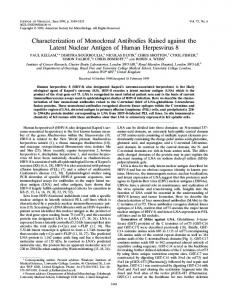
Figure 1. Comparison of the total Ig titers of seven representative mutant hybridoma clones before and after three months of storage at -120ºC. Supernatant from the culture medium of the seven hybridoma clones were assayed by ELISA against the recombinant Blo t 11 (Bt 11) and the native form from the B. tropicalis extract (Bt Ext.).Clones with asterisks (*) were used for the isolation of total RNA for the analysis of Ig gene.
reverse transcribed into first-strand cDNA using oligo-dT primers (SuperScript First Strand Synthesis System, Invitrogen) according to the manufacturer’s instructions. AID was amplified using the primers AID-F (5’-ATGGACAGC CTTCTGATGAAG-3’) and AID-R (5’-TTCAAAATCCC AACATACGAA-3’). A PCR mixture of 2 µL cDNA, 2.5 µL 10x PCR Buffer, 0.5 µL 10 mM dNTP’s, 1 µL each of forward and reverse primers, 1.5 µL MgCl2, 0.5 µL Taq polymerase (Promega, Madison, WI, USA), and 16 µL of ddwater. PCR cycling parameters were 94 ºC for 5 minutes followed by 35 cycles of 94 ºC for 45 seconds, 58 ºC for 45 seconds, 72 ºC for 90 seconds. A 10-minute final extension at 72 ºC was incorporated into the amplification parameters. A housekeeping gene, glyceraldehydes-3-phosphate dehydrogenase (GAPDH) was amplified as a control following the parameters mentioned above using the primers GAPDH-F (5’ATGGTGAAGGTCGGTGTGAA-3’) and GAPDH-R (5’TTACTCCTTGGAGGCCATGTA-3’). Amplified fragments were resolved on 1% agarose gel. RESULTS Mutant Blo t 11 hybridoma clones A total of 36 out of the 65 total Ig-positive Blo t 11 hybridoma clones after the isotyping process registered no detectable isotype by ELISA. Initial determination of total Ig titer of these hybridoma clones without isotypes showed that they secreted relatively high titers of antibody. Subsequent assays to confirm their total Ig titers after several passages and freezing, however, showed that most of these mutant hybridoma clones were unstable in terms of antibody
Vol.2 | No.1 | 2009
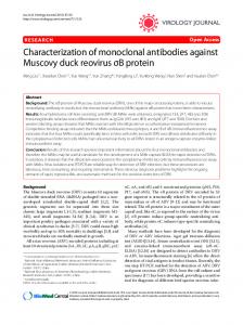
secretion as most registered almost undetectable antibody (total Ig) while others registered a significant decrease in antibody production. Figure 1 shows a summary of the comparison of total Ig titers of seven representative mutant hybridoma clones before and after three months of storage at -120ºC using recombinant FL-Blo t 11 and B. tropicalis extract as antigens. Clones P1H1, P1F10 and P1A9 registered no detectable antibody production after three months of storage while clones P1C9, P1A6, P2E1, and P2B10 registered a significant decrease in antibody production specific to both the rFL-Blo t 11 and the native Blo t 11 present in crude B. tropicalis extract. The total RNA from 4 mutant hybridoma clones (P1C9, P1A6, P2E1 and P2B10) and 2 normal hybridoma clones were isolated using the RNeasy MiniKit (Qiagen, Hilden, Germany). The isolated total RNA was used for a two-step RT-PCR using gene specific primers for the different Ig genes. RT-PCR results showed that the four mutant Blo t 11 hybridoma clones without isotypes as determined by ELISA contain RNA transcripts coding for a specific immunoglobulin isotype (Figure 2). Three of the four mutant hybridoma clones (P1A6, P2B10 and P2E1) examined showed an amplified gene fragment of about 1 kb with the IgG2a primers while the hybridoma clone P1C9 showed an amplified gene fragment of about 1 kb using the IgG3 primers. No nonspecific amplification was observed with the other sets of primers used at a PCR annealing temperature between 60-65ºC. The specificity of the primers used were confirmed with the amplification of a single discrete band with the P2E7G10 (IgG1) and Df642 (IgG2a) control hybridoma clones.
Philippine Science Letters
41
Figure 2. RT-PCR-amplified constant region of the immunoglobulin gene from the four mutant and two control hybridoma clones.
Table 1. Summary of the nucleotide sequence analysis of the 4 exons coding for the secreted form of the constant region of the immunoglobulin gene from the four mutant and two control hybridoma clones.
42
Philippine Science Letters
Vol.2 | No.1 | 2009
a
b
c
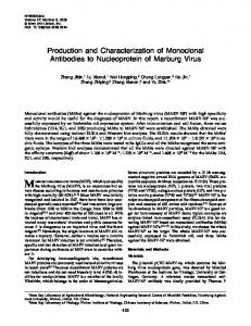
d Figure 3. Nucleotide and deduced amino acid sequence of exon 3 (a) and exon 4 (c) of the mutant hybridoma clone P1C9. The sequence alignment of the deduced amino acid of exon 3 (b) and exon 4 (d) with exons 3 and 4, respectively, of the normal IgG3 (published sequence) are shown. Stop codon and amino acid changes are shaded.
Vol.2 | No.1 | 2009
Philippine Science Letters
43
Sequence analysis of immunoglobulin exons The amplified gene fragments from the four mutant and two normal control hybridoma clones were re-amplified using high-fidelity polymerase and were sequenced. The approximately 1 kb amplified gene fragment coding for exons 1 to 4 of the constant region of the immunoglobulin gene were sequenced using gene specific primers. DNA sequences obtained were compared with the published Ig genes as well as from the DNA sequences obtained with the 2 normal control hybridoma clones. A summary of the DNA sequence analysis of the four mutant and two normal control hybridoma clones are summarized in Table 1. A total of 29, 1, 6, and 47 nucleotide mutations were detected in hybridoma clones P1C9, P2B10, P2E1 and P1A6, respectively. These nucleotide changes were specifically found only in exons 3 and 4. No mutation was observed in exons 1 and 2 of the mutant clones and in exons 1 to 4 of the two control hybridoma clones. Most of the detected mutations involved substitutions (transitions or transversions). Clone P2B10 registered a single mutation on exon 3 and no detected mutation on exon 4. Interestingly, 7 and 9 of the observed mutations in clone P1C9 and P1A6, respectively, were located on RGYW (A/G, G, C/T, A/T) and WRCY (A/T, A/G, C, C/T) hotspots where AID have been reported to occur at high frequencies. Analysis of the deduced amino acid sequences of the 4 mutant clones showed that most of the detected nucleotide mutations were active as shown in Figures 3 and 4. The 27 nucleotide mutations observed in exons 3 and 4 of clone P1C9 (Table 1) resulted in the formation of an early termination codon (TGA) at positions 246-248 in addition to the 28 amino acid substitutions and the 2 amino acid deletions noted (Figure 3). A similar early termination codon (TAG) at positions 187-189 was observed with the clone P2E1 (Figure 4) and 3 early termination codons (TGA, TGA and TAG) were observed at positions 73-75, 142-144 and 166-168 with clone P1A6 (data not shown). A total of 7 amino acid substitutions were observed before the early termination on exon 3 of clone P2E1 in addition to 38, 3 and 2 amino acid substitutions, insertions and deletions, respectively (Figure 4). There were 9 amino acid substitutions and 1 amino acid deletion observed in exon 4 of clone P2E1. A total of 3 amino acid substitutions were observed before the first early termination codon on exon 3 of clone P1A6. In addition, clone P1A6 also showed 49 amino acid substitutions and 3 amino acid deletions spanning exon 3 and exon 4 (Table 1). No early termination codon was observed with clone P2B10 but a single point mutation involving a substitution of Leucine (L) to Phenylalanine (F) was observed in exon 3 (Table 1). Epitope analysis of anti-IgG1, -IgG2a and –IgG3 monoclonal antibodies The inability of the isotyping assay using ELISA to identify the isotypes of the antibodies secreted by the mutant hybridoma clones as a result of the mutation of the epitopes recognized by the anti-IgG2a and IgG3 antibodies used was verified by epitope mapping experiments. The 3 main exons (exons 1, 3 and 4) of the constant region of IgG1, IgG2a and IgG3 mouse antibodies were expressed as GST-fusion proteins in E. coli. A total of 9 exons were expressed and affinitypurified using the Glutathione-agarose column. All exons 44
were highly expressed as soluble GST- fusion proteins. Epitope mapping by Western blot analysis showed that the anti-mouse IgG1 antibody, clone LO-MG1-2 (Serotec Ltd., Oxford, UK), reacted with the recombinant peptide encoded by exon 4 of the mouse IgG1 (Figure 5A). The anti-mouse IgG2a, clone LO-MG2a-7 (Serotec Ltd., Oxford, UK); and the anti-mouse IgG3, clone LO-MG3-7 (Serotec Ltd., Oxford, UK), both reacted with the peptide encoded by exon 4 of the mouse IgG2a (Figure 5B) and IgG3 (Figure 5C), respectively. Similar results were obtained by ELISA where the 3 monoclonal antibodies used all reacted to the recombinant peptide encoded by exon 4 of their corresponding antibodies (Figure 5D). The cross reactivity of the anti-mouse IgG3, clone LO-MG3-7, to the peptide encoded by exon 3 of the constant region of the mouse IgG2a which was observed in both Western blot analysis (Figure 5B) and ELISA (Figure 5D), might be due to the presence of similar amino acid sequences mimicking the epitope recognized by the antibody. Activation-induced cytidine deaminase transcripts in hybridomas The potential involvement of AID, an RNA and/or DNA editing enzyme implicated in somatic hypermutation, class switch recombination, and gene conversion, in inducing the observed mutations in the constant region of the immunoglobulin genes was examined by RT-PCR. AID transcripts were observed in all the hybridomas examined (Figure 6). High level of AID mRNA expression was observed in clone P1A6, the hybridoma clone exhibiting the most number of transition and transversion mutations (Table 1). Moderate to low level of AID mRNA expression was observed with three other mutant hybridoma clones. AID was also found to be expressed in clones P2E7-G10 and Df642. DISCUSSION This study reports the identification of hybridoma clones secreting functional and antigen-specific antibodies with potential mutated constant regions. The mutant monoclonal antibodies exhibited antigen-specificity against the recombinant and native Blo t 11 antigens which indicates the presence of normal and functional variable regions. The inability of the anti-IgG2a and anti-IgG3 monoclonal antibodies used to determine the subclasses of the antibodies secreted by the mutant hybridomas was supported by the identification of mutations on exons 3 and 4 of the immunoglobulin gene. This observation was further validated by the identification of the epitope locations of the anti- IgG2a and anti-IgG3 monoclonal antibodies on the constant region encoded by exon 4. To our knowledge, this is the first report on the mutations along the constant region of the mouse Ig gene. The observed mutations may be associated with faulty immunoglobulin-gene modification mechanisms (defective somatic hypermutation, class switching recombination, gene conversion, alternative splicing); myeloma cell-induced gene mutations; and DNA immunization-induced class switching abnormality. Recent studies have shown that the RNA and/or DNA editing enzyme called Activation-Induced cytidine Deaminase (AID) (Martin and Scharff 2002; Yoshikawa et al. 2002) plays an important role in the different mechanisms that diversify
Philippine Science Letters
Vol.2 | No.1 | 2009
a
b
c
d Figure 4. Nucleotide and deduced amino acid sequence of exon 3 (a) and exon 4 (c) of the mutant hybridoma clone P2E1. The sequence alignment of the deduced amino acid of exon 3 (b) and exon 4 (d) with exons 3 and 4, respectively, of the normal IgG2a (published sequence) are shown. Stop codon and amino acid changes are shaded.
Vol.2 | No.1 | 2009
Philippine Science Letters
45
Figure 5. Epitope mapping analysis of the anti-IgG1 (a), IgG2a (b), and IgG3 (c) monoclonal antibodies by Western blot analysis. The results obtained in Western blot analysis were confirmed by ELISA (d).
immunoglobulin genes including somatic hypermutation (Rada et al. 1997; Papavasiliou and Schatz 2002), gene conversion (Harris et al. 2002), and class switch recombination (Weill and Reynaud 1996; Muramatsu et al. 2000). Somatic hypermutation causes point mutations in the vicinity of the variable exon resulting in the generation of B cells with very high affinity antibodies. Expression of AID had been found to activate somatic hypermutation in hybridomas (Martin and Scharff 2002), B cells (Zhang et al. 2001), fibroblasts (Yoshikawa et al. 2002), and even in Escherichia coli (Ramiro et al. 2002), by deaminating DNA. AID appears to preferentially initiate mutations on the G and C nucleotides within RGYW (A/G, G, C/T, A/T) and WRCY (A/T, A/G, C, C/T) hotspots sequences (Martin et al. 2002). Although the activity of AID to initiate point mutations has been reported to occur in the variable region and in the sequences 1.5 kb downstream from the immunoglobulin promoter, it cannot be ruled out that it may exert its activity along the exons coding for immunoglobulin constant region under abnormal conditions. It has been previously proposed that high transcription rates alone may predispose any gene to mutation by AID (Martin and Scharff 2002). In this study, AID mRNA expression was observed in all the hybridomas examined. Of particular interest was the observation that AID mRNA is highly expressed in clone P1A6, a mutant hybridoma with the most number of mutations on exons 3 and 4. The level of AID mRNA expression with the other clones, however, shows no correlation with the frequency of mutations. Added to this is the observation that AID mRNA was found to be highly expressed in clone P2E7-G1, a clone with no mutation in the 46
constant region exons. Taking into consideration, however, that all hybridomas are the result of a series of mutational processes in their immunoglobulin genes probably under the influence of AID, the observed AID mRNA expression in all the hybridomas used in this study is not surprising. However, it is also possible that another unknown factor in addition to AID is required to induce high frequency of mutation in the constant region of Ig, therefore it is possible that such additional factor is absent in clone P2E7-G1. The process of gene conversion involves the transfer of sequence information from a pseudogene (ψV) into the variable region resulting in sequence mutations in the VDJ regions, a normal process in B cell maturation that contributes to the diversity of immunoglobulins (Papavasiliou and Schatz 2002). However, a potential faulty gene conversion downstream of the VDJ regions that could result in mutations in the constant region under abnormal conditions cannot be ruled out. Related to the above possibility, an alternative splicing mechanism of murine ICAM-1 containing 5 extracellular immunoglobulin domains has been reported resulting in transcripts missing 69 nucleotides from the 3’ terminus of the exon 6 (Mizgerd et al. 2002). Such alternative splicing could similarly happen in the upstream exons of the immunoglobulin gene that could potentially result in exon truncations or skipping of amino acids. The role of AID is not only limited to somatic hypermutation but is also implicated in class switch recombination. It has been postulated that AID edits the mRNA of the endonuclease responsible for the DNA lesions in both class switch recombination and somatic hypermutation
Philippine Science Letters
Vol.2 | No.1 | 2009
Figure 6. AID mRNA expression in four Blo t 11 hybridomas with mutated immunoglobulin constant region genes and two control hybridomas. GAPDH transcripts of each hybridoma were amplified as controls.
(Honjo et al. 2002). Given this prolific activity of AID, the possibility for the editing enzyme to form DNA lesions in the immunoglobulin exons involved in class switch recombination cannot be ruled out. Interestingly, the results presented in this paper contradict the generalizations presented by Longerich et al. that AID does not gain access to the very 5’ end and the constant region of the Ig genes thus preventing somatic hypermutations along these regions (Longerich et al. 2005). Using uracil DNA glycosylase (Ung)-deficient mice in comparison with wild-type mice, Longerich et al. observed no difference in AID activity footprint (C to G transitions) on the endogenous Ig gene of isolated B cells. Although insignificant difference of the AID-induced mutations were observed between the Ung and + Ung mice, Longerich et al. nevertheless observed a number of mutations including C to G transitions along the entire Ig gene including the constant region. Noteworthy is the observation by Longerich et al. that 64 and 98 C to G transitions were observed along the 334-2428 positions of JH2 and 1284-2428 positions of JH4 regions of the Ig gene, respectively. These mutations reported by Longerich et al. exceed the number of mutations presented in this study. It is also important to note that the mutations reported by Longerich et al. were observed in isolated normal B cells and not hybridoma cells as used in this study. Recently, AID has been implicated in the hypermutation of tumor suppressor and DNA repair genes in chronic myeloid leukemia (Klemm L et al. 2009) and in inflammation-associated carcinogenesis (Chiba T and Marusawa H 2009; Petersen-Mahrt SK 2009) thus playing an important role in tumor biology. Considering that hybridoma cells are fusion of myeloma cells and normal splenocyte, it cannot be ruled out that AID activity may be more pronounced in hybridoma cells than in normal B cells.
Vol.2 | No.1 | 2009
The use of DNA immunization in the induction of the immune response in mice could also be a potential factor involved in the observed mutations. In the conventional method of hybridoma technology, substantial amounts of antigens are introduced into mice in order to achieve a strong immune response such that a significant number of polyclonal B cells are generated for fusion. In this study, the immunization protocol was solely performed by DNA immunization with electroporation. In contrast to protein immunization, DNA immunization relies on the potentially minimal amount of in vivo expressed-antigen encoded by the immunized DNA to trigger an immune response in the host’s system. The presence of minimal amounts of antigen might have affected the ability of the generated B cells to undergo a normal class-switching mechanism thus resulted in the observed mutations. Immunoglobulin class switching involves a recombination process whereby a VHDJH segment changes its linkage from Cµ to another CH region and the event takes place in the vicinity of the switch regions, which are located 5’ of the Ch genes (Bruggemann 1987). It is well established that the process of immunoglobulin class switching is under the influence of cytokines provided by helper T cells. In connection with this, the possibility that the mainly-uncharacterized mechanisms of cytokine production in DNA immunization might have contributed to the observed mutations cannot be ruled out. Lastly, monoclonal antibody production by hybridoma technology makes use of highly unstable myeloma cells. The murine myeloma cells used for fusion with the mouse splenocytes, like any other cancer cells, possess abnormal genetic makeup that could influence the immunoglobulin gene from the fused splenocyte. The fact that the resulting hybridoma is a fusion of genetic materials from the myeloma cell and splenocyte, may result in a potentially unstable polyploid myeloma cell. As there is no literature
Philippine Science Letters
47
available regarding the observed mutations, we do not have any comparison as to whether these observed mutations are “normal” occurrences in hybridoma technology or they represent a novel observation, which may have been associated with DNA immunization that is yet to be fully elucidated and characterized.
ACKNOWLEDGMENTS The authors thank Ms. Audrey Teo Su Min for her technical assistance. This work was funded by grants from the Agency for Science, Technology and Research (A*STAR), Singapore (R-178-000-010-303), and Academic Research Fund, National University of Singapore, Singapore (R-178-000-010-112).
REFERENCES Bruggemann M. Genes encoding the immunoglobulin constant regions. In: Calabi F, Neuberger MS, Eds. Molecular genetics of immunoglobulin. New York: Elsevier Science, 1987:51-80. Chiba T, Marusawa H. A novel mechanism for inflammationassociated carcinogenesis; an important role of activation-induced cytidine deaminase (AID) in mutation induction. J Mol Med 2009; Sep 15. [Epub ahead of print]. Coker HA, Durham SR, Gould HJ. Local somatic hypermutation and class switch recombination in the nasal mucosa of allergic rhinitis patients. J Immunol 2003;171: 5602-5610. Harris RS, Sale JE, Petersen-Mahrt SK, Neuberger MS. AID is essential for immunoglobulin V gene conversion in a cultured B cell line. Current Biology 2002; 12:435-438. Honjo T, Kinoshita K, Muramatsu M. Molecular mechanism of class switch recombination: linkage with somatic hypermutation. Annu Rev Immunol 2002;20: 165-196. Klemm L, Duy C, Iacobucci I, Kuchen S, von Levetzow G, Feldhahn N, Henke N, Li Z, Hoffmann TK, Kim YM, Hofmann WK, Jumaa H, Groffen J, Heisterkamp N, Martinelli G, Lieber MR, Casellas R, Müschen M. The B cell mutator AID promotes B lymphoid blast crisis and drug resistance in chronic myeloid leukemia. Cancer Cell. 2009;16(3):232-45. Longerich S, Tahaka A, Bozek G, Nicolae D, Storb U. The very 5’ end and the constant region of the Ig genes are spared from somatic mutations because AID does not access these regions. J Exp Med 2005;1443-1454. Martin A, Bardwell PD, Woo CJ, Fan M, Shulman MJ, Scharff MD. Activation-induced cytidine deaminase turns on somatic hypermutation in hybridomas. Nature 2002;415:802-805. Martin A, Scharff MD. AID and mismatch repair in antibody diversification. Nature Rev. Immunol 2002;2:605-614. Mizgerd JP, Spieker MR, Lupa MM. Exon truncation by alternative splicing of murine ICAM-1. Physiol Genomics 2002;12:47-51. Muramatsu M, Kinoshita K, Fagarasan S, Yamada S, Shinkai Y, Honjo T. Class switch recombination and 48
hypermutation require activation-induced cytidine deaminase (AID), a potential RNA editing enzyme. Cell 2000;102:553-563. Noguchi E, Shibasaki M, Inudou M, Kamioka M, Yokouchi Y, Kobayashi KY, Hamaguchi H, Matsui A, Arinami T. Association between a new polymorphism in the activation-induced cytidine deaminase gene and atopic asthma and the regulation of total serum IgE levels. J Allergy Clin Immunol 2001;108:382-386. Papavasiliou FN, Schatz DG. Somatic Hypermutaion of immunoglobulin genes: merging mechanisms for genetic diversity. Cell 2002;109:S35-S44. Petersen-Mahrt SK, Coker HA, Pauklin S. DNA deaminases: AIDing hormones in immunity and cancer. J Mol Med 2009; 87:893-897. Rada C, Yelamos J, Dean W, Milstein C. The 5’ hypermutation boundary of chains is independent of local and neighboring sequences and related to the distance from the initiation of transcription. Eur J Immunol 1997;27:3115-3120. Ramiro AR, Stavropoulos P, Jankovic M, Nussenzweig MC. Transcription enhances AID-mediated cytidine deamination by exposing single-stranded DNA on the non-template strand. Nature Immunol 2002;4:452-456. Ramos JD, Cheong N, Lee BW, Chua KY. cDNA cloning and expression of Blo t 11, the Blomia tropicalis allergen homologous to paramyosin. Int Arch Allergy Immunol 2001;126:286-293. Ramos JD, Teo SMA, Lee BW, Cheong N, Chua KY, DNA immunization for the production of monoclonal antibodies to Blo t 11, a paramyosin homolog from Blomia tropicalis. Allergy 2004;59:539-547. Ramos JD, Teo SMA, Kuo IC, Lee BW, Cheong N, Chua KY. Production of monoclonal antibodies for immunoaffinity purification and quantitation of Blo t 1 allergen in mite and dust extracts. Clin Exp Allergy 2004;34: 604-610. Takhar P, Corrigan CJ, Smurthwaite L, O’Connor BJ, Durham SR, Lee TH, Gould HJ. Class switch recombination to IgE in the bronchial mucosa of atopic and nonatopic patients with asthma. J Allergy Clin Immunol 2007;119:213-218. Weill JC, Reynaud CA. Rearrangement/hypermutation/gene conversion: when, where and why? Immunol Today 1996;17:92-97. Wolfowicz CB, HuangFu TQ, Chua KY. Expression and immunogenicity of the major house dust mite allergen Der p 1 following DNA immunization. Vaccine 2003; 21:1195-1204. Yoshikawa K, Okazaki I, Eto T, Kinoshita K, Muramatsu M, Nagaoka H, Honjo T. AID enzyme-induced hypermutation in an actively transcribed gene in fibroblasts. Science 2002;296:2033-2036. Zhang W, Bardwell PD, Woo CJ, Poltoratsky V, Scharff MD, Martin A. Clonal instability of V region hypermutation in the Ramos Burkitt’s lymphoma cell line. Intl Immunol 2001;13:1175-1184.
Philippine Science Letters
Vol.2 | No.1 | 2009