Mar 27, 1991 - pooled human plasma using Thromboscreen Kontact reagent (Pacific. Hemostasis) was determined in the presence of synthetic peptides.
VOl. 266, No 18, Issue of June 25, pp. 11975-11979,1991 Printed in U.S.A.
THEJOURNAL OF BIOLOGICAL CHEMISTRY 8 1991 by The American Society for Biochemistry and Molecular Biology, Inc
Chimeric Antithrombin Peptide CHARACTERIZATION OF AN ARG-GLY-ASP (RGD)- AND HIRUDIN CARBOXYL TERMINUS-CONTAINING SYNTHETICPEPTIDE* (Received for publication, March 27, 1991)
Frank C. Church$, Jeanne E. Phillips, andJoan L. Woods From the The Centerfor Thrombosis and Hemostasis and Departmentsof Pathology and Medicine, University of North Carolina School of Medicine, Chapel Hill, North Carolina27599
and
We investigated the properties of an artificial chi- hirudin is required for anticoagulant activity; the minimal meric peptide that contains an Arg-Gly-Asp (RGD)- peptide length being about 12 amino acid residues to tripeptide, the versatile cell recognition signal of ex- Leu64)(4, 5). Hirudin and its fragments have different biotracellular matrix protein components, coupled to a chemical properties as potential therapeutic anticoagulants carboxyl-terminalfragment of the highlyspecific that could favor one over another based on the desired phar53-64): a-thrombin inhibitor, hirudin (residues macological characteristics. WG-SANGDFEEIPEEYL (RGD-hir~din'~-'~). HiAdhesion of blood platelets to vessel wall components and r ~ d i n ~ and ~ - ' RGD-hir~din'~-'~ ~ inhibited the fibrino- their subsequent activation is a central hemostatic event. An gen clotting activity of a-thrombin and prolonged the essential component of platelet adhesion and aggregation is activated partial thromboplastin time of human the cell surface receptor aI& (also known as glycoprotein plasma. In addition, both peptides afforded total proIIb-IIIa), which is a member of the integrin family (10-14). tection to thrombin from trypsinolysis. Neither h i r ~ d i n ' ~ - 'nor ~ R G D - h i r ~ d i n ~ dramatically ~-'~ inter- Platelet (YI& is a receptor for four adhesive proteins: fibrinfered with the thrombin-antithrombin inhibition re- ogen, fibronectin, vitronectin, and von Willebrand factor (10, action either in theabsence or presenceof added hep- 11, 13). a I I b & specifically recognizes a conserved tripeptide arin. a-Thrombin-induced platelet aggregationwas ef- Arg-Gly-Asp (RGD) sequence found in all four proteins and by inhibited fectively RGD- the carboxyl terminus of the y chain of fibrinogen (10,11,13,15). Additionally, there are h i r~ din'~ - '~Unlike . h i r ~ d i n ~ ~ - ' ~ , R G D - h i r ~ in d i n ~(HHLGGAKQAGDV) ~-'~ many other integrins found in numerous cell types that spesolution inhibited integrin-mediated endothelial cell and fibroblast cell attachment to polystyrene wells in cifically mediate both cell adhesion with substrates derived the presence of fetal bovine serum. Collectively, our from extracellular matrix and body fluids and cell-cell interresults demonstrate that RGD-hir~din'~-'~anticohas actions (10, 16). agulant/antiplatelet aggregation activity attributable There areexamples of hybrid molecules either that combine to its hirudin sequence and integrin-directed cell attwo functions or that acquire a new function. A bifunctional tachment activity due to its RGD site. Our results thrombin inhibitor has been prepared by linking (D-Phe)suggest that this chimericmotif may serve as a proto- Pro-Arg-Pro- and hirudin carboxyl-terminal fragments (17, type for a new class of anticoagulants where an inte- 18). RGD- and HHLGGAKQAGDV-containing sequences grin-specific sequence utargets" the peptide to a cell either coupled to or genetically engineered intoa carrier (ultimately through theplateletintegrintrapped amid a thrombus with ensuing proteinase inhibition. protein have integrin-specific cell binding activity similar to the parentadhesive protein (15,19-21). We hypothesized that an artificial chimeric peptide could be constructed that incorporated antithrombin activity and integrin-directed cell atHirudin is a highly specific a-thrombin inhibitor isolated tachment activity. This chimera motif is based on the finding from the salivary gland of the European bloodsucking leech that platelet phospholipid microparticles produced following platelet activation contain the components for assembly of Hirudo medicinalis (1-3). Recent structure-function studies have shown that both amino- and carboxyl-terminaldomains the prothrombinase complex and functional (Y&3 receptors of hirudin bind to thrombin, andthe isolated hirudin domains (22). Therefore, coupling these sequence types into a chimera inhibit thrombin through different mechanisms (4-9). The might provide a "targeted antithrombin agent to specifically amino-terminal hirudin domain binds to the active site of bind cells at a thrombus for inhibition of thrombin. In the present investigation, we synthesized a chimeric thrombin, whereas the carboxyl-terminal hirudin fragment peptide by adding the RGD tripeptide, aminimal cell adhesion binds to thefibrinogen recognition site (adjacentto theactive site) (4-9). Only a small portion of the carboxyl terminus of sequence from fibronectin and other adhesive proteins, to a segment of the carboxyl terminus of hirudin (termed chimeric * This work was supported in part by a grant-in-aid from the North antithrombin peptide).We report here that thechimeric antiCarolina Affiliate of the American Heart Association and by a Uni- thrombin peptide has both antithrombin and cell adhesion versity Research Council grant from The University of North Caro- activities comparable to itsindividual constituents. lina at Chapel Hill.The costs of publication of this article were defrayed in part by the payment of page charges. This article must EXPERIMENTALPROCEDURES therefore be hereby marked "aduertisement" in accordance with 18 U.S.C. Section 1734 solely to indicate this fact. 4 To whom correspondence should be addressed Campus Box 7035, Division of Hematology, 416 Burnett-Womack,University of North Carolina, Chapel Hill, NC 27599. Fax: 919-966-7639.
Materiak-All N-(9-Fluorenyl)methoxycarbonyl-aminoacid derivatives and reagents were obtained from Milligen/Cambridge Research Biochemicals. Human a-thrombin and antithrombin were purified as described (23,24). Heparin was provided by Diosynth; ~ 1 -
11975
11976
RGD-Hirudin Chimera
tosylamido-2-phenylethyl chloromethyl ketone-treated trypsin was from Cooper Biochemicals. Bovine fibrinogen was from Miles Laboratories; Chromozyme T H (tosyl-Gly-Pro-Arg-p-nitroanilide)was obtained from Boehringer Mannheim. Peptide Synthesis-Peptides were assembled using a Milligen Pepsynthesizer as described previously (25). Purity of the peptides was analyzed by reverse-phase HPLC' (24,26), andif necessary, peptides were further purified by HPLC on a preparative Vydac Cls column. All peptides were analyzed either by amino acid analysis or by primary structural analysis on an Applied Biosystems 475A Protein Sequencer (Protein Chemistry Laboratory, Department of Chemistry of this institution). An excellent correlation between expected and actual values/sequences was found for all peptides. Sequences of synthetic hirudin"" and R G D - h i r ~ d i peptides n ~ ~ ~ ~ are shown below (picomoles of amino acid yield/cycle are shown in parentheses).
unit/ml final concentration) was added and the light transmittance (Bio/Data PAP-4 Aggregometer) was recorded. These experiments were performed in triplicate four times with four different healthy volunteers and the results averaged. Cells and Cell Attachment Assays-Human dermal fibroblasts (supplied by Dr. R. A. Briggaman, Department of Dermatology of this institution) were grown in DMEM (GIBCO) supplemented with 10% FBS, 100 units/ml penicillin and 100 pg/ml streptomycin. The human endothelial cell line (EA.hy 926;supplied by Dr. C-J. S Edge11 of this institution) was grown as described previously (28). Cell adhesion activity of the synthetic peptides was determined as described (29). Briefly, -1 X 10' cells/ml of trypsinized cells were mixedwith DMEM containing 10% FBS and 0.5, 0.5, 1.0, 1.5, and 1.5 mg/ml of RGD, " RGE, h i r u d i ~ P ~ ~ , R G D - h i r ~ dor i nR ~G ~ -E~- h~i,r ~ d i n ~ ~peptides, respectively (these concentrationsprovided approximately equal molar amounts of RGD/E). Cells that attached to the microtiter plate wells after incubation for 60 min a t 37 "C and 5%CO, were quantified by staining with Crystal Violet, solubilizing the stained cells with ethylene glycol monomethyl ether, and theabsorbance a t 600 nm was compared with standard curves of serially diluted cells (30). These experiments were performed from three tosix times. RESULTS
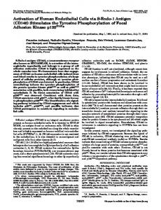
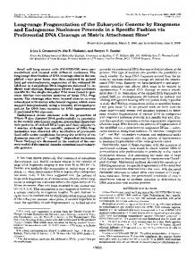
All other peptides and their sequences using the one-letter abbreviaAnticoagulant and Antithrombin Activities-Coupling the tion (shown in parentheses) were as follows: RGD-peptide cell adhesive RGD sequence (andthe inactive RGE con(GRGDSAY); RGE-peptide (YARGESA); RGE-hir~din~~"' former) to h i r ~ d i n did ~ ~not - ~ affect ~ the ability of h i r ~ d i n ~ ~ - ~ ~ (WGRGESANGDFEEIPEEYL); and HC38-66(DFHKENTVTNDto inhibit fibrinogen hydrolysis by thrombin (Fig. 1,top). The WIPEGEEDDDYLDLEKIY) (25). required for 50% inhibition (IC5,) for Anticoagulclnt and Antithrombin Assays-All experiments were concentration ~G " jD~ - h i r ~ d i n ~and ~ - ~RGE-hirudinS3'j4 ~, was -0.6 performed in a buffer that contained 20mM HEPES, 150 mM NaCI, h i r ~ d i n ~, R 0.1% (w/v) polyethylene glycol ( M , = 8000) at pH 7.4. Fibrinogen p ~ There . was also a dose-dependent increase in the aPTT clotting activity of human a-thrombin was measured in bovine serum of normal pooled human plasma (average clotting time of 40 albumin-coated microtiter plates by incubating 50 p1 of thrombin (10 1.3 s for 100% plasma) for RGD-hir~din'~-~~, nM stock) with 50 p1 of a synthetic peptide (8-20 p~ stock). After 1 min, 100 pl of fibrinogen (5 mg/ml stock) was added, briefly agitated, 100 and theabsorbance at 405 nm was measured every 5 s for 2 min in a * P VmaXkinetic microplate reader (Molecular Devices). These experiments were performed in triplicate from three tofive times, aPTT of 80 pooled human plasma using Thromboscreen Kontact reagent (Pacific Hemostasis) was determined in the presence of synthetic peptides 60 with a Fibrometer as described (27). Experiments were performed three times and the results averaged. Antithrombin inhibition assay of thrombin in the presence of a 40. 200-fold molar excess of either hirudin5344or R G D - h i r ~ d i n ~ to~ - ~ ~ thrombin was performed as described previously (27).Thrombin 20 inhibition by antithrombin-heparin in the presence of synthetic peptide was performed by incubating 1 nM thrombin with 100 nM 0 0 for 1 min, followed by 10 nM human antithrombin in the n . . . . presence of 0.05 to 500 pg/ml heparin. After 20 s, Chromozyme T H 1 .o 0.0 2.0 3.0 4.0 with polybrene (to neutralize the added heparin) was added, substrate [Synthetic Peptide] (1M) hydrolysis was stopped after 60 min by the addition of glacial acetic acid, and the absorbance at 405 nm was determined. Inhibition rate constants were calculated as detailed previously (27). These experiments were performed three times. Proteolysis of a-Thrombin by Trypsin-Trypsinolysis of a-thrombin was performed with 2.75 p~ thrombin (100 pg in 100 pl) in the or R G D - h i r ~ d i n ~in~ ?HEPES~~ presence of 125 p~ hirudin"" buffered saline, pH 7.4. After a 5-min incubation at room temperature, the reaction was initiated by the addition of 2 pg of ~l-tosylamido-2phenylethyl chloromethyl ketone-treated trypsin and stopped after 120 min by the addition of 1 mM phenylmethylsulfonyl fluoride. The extent of proteolysis was assessed by sodium dodecyl sulfate-polyacrylamide gel electrophoresis in15% slab gels without chemical reduction of samples. Silver nitrate was used to stain the polypeptides. This experiment was performed three times. 1 5 10 Platelet Aggregation Assay-Platelet aggregation assays were per[Synthetic Peptide] (pM) formed using human platelet-rich plasma (diluted to 300,000 platelets/pl) by drawing blood (9 parts) into 3.8% (w/v) sodium citrate (1 FIG. 1. Anticoagulant and antithrombotic activities of synpart) from a volunteer who had nothad any aspirinor related products thetic peptides. Top, fibrinogen clotting activity of human a-thromfor at least 14 days. Platelet aggregation was performed by adding 40 bin was measured as described under "Experimental Procedures": and RGD-hir~din'~.~'(A).R G E - h i r ~ d i n ~had ~ - ~es' pl of a synthetic peptide solution to 450 pl of platelet-rich plasma at hirudinS3-64 (0) 37 "C. After a 2-min incubation, 10 pl of a-thrombin (0.4 p M NIH sentially the same activity as shown here for h i r ~ d i n ' ~and - ~ ~RGDhir~din'"~ (data not included). A control peptide, HC39-66(O), did The abbreviations used are: HPLC, high performance liquid chro- not inhibit fibrinogen clotting activity. Bottom, aPTT assays were matography; HEPES, 4-(2-hydroxyethyl)-l-piperazineethanesulfonic performed using normal human pooled plasma as detailed under acid aPTT, activated partial thromboplastin time; DMEM, Dulbec- "Experimental Procedures." The control HC39-66peptide did not prolong the aPTT (tested at 10 p ~ ) . CO'S modified Eagle's medium; FBS, fetal bovine serum.
+
-
-
-.
.
.
.
I
RGD-Hirudin Chimera
a "
11977
b
c
d
"-
e
-
0 "
"I
.01
.1
1
10
100
1000
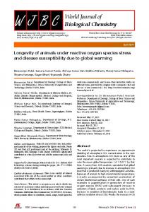
[Heparin] (pg/mL) FIG. 2. Effect of h i r ~ d i n ~ ' -on ' ~thrombin inhibition by antithrombin-heparin. Thrombininhibition by antithrombin in the presence of various amounts of heparin was performed as detailed under "Experimental Procedures" either in the absence(0)or in the presence (A) of hirudinSR-". Thrombin inhibition was determined as the second-order rate constantof inhibition ( X 10"j M" s-').
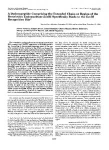
FIG. 3. Effect of hirudins3-" and RGD-hirudin6"'" on trypsinolysis of a-thrombin. Trypsinolysis of a-thrombin was performed in the absence and presence of hirudin"'-64 and RGD-hirudin":" " as described under "Experimental Procedures" with assessment of proteolysis by gel electrophoresis. Lanes: a, Bio-Rad low molecular weight standards; b, thrombin alone; c, thrombin plus trypsin; d, thrombin plus trypsin and hirudin"':"fi4;and e, thrombin plus trypsin and RGD-hirudin":"".
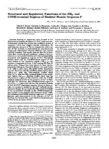
and RGE-hir~din'"'~ (Fig. 1, bottom). We examined thrombin inhibition by the plasma serpin antithrombin in the presence of h i r ~ d i n ~ ~and - ' ~ RGDhirudin"-G4. Neither h i r ~ d i n ~ " nor ' ~ RGD-hir~din~"'~ interfered with the thrombin-antithrombininhibition reaction (in TABLE I the absence of added heparin) asshown by second-order rate Inhibition of cell adhesion by synthetic oeotides constants of 1.37,1.22, and 1.34 ( X lo5M" min-') in the Cell adhesion (absorbance 600 nmY absence of peptide, and in the presence of a ZOO-fold molar Peptide Endothelial cells Fibroblasts excess of h i r ~ d i n ' ~or ' ~ RGD-hir~din"~'~ ~ to thrombin,re% spectively. We also determined the effect of h i r ~ d i n on ~ ~thrombin -~~ RGD 53 (0.121 k (0.097 0.007) 34 k 0.003) 37 (0.103 f 0.006) RGD-hir~din"-'~ 45 (0.103 f 0.007) inhibition by antithrombin-heparin. At a 100-fold molar ex79 (0.223 k 0.017) 96 (0.219 k 0.056) RGE cess of hirudin"-64 to thrombin, there was essentiallyno HirudinS3-fi4 91 (0.207 0.020) 81 (0.229 f 0.024) difference in the rateof thrombin inhibition by antithrombin RGE-hirudin"" 92 (0.209 f 0.073) 80 (0.225 f 0.010) in thepresence of various amounts of heparin (Fig. 2). , . Cell adhesion was measured by Crystal Violet staining of cells as Trypsin hydrolyzes a-thrombin at unique sites in the Bunder"Experimental Procedures." The data are from a chain to form &- and yT-thrombin derivatives. We assessed described representative experiment and they are consistent with the trend in the effect of hirudin53" and RGD-hir~din'"~~ on trypsinolysis all experiments evaluating these peptides to inhibitcell adhesion in of a-thrombin. Both RGD-hin~din"-'~ and af- the presence of FBS. The 100% cell adhesion value was arbitrarily forded essentially total protection to a-thrombin during in- assigned to thelowest dilution of cells (-1 X IO4 cells) in the standard cubation with trypsin (Fig. 3). Control experiments verified curve which gave absorbances a t 600 nm of 0.228 and 0.262 for endothelial cells and fibroblasts,respectively. that the hir~din"-~~-containing peptides had no inhibitory effect on trypsin. We examined the peptides for dose-dependent inhibitionof tion of the RGD/Esequence to hirudin""-64is not detrimental platelet aggregation in a-thrombin-stimulated human plate- to its anticoagulant and antithrombinactivities. The dataalso lets. Platelet aggregation induced by a-thrombin was inhibited suggest that hir~din":"'~ and RGD-hirudin"" bind to the most effectively by h i ~ d i n " ~ - ~ ~ , R G D - h i r and ~ d i RGEn ~ " ~ ~ ,same site on thrombin since neither influences inhibition by hirudin53-G4 (IC50 of 7 pM for each peptide),but less effectively the plasma serpin antithrombin and both protect thrombin by the RGD-peptide (IC50 -100 p ~ ) and , with no effect by during trypsinolysis. Cell Adhesion Activity-We compared each synthetic pepthe RGE-peptide (tested to 300 p ~ ) . 'Complete inhibition of a-thrombin-induced platelet aggregation was observed with tide in solution for its ability to inhibit fibroblast and endo15 PM h i r ~ d i n ~ " ~ ~ , R G D - h i r ~adni n cell attachment in the presence of FBS. Cell surface d ~R ~G -'E ~ ,- h i r ~ d i n ' ~ - ~ ~thelial . These data indicate that thefibrinogen clotting and platelet integrins will bind to RGD-containing adhesive proteins presaggregation activities of a-thrombin (ina purified or plasma- ent in serum as FBS coats the microtiter plate surface. We the were quite based assay system) are inhibited to essentially the same found that RGD-hir~din"-~~ andRGD-peptide extent by and RGD/E-hir~din'~-'~ and that addi- effective at preventing cell attachment, whereas hirudin""'", the RGE-peptide, and RGE-hir~din"'-'~ did not interferewith RGD-containing peptides prevent platelet aggregation by inhib- cell adhesion (Table I). Microscopic inspection of fibroblasts iting fibrinogen binding to activated platelets (10-14). Thus, in ad- and endothelial cells verified that hirudin"-", the RGE-pepdition to testing the hirudin site of RGD-hir~din":"~' toblock throm- tide, and RGE-hir~din"-~~ had no noticeable effect on cell bin-mediated platelet aggregation, we also compared the synthetic peptides for dose-dependent inhibition in ADP-stimulated platelets. attachment. However, RGD-hirudin"-"' and theRGD-peptide There was a similar concentration dependence for the RGD-peptide did affect adhesion in that the cells were rounded and not surface (shown for fibroblasts inFig. 4). These and RGD-hirudin""j4 to inhibit ADP-stimulated plateletaggregation attached to the data demonstrate that RGD-hir~din"-'~ functions like the (J. L.Woods and F. C. Church, unpublished observation).
*
~
11978
RGD-Hirudin Chimera
sulfation (5))in an effort toincrease its overall antithrombin/ anticoagulant potency. Ourresultsdemonstratethat RGD-hirudin":""' hasthe same cell-binding activity as the RGD-peptide alone; thus, RGD in theRGD-hirudin":""" chimera must assume anactive conformation. Many proteins have been identified that contain the RGD tripeptide sequence, but the presence of an RGD sequence does not necessarily confer cell adhesion activity (10). There is sufficient evidence to suggest that both RGD conformation and environment contribute to integrindirected cell recognition (10, 13, 20,29, 38-41). This recognition specificity (and affinity)for RGD-containing peptides/ proteins implies that a unique sequence can be "engineered" to preferentially interact with a particular integrin (for instance, by stereochemical isomerization,cyclization, ora unique next-neighbor sequence). Indeed, RGD peptide-albumin conjugates have been shown to recognize specific integrins (19, 21). F I ~ ;4.. Effect of synthetic peptides on fibroblast adhesion. curl& is the dominant fibrinogen receptor in platelets (10Inhihition o f ' filwoblast cell adhesion in the presence of' DMRM/FRS 14). Cross-linking studies have shown that RGD binds prewith and without synthetic peptides was performed as descrilml under dominantly to the p:( subunit, whereas HHLGGAKQAGDV "Experimental Procedures." u, DMEM/FRS alone: h, DMEM/FHS CYIII,subunit. There are other fibrinogen receptors andtheRGD-peptide; c, DMEM/FRSand hirudin"" "I; and d, binds to the DMEM/FRS and RGD-hirudin" I". including the a,& integrin found primarily on endothelial cells (40) and aM/3.on leukocytes(42). Comparison of the RGD-peptide in inhibition of cell adhesion,butthatthe binding specificity of all&and a& for fibrinogen showsthat hirudin.-,.llil sequence alone does not affect cell adhesion. allt,fi:3 preferentially recognizes both an RGD-peptidemodeled after Aa$I:?$15 RGD (but not AaRiS-Ri4 RGD) and y-chain400~4'1 We investigated whether RGD-hirudin":"'" could act as a "bridge" between RGD-specific cell receptors and thrombin HHLGGAKQAGDV sequences, whereas exclusively in(as a replacementfor the adhesive proteins present in FBS). teracts with an RGD-peptide modeled after theAa5i'"5i.' RGD We prepared thrombincomplexes with RGD-hirudin"""'" and sequence (40). There are many other integrins that interact hirudin.->'l 1 2 and adsorbed the thrombin-peptide complexes to withdifferentproteinsitesthanthosejust described, for polystyrene; next, fibroblasts were added in the absence of instance, leukocyte a& binds anovel fibrinogen site (neither FBS, and the number of fibroblasts attached and spread were RGD nor the carboxyl terminus of the y-chain) (42) andalpI determined. Interestingly, thrombin alone or in complex with in amelanoma cell line recognizes afibronectin sequence the peptides promoted the adhesion and spreading of fibro- consisting of X-Asp-Y (43). Therefore, itwould appear that blasts to -70% of the adhesion observedwith fibronectin appropriate peptidesequences can be designed to specifically (data not included). It should be noted that the B-chain of target the chimera to platelet alll,p:land not other integrins thrombin has an RGDsequence which apparently acts as an capable of binding fibrinogen or other proteins. adherent substrate (31). Thus, we were not able to demonPreviousstudieshave shown that RGD-and HHLGGAstrate the coordinating activitiesof cell adhesion and throm- KQAGDV-containing peptides preventfibrinogen binding to bin inhibition with our initial chimeras. This could be due to platelets and plateletaggregation and alter the conformation not only the "active" RGD adhesion sequence in thrombin for of purified platelet allI&,(10-15).? These peptide sequences fibroblasts(31)ortheactualamount of anRGD-peptide have been implicated as potential candidatesfor therapeutic necessary to support cell adhesion (10) but also possibly to the absence of an adequate"spacer" separating thetwo active antiplatelet agents (13, 14). Furthermore, a family of RGDcontainingproteins from a variety of snakevenomsand sites in this chimera. leeches has recentlybeendescribed aspotentantiplatelet compounds (44,45). Future chimeric antithrombin peptide DISCUSSION This study was undertaken to characterize a chimera com- designs for the integrin-directed site (platelet a111,pJwill incorporate unique/specific RGD sequences (such as that in the bining RGD and hirudin sequences. Our results with RGDfibrinogen Aa!K-$15 chain or that found in the snake venom hirudin,~>:i-l;.l are inaccordwithprevious observations using RGD-protein family) and non-RGD sequences (such as that hirudin carboxyl-terminal fragments in anticoagulant, antiin the fibrinogen y-chain""""'" sequence). thrombin, and plateletaggregation inhibition assays(4-9,32The possibility for an achievable targeted chimeric anti35). Our data and those of others indicate that hirudin carthrombin peptideis strengthened by Bode et al. (46), Sandberg boxyl-terminal fragments bind to the fibrinogen recognition site(anion exosite domain) of thrombin which effectively et al. (47), and more recently by the work of Sims et al. (22) in their detection of both functional prothrombinasecomplex blocks both fibrinogen clottingandthrombin-stimulated plasma componentsand allt,p:,incorporatedintoplatelet platelet aggregation activities. These hirudin fragments also membrane microparticles. We ultimately envision a chimeric do not affect thrombin inhibition by the serpin antithrombin antithrombin peptide combining al,t,~:l-specific and thrombin with or without heparin. Thus, hirudin fragments that are targeted to the anion exosite of thrombin, not the active site, anionexosite-directed active sites.Thispeptide would be within may work independently of antithrombin to regulate throm- capable of interacting with stimulated platelets trapped only blockingplatelet-fibrinogen (or other bin. Future chimeric antithrombin peptide designsfor the a thrombus and not proteins) interactions but also halting hirudin site will include variations in the sequence of the RGD-containing carboxyl-terminal fragment (36) and specific chemical modi- thrombin-mediated fibrinogen clotting and platelet aggregafication of Tyr".' (either by nitration (37), iodination (37), or tion activities. Finally, the partnership of distinct/different
RGD-Hirudin Chimera
11979
23. Church, F. C., and Whinna, H. C. (1986) Anal. Biochem. 157, 77-83 24. Griffith, M. J., Noyes, C. M., and Church, F. C. (1985) J. Biol. Chem. 260,2218-2225 Acknowledgments-We thank Professors Charlotte W. Pratt, Mau- 25. Church, F. C., Pratt, C.W., and Hoffman, M. (1991) J. Biol. reane Hoffman, and Gilbert C. White, I1 for their helpful discussions Chem. 2 6 6 , 704-709 and critical reading of the manuscript; Professors Robert A. Brigga- 26. Church, F. C., Noyes, C. M., and Griffith, M. J. (1985) Proc. Natl. man and Cora-Jean S. Edgell for providing the fibroblasts and endoAcad. Sci. U. S. A . 82,6431-6434 thelial cells, respectively; and Alicia Rico-Lazarowski (Clinical Co- 27. Church, F. C., Meade, J. B., Treanor, R. E., and Whinna, H. C. agulation Laboratory, University of North Carolina Hospitals) for (1989) J. Biol. Chem. 2 6 4 , 3618-3623 assistance in the platelet aggregation studies. 28. Edgell, C. J. S , McDonald, C. C., and Graham, J. B. (1983) Proc. Natl. Acad. Sci. U. S. A . 7 5 , 4149-4152 REFERENCES 29. Pierschbacher, M.D., and Ruoslahti, E. (1987) J. Biol. Chem. 262,17294-17298 1. Markwardt, F. (1970) Methods Enzymol. 19,924-934 30. Gonias, S . L., Pizzo, S. V., and Hoffman, M. (1988) Cancer Res. 2. Harvey, R. P., Degryse, E., Stefani, L., Schamber, F., Cazenave, 48,2021-2024 J. P., Courtney, M., Tolstoshev, P., and Lecocq, J. P. (1986) 31. Skibbens, J. E., and Church, F. C. (1991) FASEB J. 5, A1260 Proc. Natl. Acad. Sci. U. S. A. 83, 1084-1088 (Abstr. 5159) 3. Markwardt, F. (1990) Biomed. Prog. 2, 19-23 32. Jakubowski, J. A,, and Maraganore, J. M. (1990) Blood 75,3994. Mao, S. J. T., Yates, M. T., Owen, T. J., and Krstenansky, J. L. 406 (1988) Biochemistry 27,8170-8173 33. Bourdon, P.. Fenton, J. W.. 11. and Maraganore, J. M. (1990) 5. Maraganore, J. M., Chao, B., Joseph, M. L., Jablonski, J., and Biochemistry 29,6379-6384 Ramachandran, K. L. (1989) J. Biol. Chem. 2 6 4 , 8692-8698 34. Naski, M. C., Fenton, J . W., 11, Maraganore, J. M., Olson, S. T., 6. Chang, J., Ngai, P. K., Rink, H., Dennis, S., and Schlaeppi, J. and Shafer, J. A. (1990) J . Biol. Chem. 265,13484-13489 (1990) FEBS Lett. 261,287-290 35. Rydel, T. J., Ravichandran, K. G., Tulinsky, A,, Bode, W., Huber, 7. Dodt, J., Kohler, S., Schmitz, T., and Wilhelm, B. (1990) J. Biol. R., Roitsch. C.. and Fenton,J. W.. I1 (1990) Science 2 4 9 , 277Chem. 265.713-718 8. Dennis, S., Wallace, A., Hofsteenge, J., and Stone, S. R. (1990) 36. Krstenansky, J. L., Owen, T. J., Yates, M. T., and Mao, S J. T. Eur. J. Biochem. 188,61-66 (1988) Thromb. Res. 52, 137-141 9. Chang, J. (1990) J . Biol. Chem. 265, 22159-22166 37. Winant, R. C., Lazar, J , B., and Johnson, P. H. (1991) Biochem10. Ruoslahti, E., and Pierschbacher, M. D. (1987) Science 238,491istry 30, 1271-1277 497 38. Gehlsen, K. R., Argraves, W. S., Pierschbacher, M.D., and 11. Phillips, D. R., Charo, I. F., Parise, L. V., and Fitzgerald, L. A. Ruoslahti, E. (1988) J. Cell Biol. 106, 925-930 (1988) Blood 71,831-843 39. Hautanen, A,, Gailit, J., Mann, D. M., and Ruoslahti (1989) J . 12. Kunicki, T. J. (1989) Blut 5 9 , 30-34 Biol. Chem. 264,1437-1442 13. Plow, E. F., andGinsberg, M. H.(1989)Prog. Hemostasis Thromb. 40. Smith, J. W., Ruggeri, Z. M., Kunicki, T. J., and Cheresh, D. A. 9, 117-156 (1990) J. Biol. Chem. 265,12267-12271 14. Coller, B. S. (1990) N . Engl. J. Med. 322,33-42 41. D'Souza, S . E., Ginsberg, M. H., Burke, T. A., and Plow, E. F. 15. Kloczewiak, M., Timmons, S., Lukas, T. J., and Hawiger, J . (1990) J. Biol. Chem. 265,3440-3446 (1984) Biochemistry 23, 1767-1774 42. Altieri, D. C., Aghanyo, F. R., Plescia, J., Ginsberg, M. H., 16. Albelda, S. M., and Buck, C. A. (1990) F A S E B J. 4, 2868-2880 Edgington, T. S., and Plow, E. F. (1990) J. Biol. Chem. 265, 17. Maraganore, J. M., Bourdon, P., Jablonski, J., Ramachandron, 12119-12122 K. L., and Fenton,J. W., I1 (1990) Biochemistry 29,7095-7101 43. Mould, A. P., Komoriya, A,, Yamada, K. M., and Humphries, M. 18. DiMaio, J., Gibbs, B., Munn, D., Lefebvre, J., Ni, F., and Konishi, J. (1991) J. Biol. Chem. 266,3579-3585 Y. (1990) J. Biol. Chem. 265, 21698-21703 44. Dennis, M. S., Henzel, W. J., Pitti, R. M., Lipari, M. T., Napier, 19. Singer, I. I., Scott, S., Kawka, D. W., Kazazis, D. M., Gailit, J., M. A., Deisher, T. A., Bunting, S., and Lazarus, R. A. (1989) and Ruoslahti, E. (1988) J. Cell Biol. 106, 2171-2182 Proc. Natl. Acad. Sci. U. S. A . 87,2471-2475 20. Maeda, T., Oyama, R., Ichihara-Tanaka, K., Kimizuka, F., and 45. Seymour, J. L., Henzel, W. J., Nevins, B., Stults, J. T., and Sekiguchi, K. (1989) J . B i d . Chem. 2 6 4 , 15165-15168 Lazarus, R. A. (1990) J . Biol. Chem. 2 6 5 , 10143-10147 21. Danilov, Y. N., and Juliano, R.L. (1989) Exp. Cell Res. 1 8 2 , 46. Bode, A. P., Sandberg, H., Dombrose, F. A., and Lentz, B. R. 186-196 (1985) Thromb. Res. 39, 49-61 22. Sims, P. J., Wiedmer, T., Esmon, C. T., Weiss, H. J., and Shattil, 47. Sandberg, H., Bode, A. P., Dombrose, F. A., Hoechli, M., and S. J. (1989) J. Biol. Chem. 264,17049-17057 Lentz, B. R. (1985) Thromb. Res. 39, 63-79
target sites in these chimeras might support cooperative multifunctional activities.
.e""