2892
Research Article
Impaired tissue growth is mediated by checkpoint kinase 1 (CHK1) in the integrated stress response Elke Malzer1,2,*, Marie-Louise Daly1,*, Aileen Moloney2, Timothy J. Sendall2, Sally E. Thomas1, Edward Ryder2, Hyung Don Ryoo3, Damian C. Crowther1,2, David A. Lomas1 and Stefan J. Marciniak1,‡ 1
Department of Medicine, University of Cambridge, Cambridge Institute for Medical Research (CIMR), Wellcome Trust/MRC Building, Hills Road, Cambridge, CB2 0XY, UK Department of Genetics, University of Cambridge, Downing Site, Cambridge, CB2 3EH, UK 3 Department of Cell Biology, NYU Langone Medical Center, 550 First Avenue, New York, NY 1106, USA 2
*These authors contributed equally to this work ‡ Author for correspondence (
[email protected])
Journal of Cell Science
Accepted 2 June 2010 Journal of Cell Science 123, 2892-2900 © 2010. Published by The Company of Biologists Ltd doi:10.1242/jcs.070078
Summary The integrated stress response (ISR) protects cells from numerous forms of stress and is involved in the growth of solid tumours; however, it is unclear how the ISR acts on cellular proliferation. We have developed a model of ISR signalling with which to study its effects on tissue growth. Overexpression of the ISR kinase PERK resulted in a striking atrophic eye phenotype in Drosophila melanogaster that could be rescued by co-expressing the eIF2 phosphatase GADD34. A genetic screen of 3000 transposon insertions identified grapes, the gene that encodes the Drosophila orthologue of checkpoint kinase 1 (CHK1). Knockdown of grapes by RNAi rescued eye development despite ongoing PERK activation. In mammalian cells, CHK1 was activated by agents that induce ER stress, which resulted in a G2 cell cycle delay. PERK was both necessary and sufficient for CHK1 activation. These findings indicate that non-genotoxic misfolded protein stress accesses DNA-damage-induced cell cycle checkpoints to couple the ISR to cell cycle arrest. Key words: CHK1, Cell cycle, Endoplasmic reticulum stress, PERK
Introduction Secreted proteins are folded, modified and assembled into multiprotein complexes in the endoplasmic reticulum (ER) (Ron and Walter, 2007). When impaired ER protein folding threatens to overwhelm chaperone reserves, a homeostatic mechanism called the unfolded protein response (UPR) is activated. The UPR combines transient attenuation of protein synthesis with a transcriptional program that increases protein-folding capacity. The ER-resident kinase PERK (eukaryotic translation initiation factor 2 kinase, also known as PEK) phosphorylates the translation initiation factor eIF2 to induce the translational attenuation (Harding et al., 2001; Harding et al., 1999). Protein translation is subsequently restored by induction of GADD34, an eIF2 phosphatase (Brush et al., 2003; Ma and Hendershot, 2003; Novoa et al., 2001). This translational response can be invoked by other stresses, each with its own cognate kinase, and has therefore been termed the ‘integrated stress response’ (ISR) (Harding et al., 2003). eIF2 phosphorylation is therefore a necessary adaptation to ER and other stresses, but it has become apparent that it might also contribute to cell death and tissue dysfunction if sustained (Harding et al., 2000a; Harding et al., 2001; Kumar et al., 2001; Lin et al., 2009; Marciniak et al., 2004; Srivastava et al., 1998; Zinszner et al., 1998). In the mammary epithelium, PERK limits growth during acinar morphogenesis and dominant-negative PERK constructs are tumorigenic in breast-cancer-derived cells, suggesting that PERK negatively regulates growth of this tissue (Sequeira et al., 2007). However, solid tumours deficient in PERK grow poorly in hypoxic conditions, revealing a role for the ISR in enabling tissues to match cell proliferation with oxygen and nutrient supply (Bi et al., 2005). Indeed, regulation of the rate of protein translation in response to nutrient levels is an important factor in determining cell growth
and helps to integrate stress signalling from other kinases, such as AMPK and mTOR (Liu et al., 2006). In disease states characterised by ER stress, it is likely that impaired cellular proliferation caused by PERK manifests as pathology in tissues where continued proliferation is required, for example in pancreatic -cells. However, the mechanisms by which ER stress affects the cell cycle remain poorly understood. We therefore, aimed to perform a screen for modifiers of PERK-induced attenuation of tissue development to dissect these complex phenomena. Here, we report the generation of a Drosophila model of PERK activation that we used in an unbiased forward genetic screen for PERK effectors. Among those effectors identified was a transposable element insertion in the grapes gene. CHK1, the mammalian orthologue of grapes, was rapidly activated in cultured mammalian cells in response to ER stress. PERK activation was both necessary and sufficient for this to occur. During genotoxic stress, CHK1-mediated degradation of CDC25A arrests the cell cycle (Sanchez et al., 1997). We found that during ER stress, CDC25A was rapidly degraded in cultured cells, but could be partially stabilised by knockdown of CHK1. These findings indicate that non-genotoxic misfolded protein stress accesses DNA-damageinduced cell cycle checkpoints to couple ER stress to cell cycle arrest. Results Generation of a Drosophila model of prolonged PERK activation
Human and Drosophila PERK proteins share 32% identity (Sood et al., 2000). We generated UAS-PERK that allows full-length Drosophila PERK overexpression through the Gal4-UAS gene system (Brand and Perrimon, 1993). When PERK expression was
ISR and CHK1 activation
Journal of Cell Science
driven posterior to the morphogenetic furrow by GMR-Gal4 (hereafter GMR>PERK), four out of five lines showed pupal lethality (>40 necrotic pupae visualised in each line). This probably reflects leaky PERK expression in tissues outside the eye (Freeman, 1996). When flies were grown at 18°C, it was possible to isolate a single line with a visible defect in eye morphogenesis, but without significant lethality (Fig. 1A). However, at 25°C, the eye phenotype was more severe and GMR>PERK flies failed to eclose at the expected mendelian ratios (supplementary material Fig. S1A,B). The sensitivity of this system to small changes in PERK expression
2893
suggested it would be suitable to screen for PERK modifiers. The eye phenotype was dependent upon PERK kinase activity, because flies expressing the K671R-PERK mutant (kinase dead) that lacks kinase activity in vitro (Pomar et al., 2003) showed normal eye development despite at least equivalent levels of transgene expression (Fig. 1A,B). During activation, PERK undergoes transautophosphorylation, which results in retarded mobility on SDSPAGE (Harding et al., 1999; Marciniak et al., 2006). To detect PERK expression, antisera to a recombinant PERK-GST were generated. Overexpressed PERK migrated as two species on SDS-
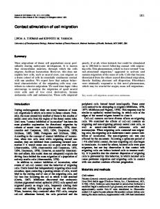
Fig. 1. Overexpression of PERK impairs eye development in Drosophila melanogaster. (A)Representative photomicrographs (top) and electron micrographs (bottom) of Drosophila eyes. Driver control (GMR-Gal4), kinase-dead PERK (GMR-Gal4>UAS-PERK-KR), PERK (GMR-Gal4>UAS-PERK-WT), GADD34 (GMR-Gal4>UAS-GADD34), PERK ⫻ GADD34 (GMR-Gal4>UAS-PERK; GADD34). (B)Immunoblot analysis of fly heads (three per lane) from indicated genotypes. Black arrowhead indicates upper hyperphosphorylated PERK band, open triangle indicates unphosphorylated inactive PERK. Blotting for actin provided the loading control. Note the increased level of PERK-WT immunoreactivity in GADD34-rescued flies. This reflects the preservation of PERKexpressing retinal tissue. (C)Fluorescence microscopy images of representative eye imaginal discs labelled for DNA synthesis with BrdU (red) and for PERK expression (green). All images aligned anterior (right) to posterior (left). Note the stripe of BrdU labelling indicative of DNA synthesis (S phase) immediately posterior to the morphogenetic furrow (arrowheads). GMR>PERK and GMR>PERK ⫻ GADD34 discs show no differences. (D)Fluorescence microscopy images of representative eye imaginal discs stained for phospho-histone H3 (phospho-H3, green) and ELAV expression (red). Note the stripe of phospho-H3 labelling, indicative of mitosis (M phase) posterior to the morphogenetic furrow (arrowheads). In the GMR>PERK eye imaginal discs, this stripe is markedly broadened. (E)Fluorescence microscopy images of representative eye imaginal discs in which PERK expression (blue) was driven in mosaic clones (GFP absent) by the tubulin promoter using the Gal4– Gal80 temperature-sensitive system. Top panel shows the kinase-dead PERK-expressing discs, whereas the bottom panel shows those expressing wild-type PERK. Phospho-histone H3 staining (red) was used to mark mitosis. Note the absent second mitotic wave in the clones lacking GFP expression (lower panel). Genotype: ey-flp; tubulin>Gal80ts; tubulin>FRT, GFP, stop, FRT, Gal4/ uasPERK.
Journal of Cell Science
2894
Journal of Cell Science 123 (17)
PAGE and immunoblot, with an upper active band (Fig. 1B, black triangle) and an inactive lower band (Fig. 1B, white triangle). In unstressed conditions, endogenous PERK is held inactive through interaction of the ER chaperone BiP with its luminal stress-sensing domain (Bertolotti et al., 2000). Overexpression of PERK probably overwhelms the capacity of BiP to maintain all PERK molecules in their inactive state resulting in autoactivation. To confirm that the eye phenotype was mediated by phosphorylation of eIF2, the physiological target of PERK, we generated fly lines expressing murine GADD34, a regulatory subunit of protein phosphatase 1 (PP1) that directs phosphatase activity specifically towards eIF2 (Novoa et al., 2001). When expressed at low levels, GADD34 failed to affect eye development (Fig. 1A). However, GADD34 rescued the developmental abnormalities caused by PERK overexpression (Fig. 1A). This was not through trivial effects on transgene expression, because levels of PERK protein were preserved in the double transgenic animals (Fig. 1B and Fig. 2B). We reasoned that the eye phenotype might reflect activation of pro-apoptotic pathways. However, when GMR-Gal4 was used to drive PERK in combination with the caspase antagonist p35, synthetic pupal lethality was observed (n3, data not shown). By contrast, expression of either caspase antagonist alone had no detectable toxic phenotype. This raised the possibility that apoptotic death of a subset of cells is necessary for survival of the PERKexpressing flies; alternatively, forced overexpression of PERK might have revealed a survival benefit of caspase activation during ISR signalling. To determine whether PERK expression posterior to the morphogenetic furrow affected cell cycle progression, eye imaginal discs were dissected from third instar larvae and stained for markers of S-phase (BrdU) or mitosis (phospho-histone H3) (Fig. 1C,D). The region of retinal differentiation posterior to the morphogenetic furrow was demonstrated either by staining for PERK overexpression (Fig. 1C) or expression of ELAV (Fig. 1D). BrdU incorporation was no different between PERK-expressing animals and controls expressing both PERK and GADD34 (Fig. 1C) or driver controls (data not shown). By contrast, the second mitotic wave was less well defined in the GMR>PERK animals compared with driver controls (Fig. 1D, arrowhead and supplementary material Fig. S1C). We tested this further by driving expression of either the wild-type PERK or the kinase-dead mutant under the tubulin promoter for 14 hours in mosaic clones using the conditional Gal4–Gal80 temperature-sensitive system (McGuire et al., 2003) (Fig. 1E). Expression of the inactive mutant had no effect on the second mitotic wave identified by phospho-histone H3 staining; by contrast expression of wild-type PERK markedly reduced phosphohistone H3 staining. These results suggested that, surprisingly, PERK expression did not affect G1–S progression in the developing eye, but was associated with impaired G2–M progression. Genetic screen for suppressors of the PERK eye phenotype
The GMR>PERK model was then used in an unbiased screen for genetic modifiers. Virgin female PERK-expressing flies were crossed with males from a library of 3000 Gene Search (GS)element insertions (Rival et al., 2009; Toba et al., 1999). GS elements are modified transposons that insert pseudo-randomly into the fly genome, resulting either in up- or downregulation of neighboring genes. We selected offspring that expressed both PERK and the GS-element and screened them for suppression of the
small, depigmented PERK eye. Eighty-three lines (2.7%) rescued eye development, which yielded 32 unique suppressor loci. Twentyfour of these suppressors still displayed overexpression of PERK when fly heads were assessed by SDS-PAGE and immunoblot analysis. Among these suppressors was an intronic insertion in the grapes gene (2L:16,684,855) that is likely to disrupt transcription (Fig. 2A). Preserved PERK expression in the rescued animals suggested that the rescue represented a bone fide genetic interaction and not a consequence of impaired transgene expression (Fig. 2B). Grapes is the Drosophila orthologue of mammalian checkpoint kinase 1 (CHK1), which is required for the G2–M DNA-damage checkpoint (Fogarty et al., 1997; Liu et al., 2000; Zachos et al., 2003). In eukaryotes, CHK1 activation requires the interaction of a complex containing RAD9, RAD1 and HUS1 with damaged DNA and subsequent recruitment of the kinase ATR (Martinho et al., 1998; Takai et al., 2000; Weiss et al., 2002). CHK1 activation, in turn, leads to cell cycle arrest both by activation of the tumour suppressor p53 and inactivation of the dual specificity phosphatase CDC25 (Roos and Kaina, 2006; Sanchez et al., 1997).
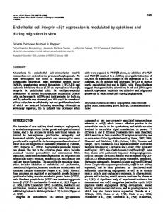
Fig. 2. Rescue of eye development by transposon insertion in the grapes gene. (A)Representative photomicrographs and eyes from animals expressing PERK without (GMR-Gal4>UAS-PERK-WT) and with a transposon in the grapes gene (GMR-Gal4>UAS-PERK-WT; grp+/–) (top). Corresponding electron micrographs are shown below. (B)SDS-PAGE and western blot analysis to assess the expression of PERK in the eyes rescued by transposable element insertion within the grapes gene. Note the elevated levels of transgenic PERK in eyes rescued by the transposable elements and by GADD34. (C)Representative photomicrographs of GMR>PERK-WT, GMR>PERK-WT ⫻ grapes RNAi. The crosses were repeated on at least three independent occasions. A minimum of three independent repeats were performed for each cross. Images of typical progeny are shown.
ISR and CHK1 activation The grapes insertion line (2L:16,684,855) successfully complemented the maternal lethality phenotype of grp1 (2L:16,685,378) (Fogarty et al., 1997), a well-characterised null allele, indicating that the line that rescued the eye phenotype in flies expressing PERK was not a complete null. A grapes siRNA (v12680, Vienna Drosophila RNAi Center VDRC, Austria) rescued the PERK eye phenotype (Fig. 2C), although less completely than the P-element insertion. This probably reflects the sensitivity of cell cycle progression to grapes protein levels. Consistent with this, grapes has been shown to have a crucial role during the late nuclear cycles of Drosophila embryos (Fogarty et al., 1997; Fogarty et al., 1994), perhaps through effects on altered chromosomal condensation (Yu et al., 2000), cyclin A degradation (Su et al., 1999) or cyclin B localisation (Royou et al., 2008). Similarly, CHK1 is essential for mammalian embryogenesis (Liu et al., 2000; Takai et al., 2000; Zachos et al., 2003), but also behaves as a haploinsufficient tumour suppressor, with tumours failing to lose both alleles (Lam et al., 2004; Liu et al., 2000).
Journal of Cell Science
CHK1 undergoes transient activation during ER stress mediated by PERK
The relevance of CHK1 to PERK signalling was then assessed in cultured HCT116 human colon carcinoma cells. These cells were chosen because they possess intact cell cycle checkpoints (Zhang
Fig. 3. CHK1 is phosphorylated during ER stress. (A)Immunoblot of whole cell lysate from HCT116 cells treated for the indicated times with thapsigargin (500 nM). UV indicates irradiation with 150 J/m2 UV used as the positive control. (B)Graphical representation of three replicates of experiment A showing P317-CHK1 band densitometry normalised to UV control (mean ± s.e.m.). (C)Immunoblot of mouse embryonic fibroblasts (MEFs) untreated (UT) or treated with thapsigargin 500 nM for 1 hour (Tg), tunicamycin 2.5g/ml for 2 hours (Tm) or DTT 1 mM for 1 hour (DTT). The gels are representative of three repeats.
2895
et al., 2006). Treatment with thapsigargin inhibits the SERCA calcium pump and rapidly induces ER stress. This resulted in phosphorylation of CHK1 on Ser317 (Fig. 3A,B). Similar results were obtained with murine embryonic fibroblasts (MEFs) (Fig. 3C). Phosphorylation of Ser317 by the kinase ATR is required for CHK1 activation during genotoxic stress, for example UV irradiation (Martinho et al., 1998; Weiss et al., 2002; Zhao and Piwnica-Worms, 2001). This provided a positive control. Similar CHK1 Ser317 phosphorylation was seen with other ER-stressinducing agents, including tunicamycin and DTT (Fig. 3C). The time-course of CHK1 phosphorylation was similar to that previously described for eIF2 phosphorylation by PERK in response to ER stress (Harding et al., 1999). We therefore tested the effect of PERK genotype on ER-stress-induced CHK1 phosphorylation (Fig. 4). There was a rapid but transient phosphorylation of CHK1 in Perk+/+ MEFs (Harding et al., 2000b), but this was absent in Perk –/– MEFs (Fig. 4A). Several parallel signalling pathways are activated during ER stress (Ron and Walter, 2007). We therefore assessed whether PERK was sufficient for activation of CHK1 by using the Fv2E-PERK chimeric kinase, wherein the stress-responsive ER luminal domain of PERK is replaced by the Fv2E dimerisation motif (Fig. 4B). This protein is
Fig. 4. CHK1 activation during ER stress is mediated by PERK phosphorylation of eIF2. (A)Immunoblot of Perk+/+ and Perk–/– MEFs treated with thapsigargin 500 nM for the indicated times. (B)Immunoblot of CHO cells stably expressing Fv2E-PERK treated for indicated times with 100 nM AP20187 dimerisation compound. Salt-extracted nuclear proteins (nuclear) are shown in panels 2 and 3, all other panels are of soluble post-nuclear supernatant proteins. (C)Immunoblot of eIF2SS and eIF2AA MEFs treated for the indicated times with thapsigargin 500 nM (Tg) or cycloheximide 50g/ml (Cyc). The samples were run on a single gel but have been separated for clarity. The gels shown are representative of three repeats.
Journal of Cell Science
2896
Journal of Cell Science 123 (17)
normally inactive, but undergoes dimerisation upon treatment of cells with AP20187 (Lu et al., 2004b). This causes activation by trans-autophosphorylation of the kinase domain, which is detectable as an upward size shift on SDS-PAGE (Harding et al., 1999; Lu et al., 2004b; Marciniak et al., 2006). There was rapid phosphorylation of Ser317 in CHK1 when cells were treated with AP20187 (Fig. 4B). This occurred in both the cytosolic and nuclear fractions of CHK1, again displaying a transient nature despite sustained activation of Fv2E-PERK (Fig. 4B). The rescue by GADD34 of the GMR>PERK model demonstrated that eIF2 phosphorylation was responsible for the eye phenotype (Fig. 1A). We wished to determine whether CHK1 phosphorylation was also mediated by eIF2 phosphorylation or was an unrelated consequence of PERK activation. PERK phosphorylates eIF2 on Ser51, but cells homozygous for an alanine substitution of this residue are immune to the translation attenuation effects of PERK (Scheuner et al., 2001). We treated wild-type eIF2SS and mutant eIF2AA cells with thapsigargin to induce ER stress and assessed the degree to which CHK1 was phosphorylated (Fig. 4C). In wildtype eIF2SS cells, the expected phosphorylation of CHK1 on residue 317 was observed, but when mutant eIF2AA cells were similarly treated, no CHK1 phosphorylation was detected. CHK1 phosphorylation is therefore a response to eIF2 phosphorylation rather than CHK1 being a PERK substrate. Since eIF2 phosphorylation attenuates protein synthesis, we tested the effect of direct inhibition of protein translation with cycloheximide (Fig. 4C). This agent induced rapid phosphorylation of CHK1 but, as expected, was insensitive to the serine-to-alanine substitution because cycloheximide acts directly upon the ribosome. Taken together, these data demonstrate that in mammalian cells, PERK-mediated phosphorylation of eIF2 and the consequent inhibition of protein translation result in phosphorylation of CHK1 on Ser317. CHK1 impairs G2 cell cycle progression during ER stress
Next, we investigated the effect of ER stress and isolated PERK activation on cell cycle progression in mammalian cells. Treatment of asynchronous CHO cells with either tunicamycin or thapsigargin caused a progressive G1 cell cycle arrest at 24 and 36 hours (Fig. 5A, left and middle panels). A similar response has previously been described in mammalian cells and suggested to involve PERKmediated loss of cyclin D1 (Brewer and Diehl, 2000; Brewer et al., 1999; Raven et al., 2008). It was therefore surprising that treatment of CHO cells stably expressing Fv2E-PERK with AP20187 for up to 36 hours caused no G1 cell cycle arrest (Fig. 5A, right panel). To determine whether this effect was dependent upon the degree of PERK activation, cells were treated with 0 to 100 nM AP20187 and still no evidence of G1 arrest was observed, despite activation of Fv2E-PERK and loss of cyclin D1 (supplementary material Fig. S2A,B). It was important to establish whether CHK1 activity is relevant to cell cycle progression during ER stress. HCT116 cell lines have previously been generated that express CHK1 siRNA or a scrambled control siRNA under the control of a tetracycline-responsive promoter (Ganzinelli et al., 2008). When cultured in the presence of doxycycline, levels of CHK1 protein were knocked down by over 75% as estimated by immunoblot (Fig. 5B). Remarkably, when asynchronous cultures of HCT116 cells were treated with thapsigargin, an early increase in G2 phase cells was observed that was completely abrogated by CHK1 siRNA (Fig. 5C–E). In addition, after 24 hours, control cells demonstrated substantial
Fig. 5. Transient G2 cell cycle delay during ER stress is mediated by CHK1. (A)Asynchronous cultures of CHO cells stably expressing Fv2EPERK were treated for the indicated times with tunicamycin (2.5 mg/ml), thapsigargin (500 nM) or AP20187 (100 nM). One million cells were fixed in ethanol and DNA content labelled with propidium iodide was determined by FACS analysis. The positions of cell populations with 2N and 4N DNA content are illustrated. (B)HCT116 cells stably transfected with a Tet-ON CHK1 siRNA or control scrambled sequence were treated for 48 hours with 1 mg/ml doxycycline. Proteins were subjected to immunoblot for CHK1 and actin. (C)Asynchronous cultures of HCT116 Tet-ON CHK1 siRNA or control cells treated with 500 nM thapsigargin and subjected to FACS analysis of the cell cycle. Representative results from four independent repeats are shown. (D)Pooled data from experiment in C are shown as mean ± s.e.m. (E)Data from C expressed as ratio of G2/G1 phase, mean ± s.e.m. (F)Data from C for subG1 phase, mean ± s.e.m. *PPERK eyes rescued by cyclin E expression. Cyclin E (GMR-Gal4>UAS-cyc E), PERK (GMRGal4>UAS-PERK-WT), PERK ⫻ cyclin E (GMR-Gal4>UAS-PERK-WT, cyc E). A minimum of three independent repeats were performed for each cross. Images of typical progeny are shown. (D)Schematic pathway to show PERK-mediated phosphorylation of eIF2 leads to translation attenuation during ER stress. Inhibition of protein translation induces CHK1 phosphorylation, which in turn impairs cell cycle progression in part by depletion of cdk2–cyclin-E activity.
cycle. CDC25A is a short-lived protein that has been observed to decay on inhibition of protein translation with cycloheximide (Molinari et al., 2000). We therefore tested the effect of ER stress on CDC25A and observed a rapid decline with thapsigargin treatment (Fig. 6A,B). When CHK1 was partially depleted by RNA interference (Ganzinelli et al., 2008), stabilisation of CDC25A was observed (Fig. 6A,B). Previous studies have identified loss of cyclin D1 as a potential mediator of PERK-dependent cell cycle arrest (Brewer and Diehl, 2000; Brewer et al., 1999). However, when we tested the effect of three UAS–cyclin-D lines on eye development in GMR>PERK flies, each failed to rescue eye morphogenesis (data not shown and supplementary material Fig. S4). By contrast, forced overexpression of cyclin E partially rescued eye development (Fig. 6C). This effect did not reflect a generalised positive effect on eye development, because cyclin E failed to rescue other rough-eye models including overexpression of wild-type Tau, R406W Tau or dronc (supplementary material Fig. S4B,C). Indeed, in some cases, the rough eye phenotype was worsened by cyclin E (supplementary material Fig. S4B,C). Taken together, these data provide evidence for the involvement of CHK1 in cell cycle responses during PERK activation via a pathway illustrated in Fig. 6D. This pathway provides a mechanistic explanation for some of the impairment of cell cycle progression seen during ER stress. It also identifies a site of interaction between DNA damage response networks and the integrated stress response. Discussion Cells and animals deficient in PERK are hypersensitive to ER stress (Harding et al., 2001; Harding et al., 2000b), but prolonged signalling through PERK impairs cell growth and triggers cell death (Harding et al., 2009; Lin et al., 2009). This suggests that the utility curve of translation attenuation is biphasic. Our study has shed light on the descending limb of this curve by identifying how
it impacts on cell cycle components at several levels. In a screen for effectors of PERK signalling, we identified grapes, a component of the DNA damage response. Subsequent analysis of cultured mammalian cells revealed ER-stress-induced phosphorylation of CHK1 to be a consequence of eIF2 phosphorylation and subsequent translational repression. We also observed that ER stress destabilises CDC25A in a CHK1-dependent manner to cause G2 cell cycle delay. The integrated stress and DNA damage responses are known to share components. For example, cell cycle arrest after UV irradiation is mediated in part by phosphorylation of eIF2 by GCN2 or PERK, although the mechanism of kinase activation is unknown (Jiang and Wek, 2005; Wu et al., 2002). The eIF2 phosphatase GADD34 was first studied in the context of the DNA damage response, accordingly its name derives from ‘growth arrest and DNA damage 34’ (Fornace et al., 1989; Hollander et al., 1997; Zhan et al., 1994). Only later was it found to affect ER stress signalling (Novoa et al., 2001). Similarly, CHOP, a transcription factor induced by ER stress, is identical to GADD153, which is induced by numerous genotoxins (Barone et al., 1994; Friedman, 1996; Gately et al., 1994; Gujuluva et al., 1994; McCullough et al., 2001). However, our study is the first to implicate CHK1 in the cell cycle effects of the ISR; although, transcriptional profiling previously identified a factor involved in CHK1 activation, RAD1, to be induced during ER stress (Marciniak et al., 2004). It has been shown that PERK activation causes loss of cyclin D1 and G1 cell cycle arrest either through inhibition of cyclin D1 translation or increased degradation (Brewer and Diehl, 2000; Brewer et al., 1999; Raven et al., 2008). In parallel, ribosomal biogenesis is rapidly repressed by PERK (DuRose et al., 2009) and subsequent binding of free ribosomal proteins to the E3 ligase MDM2 appears to stabilise p53 and cause G1 arrest (Zhang et al., 2006). However, PERK-dependent activation of the GSK3 during ER stress has been shown to destabilise p53 (Baltzis et al., 2007;
Journal of Cell Science
2898
Journal of Cell Science 123 (17)
Qu et al., 2004). Moreover, growth arrest is only attenuated, not abolished in Perk–/– cells, suggesting additional regulation (Zhang et al., 2006). Our observations confirmed the slow onset of G1 arrest during ER stress, but surprisingly this was not reproduced by isolated PERK activation. Therefore, current models in which PERK mediates cyclin D1 loss to cause G1 arrest appear to be incomplete. G2 delay might be an ancient response to ER stress, predating the evolution of PERK. In yeast, which lack PERK, a delay in G2–M phase has been described following treatment with ER-stressinducing agents (Bonilla and Cunningham, 2003). Interestingly, in yeast, this requires active Swe1p kinase, which is responsible for inhibitory phosphorylation of the cyclin-dependent kinase Cdc28p and is functionally antagonised by Mih1p/CDC25 (Russell et al., 1989). It is important to note that PERK expression generated an obvious phenotype in the fly eye, but isolated PERK activation in CHO-K1 cells had little effect on cell cycle progression. Primary retinal cells have intact cell cycle checkpoints, whereas CHO-K1 cells are defective in some aspects of checkpoint signalling, for example via p53 (Hu et al., 1999). However, in HCT116 cells, which express wild-type p53, an ER stress-induced G2–M checkpoint was seen that was dependent upon CHK1. In addition, the eye phenotype is likely to integrate the effects of PERK signalling on the cell cycle, cell death and developmental programmes, whereas the CHO-K1 cell experiments were designed primarily to test effects of CHK1 phosphorylation and cell cycle phase. The relevance of p53 signalling to the phenotypes observed in each system is unclear, because the p53 orthologue of Drosophila regulates apoptosis in a manner analogous to its mammalian counterpart, but it lacks an effect on cell cycle progression. It has been proposed that cell cycle regulation by p53 might have evolved subsequent to the divergence of vertebrates and arthropods (Steller, 2000). Since CHK1 is able to phosphorylate and activate p53 (Shieh et al., 2000), it will be of great interest to determine whether any of the grapes or CHK1-mediated effects observed in our study are mediated by p53. Our observation that CHK1 has a role in the loss of CDC25A during ER stress suggests an additional mechanism by which PERK contributes to G1 arrest, because CDC25A is required to maintain the activity of cdk2–cyclin-E and allow entry into S phase (Malumbres and Barbacid, 2009). However, since isolated PERK activation does not induce G1 arrest, it appears likely that additional signals are required. In addition, our finding that CHK1 mediates an earlier G2 delay is consistent with previous work showing that disruption of the CHK1–CDC25A pathway abrogates S and G2 checkpoints following exposure to ionising radiation (Zhao et al., 2002). A CHK1-dependent increase in sub-G1 late apoptotic HCT116 cells was noted during ER stress. This was remarkable, because in the context of DNA damage, CHK1 signalling is anti-apoptotic (Myers et al., 2009). When, as a result of defective CHK1 signalling, cells enter mitosis with damaged DNA, the result is mitotic catastrophe and cell death (Francesconi et al., 1997; Huang et al., 2005). The absence of such a response during ER stress suggests that the G2–M checkpoint does not serve to protect the cell from DNA damage accompanying ER stress. Instead, it is plausible that cell cycle progression is delayed during ER stress so that daughter cells are not faced immediately with impaired ER function following mitosis. This might, for
example, provide additional information regarding nutritional status to the cell cycle machinery. Further work will be needed to determine whether inhibition of CHK1 will be anti-apoptotic during ER stress. In diseases characterised by ER-stress-induced cell death this might prove therapeutically useful. However, because CHK1 inhibition is currently being pursued as a chemotherapeutic option, it will be necessary to ensure that CHK1 inhibitors do not enable ER-stressed tumour cells to survive hypoxic environments more efficiently. The current study does not address directly the mechanism by which CHK1 is activated by translational repression. A number of possibilities exist. Turnover rates vary dramatically between proteins and so inhibition of their synthesis can rapidly lead to changes in the ratios of effector and inhibitory factors. For example, activation of the transcription factor NFB during ER stress involves the rapid decay of its antagonist IKB (Deng et al., 2004). It is plausible that translational attenuation might similarly lead to the loss of a CHK1 inhibitor. Alternatively, a ribosomal stress signal might activate the CHK1 pathway in addition to stabilising p53 (Zhang et al., 2006). Our study demonstrates that CHK1 is activated during ER stress by PERK repression of protein translation. It is plausible that activation of CHK1 mediates some of the growth inhibitory effects seen during ER stress. However, it is likely that the ISR interfaces with the cell cycle at several levels owing to the short life of many cell cycle regulatory proteins. Materials and Methods Expression plasmids
The cDNAs for PERK and KR PERK were kindly provided by Cesar de Haro, Centro do Biologia Molecular ‘Severo Ochoa’, Madrid, Spain (Pomar et al., 2003). Both cDNAs were inserted downstream of Gal4 UASs (UAS-PERK and UASPERK-KR) in the pUAST plasmid by directional cloning following BglII and NotI digestion. Tobacco etch virus (TEV)-cleavable GST-PERK fusion protein was generated by transfer of the PERK coding sequence into pGV67 (a gift from Brad Nolan, Yale University, New Haven, CT). UAS-GADD34 was generated by directional cloning of mGADD34 cDNA (Novoa et al., 2001) into pUAST digested with BglII and XhoI. Drosophila stocks Transgenic lines were generated by embryonic injection of each pUAST construct (EMBL Injection Service, Heidelberg, Germany) and progeny were balanced using classical genetics. The UAS-PERK-WT line used herein was an insert on chromosome III. GMR-Gal4/CyO; UAS-PERK/TM6b stocks were generated for the genetic screen and crossed with UAS-GADD34/CyO to confirm the phenotype as an ‘on’ pathway effect. w[*]; P{w[+mC]UAS-CycE.L}ML1, w[*]; P{w[+mC]GAL4ninaE.GMR}12, P{UAS-DIAP1.H} were from Bloomington Stock Center. Grp siRNA-expressing flies (v12680) were from the Vienna Drosophila RNAi Center. The following stocks were generous gifts: w*;P[w+,UAS-CycD]II.1/P[w+,UASCycD]II.1 and w*;P[w+,UAS-CycD]III.1/P[w+,UAS-CycD]III.1 were from Christian Lehner (University of Zurich, Zurich, Switzerland); UAS-dronc (Leulier et al., 2006), UAS-Tau and UAS-R406W Tau (Wittmann et al., 2001) gifts from Sara Imarisio (University of Cambridge, Cambridge, UK). grp1/CyO-LacZ (Fogarty et al., 1997) and grp209/Cyo-GFP flies (LaRocque et al., 2007) were from Tin Tin Su (University of Colorado, Boulder, CO). The GS-element (Toba et al., 1999) library used for the genetic screen has previously been described (Rival et al., 2009). All stocks had the w1118 genetic background and were maintained at 18°C using standard techniques unless otherwise stated. To express PERK conditionally in mosaic clones, a combination of FRT-mediated flip-out system (Zecca and Struhl, 2002) and the Gal4–Gal80 gene expression strategy (McGuire et al., 2003) were used. In brief, flp recombinase was expressed using the eye specific promoter to flip-out a GFP-stop cassette, thereby activating Gal4 expression. In the background, Gal80ts was expressed to block Gal4-driven PERK expression. To activate PERK expression conditionally, the temperature was shifted to 25°C, which inactivated Gal80ts and allowing Gal4-driven UAS-PERK expression. Genetic screen
Males from the GS library were crossed to virgin females expressing the PERK transgene and GMR-Gal4 driver. The eye morphology of offspring was scored independently by two microscopists. GS lines that strongly rescued eye
ISR and CHK1 activation morphogenesis were retested to confirm the effect and then the transposon insertion sites were mapped by inverse PCR (iPCR). Inverse PCR
Genomic DNA was extracted from each GS insertion line and digested with the restriction enzyme Sau3AI, which cut in the middle of the GS element as well as regularly throughout the Drosophila genome. Digested DNA was then circularised using T4 DNA ligase to provide a template for the inverse PCR reaction. The 5⬘ and 3⬘ ends of the GS element and the contiguous fragments of genomic DNA were amplified using PCR and then sequenced. The resulting sequences were checked using the BLAST algorithm on FlyBase to view the site of integration (http://flybase.bio.indiana.edu/blast). Cell culture and transfection
Stable clones of CHO cells expressing Fv2E-PERK have been described previously (Lu et al., 2004a). These were treated with AP20187 as indicated (ARIAD; www.ariad.com/regulation kits). Lysates were prepared in 0.5% Triton X-100, 100 mM NaCl, 20 mM Tris-HCl, pH 7.4, 1 mM DTT, 1 mM PMSF, 4 g/ml aprotinin, 2 g/ml pepstatin A, 10 mM tetrasodium pyrophosphate, 15.5 mM glycerophosphate, 100 mM NaF. Perk+/+ and Perk–/– MEFs have been described previously (Harding et al., 2000b) eIF2AA and eIF2SS MEFs were a gift from Randal J. Kaufman (Howard Hughes Medical Institute, Chevy Chase, MD) and HCT116 cells were a gift from Paul Lehner (Cambridge Institute for Medical Research, Cambridge, UK). HCT116 cells stably transfected with inducible CHK1 siRNA or scrambled control siRNA were a kind gift from Giovanna Damia (Istituto Mario Negri, Milan, Italy) (Ganzinelli et al., 2008).
Journal of Cell Science
Immunoblot and immunohistochemistry
Mammalian PERK, CHOP and GADD34 were detected with polyclonal antisera as described previously (Marciniak et al., 2004). Drosophila PERK was detected using a polyclonal antiserum raised against the kinase domain of recombinant PERK. Briefly, a GST-PERK kinase domain was expressed in BL21 E. coli and purified using Glutathione affinity beads (Amersham Biosciences, Sweden). The kinase domain was cleaved from the GST using TEV protease. The protease was then removed by Ni-NTA affinity matrix (Qiagen, Hilden, Germany) and the purified PERK kinase domain used to immunise two rabbits (Cambridge Research Biochemicals, Cleveland, UK). Anti-PERK antiserum was used at a dilution of 1:1000. Commercial antibodies used were CHK1 (#2345, Cell Signaling), phosphoserine 317 CHK1 (#2344, Cell Signaling), cyclin D1 (mouse monoclonal DCS6, #2926, Cell Signaling) and CDC25A (mouse monoclonal F-6, sc-7389, Santa Cruz). Imaginal discs were stained using standard techniques. Antibodies used were 1:50 anti-BrdU antibody (BD Pharmingen), 1:500 polyclonal rabbit anti-phospho Histone H3 (Ser10) antibody (Millipore, Billerica, MA), and 1:200 mouse monoclonal antiELAV (clone 9F8A9, Developmental Studies Hybridoma Bank at University of Iowa). The anti-PERK antiserum was affinity-purified against the same epitope before use.
This work was supported by the MRC (UK), EPSRC and Addenbrooke’s Charitable Trust. S.J.M. is an MRC Clinician Scientist (G0601840). We are grateful to Andrew Spackman for excellent technical support. Deposited in PMC for release after 6 months. Supplementary material available online at http://jcs.biologists.org/cgi/content/full/123/17/2892/DC1
References Baltzis, D., Pluquet, O., Papadakis, A. I., Kazemi, S., Qu, L. K. and Koromilas, A. E. (2007). The eIF2alpha kinases PERK and PKR activate glycogen synthase kinase 3 to promote the proteasomal degradation of p53. J. Biol. Chem. 282, 31675-31687. Barone, M. V., Crozat, A. Y., Tabaee, A., Philipson, L. and Ron, D. (1994). CHOP (GADD153) and its oncogenic variant, TLS-CHOP, differ in their ability to induce G1/S arrest. Genes Dev. 8, 453-464. Bertolotti, A., Zhang, Y., Hendershot, L., Harding, H. and Ron, D. (2000). Dynamic interaction of BiP and the ER stress transducers in the unfolded protein response. Nat. Cell Biol. 2, 326-332. Bi, M., Naczki, C., Koritzinsky, M., Fels, D., Blais, J., Hu, N., Harding, H., Novoa, I., Varia, M., Raleigh, J. et al. (2005). ER stress-regulated translation increases tolerance to extreme hypoxia and promotes tumor growth. EMBO J. 24, 3470-3481. Bonilla, M. and Cunningham, K. W. (2003). Mitogen-activated protein kinase stimulation of Ca(2+) signaling is required for survival of endoplasmic reticulum stress in yeast. Mol. Biol. Cell 14, 4296-4305. Boutros, R., Lobjois, V. and Ducommun, B. (2007). CDC25 phosphatases in cancer cells: key players? Good targets? Nat. Rev. Cancer 7, 495-507. Brand, A. H. and Perrimon, N. (1993). Targeted gene expression as a means of altering cell fates and generating dominant phenotypes. Development 118, 401-415. Brewer, J. W. and Diehl, J. A. (2000). PERK mediates cell-cycle exit during the mammalian unfolded protein response. Proc. Natl. Acad. Sci. USA 97, 12625-12630.
2899
Brewer, J. W., Hendershot, L. M., Sherr, C. J. and Diehl, J. A. (1999). Mammalian unfolded protein response inhibits cyclin D1 translation and cell-cycle progression. Proc. Natl. Acad. Sci. USA 96, 8505-8510. Brush, M. H., Weiser, D. C. and Shenolikar, S. (2003). Growth arrest and DNA damageinducible protein GADD34 targets protein phosphatase 1alpha to the endoplasmic reticulum and promotes dephosphorylation of the alpha subunit of eukaryotic translation initiation factor 2. Mol. Cell. Biol. 23, 1292-1303. Deng, J., Lu, P. D., Zhang, Y., Scheuner, D., Kaufman, R. J., Sonenberg, N., Harding, H. P. and Ron, D. (2004). Translational repression mediates activation of Nuclear Factor kappa B by phosphorylated translation initiation factor 2. Mol. Cell. Biol. 24, 10161-10168. DuRose, J. B., Scheuner, D., Kaufman, R. J., Rothblum, L. I. and Niwa, M. (2009). Phosphorylation of eukaryotic translation initiation factor 2alpha coordinates rRNA transcription and translation inhibition during endoplasmic reticulum stress. Mol. Cell. Biol. 29, 4295-4307. Fogarty, P., Kalpin, R. F. and Sullivan, W. (1994). The Drosophila maternal-effect mutation grapes causes a metaphase arrest at nuclear cycle 13. Development 120, 21312142. Fogarty, P., Campbell, S. D., Abu-Shumays, R., Phalle, B. S., Yu, K. R., Uy, G. L., Goldberg, M. L. and Sullivan, W. (1997). The Drosophila grapes gene is related to checkpoint gene chk1/rad27 and is required for late syncytial division fidelity. Curr. Biol. 7, 418-426. Fornace, A. J., Neibert, D. W., Hollander, M. C., Luethy, J. D., Papathanasiou, M., Fragoli, J. and Holbrook, N. J. (1989). Mammalian genes coordinately regulated by growth arrest signals and DNA-damaging agents. Mol. Cell. Biol. 9, 4196-4203. Francesconi, S., Grenon, M., Bouvier, D. and Baldacci, G. (1997). p56(chk1) protein kinase is required for the DNA replication checkpoint at 37 degrees C in fission yeast. EMBO J. 16, 1332-1341. Freeman, M. (1996). Reiterative use of the EGF receptor triggers differentiation of all cell types in the Drosophila eye. Cell 87, 651-660. Friedman, A. D. (1996). GADD153/CHOP, a DNA damage-inducible protein, reduced CAAT/enhancer binding protein activities and increased apoptosis in 32D c13 myeloid cells. Cancer Res. 56, 3250-3256. Ganzinelli, M., Carrassa, L., Crippa, F., Tavecchio, M., Broggini, M. and Damia, G. (2008). Checkpoint kinase 1 down-regulation by an inducible small interfering RNA expression system sensitized in vivo tumors to treatment with 5-fluorouracil. Clin. Cancer Res. 14, 5131-5141. Gately, D., Jones, J., Christen, R., Barton, R., Los, G. and Howell, S. (1994). Induction of the growth arrest and DNA damage-inducible gene GADD153 by cisplatin in vitro and in vivo. Br. J. Cancer 70, 1102-1106. Gujuluva, C., Baek, J.-H., Shin, K.-H., Cherrick, H. and Park, N.-O. (1994). Effect of UV-irradiation on cell cycle, viability and the expression of p53, gadd153 and gadd45 genes in normal and HPV-immortalized oral keratinocytes. Oncogene 9, 18191827. Harding, H., Zhang, Y. and Ron, D. (1999). Translation and protein folding are coupled by an endoplasmic reticulum resident kinase. Nature 397, 271-274. Harding, H., Novoa, I., Zhang, Y., Zeng, H., Wek, R. C., Schapira, M. and Ron, D. (2000a). Regulated translation initiation controls stress-induced gene expression in mammalian cells. Mol. Cell 6, 1099-1108. Harding, H., Zhang, Y., Bertolotti, A., Zeng, H. and Ron, D. (2000b). Perk is essential for translational regulation and cell survival during the unfolded protein response. Mol. Cell 5, 897-904. Harding, H., Zeng, H., Zhang, Y., Jungreis, R., Chung, P., Plesken, H., Sabatini, D. and Ron, D. (2001). Diabetes Mellitus and excocrine pancreatic dysfunction in Perk–/– mice reveals a role for translational control in survival of secretory cells. Mol. Cell 7, 1153-1163. Harding, H., Zhang, Y., Zeng, H., Novoa, I., Lu, P., Calfon, M., Sadri, N., Yun, C., Popko, B., Paules, R. et al. (2003). An integrated stress response regulates amino acid metabolism and resistance to oxidative stress. Mol. Cell 11, 619-633. Harding, H. P., Zhang, Y., Scheuner, D., Chen, J. J., Kaufman, R. J. and Ron, D. (2009). Ppp1r15 gene knockout reveals an essential role for translation initiation factor 2 alpha (eIF2alpha) dephosphorylation in mammalian development. Proc. Natl. Acad. Sci. USA 106, 1832-1837. Hollander, M. C., Zhan, Q., Bae, I. and Fornace, A. J., Jr (1997). Mammalian GADD34, an apoptosis- and DNA damage-inducible gene. J. Biol. Chem. 272, 13731-13737. Hu, T., Miller, C. M., Ridder, G. M. and Aardema, M. J. (1999). Characterization of p53 in Chinese hamster cell lines CHO-K1, CHO-WBL, and CHL: implications for genotoxicity testing. Mutat. Res. 426, 51-62. Huang, X., Tran, T., Zhang, L., Hatcher, R. and Zhang, P. (2005). DNA damageinduced mitotic catastrophe is mediated by the Chk1-dependent mitotic exit DNA damage checkpoint. Proc. Natl. Acad. Sci. USA 102, 1065-1070. Jiang, H. Y. and Wek, R. C. (2005). GCN2 phosphorylation of eIF2alpha activates NFkappaB in response to UV irradiation. Biochem. J. 385, 371-380. Kumar, R., Azam, S., Sullivan, J., Owen, C., Cavener, D., Zhang, P., Ron, D., Harding, H., Chen, J., Han, A. et al. (2001). Brain ischemia and reperfusion activates the eukaryotic initiation factor 2a kinase, PERK. J. Neurochem. 77, 14181421. Lam, M. H., Liu, Q., Elledge, S. J. and Rosen, J. M. (2004). Chk1 is haploinsufficient for multiple functions critical to tumor suppression. Cancer Cell 6, 45-59. LaRocque, J. R., Jaklevic, B., Su, T. T. and Sekelsky, J. (2007). Drosophila ATR in double-strand break repair. Genetics 175, 1023-1033. Leulier, F., Ribeiro, P. S., Palmer, E., Tenev, T., Takahashi, K., Robertson, D., Zachariou, A., Pichaud, F., Ueda, R. and Meier, P. (2006). Systematic in vivo RNAi
Journal of Cell Science
2900
Journal of Cell Science 123 (17)
analysis of putative components of the Drosophila cell death machinery. Cell Death Differ. 13, 1663-1674. Lin, J. H., Li, H., Zhang, Y., Ron, D. and Walter, P. (2009). Divergent effects of PERK and IRE1 signaling on cell viability. PLoS ONE 4, e4170. Liu, L., Cash, T. P., Jones, R. G., Keith, B., Thompson, C. B. and Simon, M. C. (2006). Hypoxia-induced energy stress regulates mRNA translation and cell growth. Mol. Cell 21, 521-531. Liu, Q., Guntuku, S., Cui, X. S., Matsuoka, S., Cortez, D., Tamai, K., Luo, G., Carattini-Rivera, S., DeMayo, F., Bradley, A. et al. (2000). Chk1 is an essential kinase that is regulated by Atr and required for the G(2)/M DNA damage checkpoint. Genes Dev. 14, 1448-1459. Lu, P. D., Harding, H. P. and Ron, D. (2004a). Translation re-initiation at alternative open reading frames regulates gene expression in an integrated stress response. J. Cell Biol. 167, 27-33. Lu, P. D., Jousse, C., Marciniak, S. J., Zhang, Y., Novoa, I., Scheuner, D., Kaufman, R. J., Ron, D. and Harding, H. P. (2004b). Cytoprotection by pre-emptive conditional phosphorylation of translation initiation factor 2. EMBO J. 23, 169-179. Ma, Y. and Hendershot, L. M. (2003). Delineation of a negative feedback regulatory loop that controls protein translation during endoplasmic reticulum stress. J. Biol. Chem. 278, 34864-34873. Malumbres, M. and Barbacid, M. (2009). Cell cycle, CDKs and cancer: a changing paradigm. Nat. Rev. Cancer 9, 153-166. Marciniak, S. J., Yun, C. Y., Oyadomari, S., Novoa, I., Zhang, Y., Jungreis, R., Nagata, K., Harding, H. P. and Ron, D. (2004). CHOP induces death by promoting protein synthesis and oxidation in the stressed endoplasmic reticulum. Genes Dev. 18, 3066-3077. Marciniak, S. J., Garcia-Bonilla, L., Hu, J., Harding, H. P. and Ron, D. (2006). Activation-dependent substrate recruitment by the eukaryotic translation initiation factor 2 kinase PERK. J. Cell Biol. 172, 201-209. Martinho, R. G., Lindsay, H. D., Flaggs, G., DeMaggio, A. J., Hoekstra, M. F., Carr, A. M. and Bentley, N. J. (1998). Analysis of Rad3 and Chk1 protein kinases defines different checkpoint responses. EMBO J. 17, 7239-7249. McCullough, K. D., Martindale, J. L., Klotz, L. O., Aw, T. Y. and Holbrook, N. J. (2001). Gadd153 sensitizes cells to endoplasmic reticulum stress by down- regulating Bcl2 and perturbing the cellular redox state. Mol. Cell. Biol. 21, 1249-1259. McGuire, S. E., Le, P. T., Osborn, A. J., Matsumoto, K. and Davis, R. L. (2003). Spatiotemporal rescue of memory dysfunction in Drosophila. Science 302, 1765-1768. Molinari, M., Mercurio, C., Dominguez, J., Goubin, F. and Draetta, G. F. (2000). Human Cdc25 A inactivation in response to S phase inhibition and its role in preventing premature mitosis. EMBO Rep. 1, 71-79. Myers, K., Gagou, M. E., Zuazua-Villar, P., Rodriguez, R. and Meuth, M. (2009). ATR and Chk1 suppress a caspase-3-dependent apoptotic response following DNA replication stress. PLoS Genet. 5, e1000324. Novoa, I., Zeng, H., Harding, H. and Ron, D. (2001). Feedback inhibition of the unfolded protein response by GADD34-mediated dephosphorylation of eIF2a. J. Cell Biol. 153, 1011-1022. Pomar, N., Berlanga, J. J., Campuzano, S., Hernandez, G., Elias, M. and De Haro, C. (2003). Functional characterization of Drosophila melanogaster PERK eukaryotic initiation factor 2alpha (eIF2alpha) kinase. Eur. J. Biochem. 270, 293-306. Qu, L., Huang, S., Baltzis, D., Rivas-Estilla, A. M., Pluquet, O., Hatzoglou, M., Koumenis, C., Taya, Y., Yoshimura, A. and Koromilas, A. E. (2004). Endoplasmic reticulum stress induces p53 cytoplasmic localization and prevents p53-dependent apoptosis by a pathway involving glycogen synthase kinase-3beta. Genes Dev. 18, 261277. Raven, J. F., Baltzis, D., Wang, S., Mounir, Z., Papadakis, A. I., Gao, H. Q. and Koromilas, A. E. (2008). PKR and PKR-like endoplasmic reticulum kinase induce the proteasome-dependent degradation of cyclin D1 via a mechanism requiring eukaryotic initiation factor 2alpha phosphorylation. J. Biol. Chem. 283, 3097-3108. Rival, T., Page, R. M., Chandraratna, D. S., Sendall, T. J., Ryder, E., Liu, B., Lewis, H., Rosahl, T., Hider, R., Camargo, L. M. et al. (2009). Fenton chemistry and oxidative stress mediate the toxicity of the beta-amyloid peptide in a Drosophila model of Alzheimer’s disease. Eur. J. Neurosci. 29, 1335-1347. Ron, D. and Walter, P. (2007). Signal integration in the endoplasmic reticulum unfolded protein response. Nat. Rev. Mol. Cell Biol. 8, 519-529. Roos, W. P. and Kaina, B. (2006). DNA damage-induced cell death by apoptosis. Trends Mol. Med. 12, 440-450.
Royou, A., McCusker, D., Kellogg, D. R. and Sullivan, W. (2008). Grapes(Chk1) prevents nuclear CDK1 activation by delaying cyclin B nuclear accumulation. J. Cell Biol. 183, 63-75. Russell, P., Moreno, S. and Reed, S. I. (1989). Conservation of mitotic controls in fission and budding yeasts. Cell 57, 295-303. Sanchez, Y., Wong, C., Thoma, R. S., Richman, R., Wu, Z., Piwnica-Worms, H. and Elledge, S. J. (1997). Conservation of the Chk1 checkpoint pathway in mammals: linkage of DNA damage to Cdk regulation through Cdc25. Science 277, 1497-1501. Scheuner, D., Song, B., McEwen, E., Gillespie, P., Saunders, T., Bonner-Weir, S. and Kaufman, R. J. (2001). Translational control is required for the unfolded protein response and in-vivo glucose homeostasis. Mol. Cell 7, 1165-1176. Sequeira, S. J., Ranganathan, A. C., Adam, A. P., Iglesias, B. V., Farias, E. F. and Aguirre-Ghiso, J. A. (2007). Inhibition of proliferation by PERK regulates mammary acinar morphogenesis and tumor formation. PLoS ONE 2, e615. Shieh, S. Y., Ahn, J., Tamai, K., Taya, Y. and Prives, C. (2000). The human homologs of checkpoint kinases Chk1 and Cds1 (Chk2) phosphorylate p53 at multiple DNA damage-inducible sites. Genes Dev. 14, 289-300. Sood, R., Porter, A. C., Ma, K., Quilliam, L. A. and Wek, R. C. (2000). Pancreatic eukaryotic initiation factor-2alpha kinase (PEK) homologues in humans, drosophila melanogaster and caenorhabditis elegans that mediate translational control in response to endoplasmic reticulum stress. Biochem. J. 346, 281-293. Srivastava, S. P., Kumar, K. U. and Kaufman, R. J. (1998). Phosphorylation of eukaryotic translation initiation factor 2 mediates apoptosis in response to activation of the double-stranded RNA-dependent protein kinase. J. Biol. Chem. 273, 2416-2423. Steller, H. (2000). Drosophila p53: meeting the Grim Reaper. Nat. Cell Biol. 2, E100E102. Su, T. T., Campbell, S. D. and O’Farrell, P. H. (1999). Drosophila grapes/CHK1 mutants are defective in cyclin proteolysis and coordination of mitotic events. Curr. Biol. 9, 919922. Takai, H., Tominaga, K., Motoyama, N., Minamishima, Y. A., Nagahama, H., Tsukiyama, T., Ikeda, K., Nakayama, K. and Nakanishi, M. (2000). Aberrant cell cycle checkpoint function and early embryonic death in Chk1(–/–) mice. Genes Dev. 14, 1439-1447. Toba, G., Ohsako, T., Miyata, N., Ohtsuka, T., Seong, K. H. and Aigaki, T. (1999). The gene search system. A method for efficient detection and rapid molecular identification of genes in Drosophila melanogaster. Genetics 151, 725-737. Weiss, R. S., Matsuoka, S., Elledge, S. J. and Leder, P. (2002). Hus1 acts upstream of chk1 in a mammalian DNA damage response pathway. Curr. Biol. 12, 73-77. Wittmann, C. W., Wszolek, M. F., Shulman, J. M., Salvaterra, P. M., Lewis, J., Hutton, M. and Feany, M. B. (2001). Tauopathy in Drosophila: neurodegeneration without neurofibrillary tangles. Science 293, 711-714. Wu, S., Hu, Y., Wang, J. L., Chatterjee, M., Shi, Y. and Kaufman, R. J. (2002). Ultraviolet light inhibits translation through activation of the unfolded protein response kinase PERK in the lumen of the endoplasmic reticulum. J. Biol. Chem. 277, 1807718083. Yu, K. R., Saint, R. B. and Sullivan, W. (2000). The Grapes checkpoint coordinates nuclear envelope breakdown and chromosome condensation. Nat. Cell Biol. 2, 609-615. Zachos, G., Rainey, M. D. and Gillespie, D. A. (2003). Chk1-deficient tumour cells are viable but exhibit multiple checkpoint and survival defects. EMBO J. 22, 713-723. Zecca, M. and Struhl, G. (2002). Subdivision of the Drosophila wing imaginal disc by EGFR-mediated signaling. Development 129, 1357-1368. Zhan, Q., Lord, K. A., Alamo, I., Jr, Hollander, M. C., Carrier, F., Ron, D., Kohn, K. W., Hoffman, B., Liebermann, D. A. and Fornace, A. J., Jr (1994). The gadd and MyD genes define a novel set of mammalian genes encoding acidic proteins that synergistically suppress cell growth. Mol. Cell. Biol. 14, 2361-2371. Zhang, F., Hamanaka, R. B., Bobrovnikova-Marjon, E., Gordan, J. D., Dai, M. S., Lu, H., Simon, M. C. and Diehl, J. A. (2006). Ribosomal stress couples the unfolded protein response to p53-dependent cell cycle arrest. J. Biol. Chem. 281, 30036-30045. Zhao, H. and Piwnica-Worms, H. (2001). ATR-mediated checkpoint pathways regulate phosphorylation and activation of human Chk1. Mol. Cell. Biol. 21, 4129-4139. Zhao, H., Watkins, J. L. and Piwnica-Worms, H. (2002). Disruption of the checkpoint kinase 1/cell division cycle 25A pathway abrogates ionizing radiation-induced S and G2 checkpoints. Proc. Natl. Acad. Sci. USA 99, 14795-14800. Zinszner, H., Kuroda, M., Wang, X., Batchvarova, N., Lightfoot, R. T., Remotti, H., Stevens, J. L. and Ron, D. (1998). CHOP is implicated in programmed cell death in response to impaired function of the endoplasmic reticulum. Genes Dev. 12, 982-995.