From the Cancer Research Center, La Jolh Cancer Research Foundation, La Jolh, California 92037 .... have been described previously (40), and the rat yolk sac tumor (L2) proteoglycan was purified ...... Bitter, T., and Muir, H. M. (1962) Anal.
THEJOURNAL OF BIOLOGICAL CHEMISTRY 0 1984 by The American Societyof Biological Chemists, Inc.
Vol. 259,No. 22,Issue of November 25,pp. 13742-13750,1384 Printed in U.S.A.
Chondroitin/Dermatan Sulfate Proteoglycan in Human Fetal Membranes DEMONSTRATION OF AN ANTIGENICALLY SIMILAR PROTEOGLYCAN IN FIBROBLASTS* (Received for publication, December 21, 1983)
Michael J. BrennanS, Ake Oldberg,Michael D. Pierschbacher, and Erkki Ruoslahtig From the Cancer Research Center, La Jolh Cancer Research Foundation, La Jolh, California 92037
A proteoglycan was isolated from fetal membranes whichhad been separated from human postpartum placenta. The glycosaminoglycanside chains (M, = 55,000) were found to be composed of 75% chondroitin sulfate and 23% dermatan sulfate as determined by chondroitinase ABC or AC I1 digestion. NHa-terminal microsequencing of the intactproteoglycan revealed a single amino acid sequence of Asp-Glu-Ala-?-Gly-Ile-
opmental anomalies are accompanied by alterations in the synthesis of specific proteoglycans (7,8) and changes in these macromolecules are seen in embryogenesis (9, 10) and neoplastictransformation (11-13). Therefore, the isolation of proteoglycans and thestudy of their structures andfunctions is necessary to determine the individual role each plays. The structure of the major cartilage chondroitin sulfate proteoglycan has been studied in detail (14, 15). Recently, Gly-Pro-Glu-Val-Pro-Asp-Asp-Arg-Asp-Phe-Glusimilar proteoglycans that interactwith hyaluronic acid have Pro-?-Leu-Gly-(Pro)-Val. A rabbit antiserum raised been isolated from aorta (16) as well as from cultured cells against the intact proteoglycan reacted insodium dodecyl sulfate-polyacrylamide gel electrophoresis im- including glia1 (17) and smooth muscle cells (18,19). However, munoblotting with M, = 45,000 and 43,000 core poly- cartilaginous tissuealso contains other species of chondroitin/ dermatan sulfate proteoglycans that differ structurally from peptidesfromchondroitinase-treatedproteoglycan. Affinity-purified antibodies from. this antiserum pre- the major cartilage proteoglycan (20-22). These other species have smaller core proteins which may be encoded by different cipitatedfromhumanembryonicfibroblastculture fluid a proteoglycan which has an approximateM, = genes (7, 23), have glycosaminoglycan side chains which are 120,000 in sodium dodecyl sulfate-polyacrylamide gel larger, and do not interact with hyaluronic acid. Such proteoelectrophoresis. This proteoglycan hason the average glycans may be ubiquitous since they have been identified in various tissues including aorta (16,24),colon (12), cornea(25, two polysaccharide side chains. As defined by chondroitinase digestion, these chains consist of 66%der- 26), ovary (27), sclera (28), skin (29),and cervix (30), and are matan sulfate and20% chondroitin sulfate. Digestion synthesized by mesenchymal cells (18, 31). In addition, lowof the glycosaminoglycan with chondroitinase ABC molecular weight chondroitin/dermatan sulfateproteoglycans converted the proteoglycan to a M, = 45,000 major occur in serum and act ascarriers for platelet factor IV (32) and a M, = 43,000 minor core polypeptide. Tissue and asinhibitors of Clq (33). immunofluorescence localized the proteoglycan to inAlthough these proteoglycans are common to a wide variety terstitial matrices, suggesting that it is a product of of tissues, large variations exist in their protein and polysacmesenchymal celis. The methods we have devised for charide components. A better understanding of the number the purificationof the fetal membrane proteoglycan in of different core proteins and their relation to one another is chemical amountsand the antibodies we have prepared required before the function of these macromolecules can be func- determined. against it will allowstudies on the structural and tional propertiesof the proteoglycan and on the expres- We have used humanfetal membranes as a source of sion of immunologically cross-reactive proteoglycans proteoglycan. This tissue has been shown to contain both by various cells and tissues. ubiquitous connective tissue molecules (34, 35) and unique basement membrane components (36) and serves as a model for studies concerning the interaction of normal cells (37) and Proteoglycans appear not only to participate in the main- tumor cells (38) with the extracellular matrix. It also serves tenance of structural integrity of tissues, but they may also as a source for some of the amniotic fluid components (34, affect cellular behavior (1). They play a role in cellular adhe- 39). We describe in this report the isolation, partial characsion (2,3),locomotion (3), and growth (4) as well as contribute terization, and immunological localization of a major proteoto themaintenance and function of specialized matrices such glycan present in this human tissue. Using immunochemical as cartilage (5) and basement membranes (6). Specific devel- and radiolabeling techniques, we demonstrate that a related proteoglycan is synthesized by human embryonic fibroblasts * This work was supported by Cancer Center Support Grant CA in culture. 30199 from the Netional Cancer Institute, Department of Health and Human Services. The costs of publication of this article were defrayed in part by the payment of page charges. This article must therefore be hereby marked ‘‘advertisement” in accordance with 18 U.S.C. Section 1734 solelyto indicate this fact. $ Present address, Department of Microbiology and Immunology, National Institutes of Dental Research, National Institutesof Health, Bethesda, MD 20205. Q Recipient of Grant CA 28101 from the National Cancer Institute, Department of Health and Human Services.
EXPERIMENTALPROCEDURES
Materials Papain (twice crystallized), cesium chloride, pepstatin A, and protein A-Sepharose 4B were obtained from Sigma. Guanidine HCl was purchased from Eastman Kodak Company (Rochester, NY) and DE52 cellulose from Whatman. Sephadex and Sepharose were from Pharmacia Fine Chemicals (Uppsala, Sweden) and nitrocellulose filters (typeHA) from Millipore (South San Francisco, CA). Fluores-
13742
Fetal Membrane Fibroblast and cein and alkaline phosphatase conjugated goat anti-rabbit IgG were obtained from Cappel Laboratories (Cochranville, PA) and goat antirabbit IgG from Scantibodies Inc. (Lakeside, CA). Sodium [s6S]sulfate (300 mCi/mmol, carrier-free) and sodium [3H]borohydride (5 Ci/ mmol) were obtained from New England Nuclear. [3H]Leucine (146 Ci/mmol) was from New England Nuclear. Chondroitinase ABC and AC-I1 were purchased from Miles Laboratories (Elkhart, IN). The source and characteristics of dermatan sulfate and heparan sulfate have been described previously (40), and the ratyolk sac tumor (L2) proteoglycan was purified as described (41). Fibronectin was purified from human plasma by affinity chromatography on gelatin-Sepharose (42) and collagens were gifts from Dr. E. J. Miller (University of Alabama) and Dr. E. Engvall of this institute. Methods Cells-Human embryonic lung fibroblasts(JMR-90) were obtained from American Type Culture Collection. The cells (passage number 8-20) were cultured in Dulbecco's minimum essential medium supplemented with 10% fetal bovine serum (Tissue Culture Biologicals, Tulare, CA), 100 units/ml penicillin and 100 pg/ml streptomycin (Flow Laboratories, Irvine, CA). Confluent cultures of fibroblasts were radiolabeled for 48 h with [%]sulfate (20 pCi/ml) in serum-free medium or with [3H]leucine in leucine-free medium (Armour Pharmaceuticals Co., Tarrytown, NY) supplemented with dialyzed fetal bovine serum. The culture fluid was collected, centrifuged, precipitated with 5 volumes of 95% ethanol and theprecipitate was dialyzed against NaCI/Pi (137 mM NaC1, 1.7 mMKC1, 8.1 mM Na2HP04,1.5 mM KHzP04, pH7.3). Purification of Proteoglycan-Fresh frozen human postpartum placenta was thawed in a 37 "C water bath, washed briefly with cold distilled water, and the fetal membranes were separated from the placenta. The membranes were washed thoroughly in cold distilled water, minced, and homogenized in 8 M urea, containing 1 M NaC1, 0.5%deoxycholate, 10 mM EDTA, and 0.1 mM phenylmethylsulfonyl fluoride in 50 mM Tris buffer, pH 8.0. Four ml of the solution were used per g of tissue, and theextraction was allowedto proceed for 48 h a t 6 "C with constant mixing. The homogenate was centrifuged (2000 X g, 10 min) andthe supernatant was diluted with 4 volumes 50 mM Tris-HC1, pH 8.0. A slurry of DE52 cellulose was added using 0.5 ml of packed resin/ml of extract and the mixture was stirred a t 6 "C. The gel was then washed on a filter with 4 M urea, 0.2 M NaCl in 50 mM Tris buffer, pH 8.0, and with 4 M urea, 0.2 M NaCl in 50 mM acetate buffer, pH 4.0, using 1 liter of each buffer per 200 ml of DE52 cellulose. The DE52 cellulose was eluted with 1 M NaCl in 50 mM acetate buffer, pH 4.0, and 50-ml fractions were collected and analyzed for uronic acid. The uronate-enriched fractionswere pooled and precipitated by incubating for 48 h a t -20 "C with 5 volumes of 95% ethanol containing 0.2% saturated sodium acetate. The precipitate was collected by centrifugation, resuspended in 5 ml of 4 M urea, 1.0 M NaC1, and applied to a column of Sepahrose CL-4B (2.5 X 110 cm). The column was eluted with 4 M urea, 1.0 M NaCl, and uronic acid-containing fractions were pooled, diluted to 0.2 M NaCl in 50 mM acetate, pH 4.0, and applied to a column of DE52 cellulose (0.5 X 8 cm). The column was washed and eluted with a 300-ml linear gradient from 0.2 to 1.0 M NaCl in the acetate buffer. Uronic acid-containing fractions were pooled,dialyzed against distilled water, and lyophilized. Cesium chloride density gradient centrifugation in 4 M guanidine HC1 was performed in a Beckman type 70.1 Ti rotor at 15 "C, 100,000 X g, for 48 h. The initial density of the gradient was 1.4 g/ml. Oneml fractions were collected from the bottom, dialyzed against distilled water, and analyzed for uronic acid and protein content. Usually the bottom two fractions 0,> 1.46 g/ml) were pooled and used for further characterization. Preparation of Antiserum-A rabbit was givensubcutaneous injections of 1 mg of proteoglycan (approximately 0.3 mg of protein) in 0.5 ml of NaC1/Pi emulsified with an equal amount of Freund's complete adjuvant a t 15-day intervals. Serum was collected before injections began (preimmune serum) and 10 days after the third and each subsequent injection. The reactivity of the antiserum with purified proteoglycan and proteoglycan digested with chondroitinase ABC was demonstrated using enzyme-linked immunosorbent assay (43). The antiserum did not react with collagens type I, 11, IV, or V in this assay, but some preparations showed reactivity with fibronectin. All antisera were, therefore, absorbed by passage through a column of fibronectinSepharose. Affinity-purified antibodies were prepared using an ad-
Proteoglycan
13743
sorbent prepared by coupling fetal membrane proteoglycan to Sepharose 4B as previously described (41). Immunoprecipitation and Immunoassays-The specificity of antiproteoglycan sera was studied using immunoprecipitation of culture medium from IMR-90 fibroblasts labeled with [%]sulfate or [3H] leucine. The leucine-labeled culture medium was treated with gelatinSepharose to remove most of the fibronectin, because in preliminary experiments it was nonspecifically immunoprecipitated to a similar degree by the antiserum and nonimmune serum. Samples of the labeled media were then made 0.05% with Tween 20 and precipitated with 10 pg of antibody and 100 fig of normal rabbit IgG or normal rabbit IgG only in an overnight incubation at 4 "C using 200 pl of a 50% protein A-Sepharose (Pharmacia) suspension to isolate antibody-bound radioactivity. The particles were washed with 0.9% NaCl solution containing 0.05% Tween 20 and extracted with 2 ml of 0.1% SDS' in water. Extracts were lyophilized and analyzed by SDSPAGE. In some experiments the immune complexes bound to the protein A-Sepharose were treated with chondroitinase prior to extraction with SDS (41). The antibody-bound radioactivity was corrected for the background obtained with nonimmune serum. Radioimmunoassay was performed by incubating serial dilutions of purified fetal membrane proteoglycan with [36S]sulfate-labeled IMR-90 fibroblast-conditioned medium (20,000-30,000 cpm) and an antibody dilution giving half-maximal binding of the label in the absence of unlabeled proteoglycan. Antibody-bound radioactivity was precipitated using goat anti-rabbit IgG as a second antibody as previously described (23). For analysis by enzyme digestion, the immunoprecipitate was resuspended in 50mM acetate buffer, pH 4.0, containing 4 M urea and theradiolabeled material was separated from the antibodies on DE52 cellulose as described above for the fetal membrane proteoglycan. Immunoblotting assays were carried out on polypeptides separated by SDS-PAGE using a 4% stacking and a 10% separating polyacrylamide gel according to Laemmli (44). The macromolecules were transferred to nitrocellulose filters by diffusion, incubated with antiproteoglycan antiserum diluted 1:20 followed by horseradish peroxidase-conjugated goat anti-rabbit antibodies, and visualization of bands was carried out aspreviously described (45). Immunofluorescence-Immunofluorescencewasused to localize the proteoglycan in tissues and in cell cultures. Frozen sections of human fetal membrane or placenta (6 pm thick) were fixed in cold acetone, and human fibroblasts (IMR-90) cultured on glass coverslips were fixed with 3% paraformaldehyde. The sections or cells were incubated with affinity-purified anti-proteoglycan antibodies and fluorescein-conjugated goat anti-rabbit IgG as previously described (23). Substitution of the antibodies with preimmune rabbit serum was used as a control. Fluorescence was examined using a Zeiss microscope equipped with epifluorescent optics. Analytical Procedures-Proteoglycans were iodinated by the chloramine-?' method and repurified on DE52cellulose as previously described (41). Uronic acid analysis was performed according to Bitter and Muir (46) and protein as described by Lowry et al. (47). Digestion of proteoglycans with chondroitinase ABC or AC-11 and theglycosaminoglycan analysis of tissue samples were performed as previously described (41) in the presence of a mixture of protease inhibitors (48). Alkali treatment of the proteoglycan was carried out in 0.5 M NaOH, 0.1 M NaBH4 for 24 h at 25 "C. Polysaccharide chains were tritiated by &elimination of the proteoglycan in the presence of [3H] NaBH4 (2.5 mCi) and 0.5 M NaOH for 2 h. The material was repurified on DE52 cellulose. Amino acid and amino sugar analyses were kindly performed by Dr. E. Miller (University of Alabama). Proteoglycan samples were hydrolyzed in 4 M HC1 a t 100 "C for 4 h to quantitate amino sugars and in 6 M HCI at 110 "C for 24 h to quantitate amino acids. The samples were analyzed in a Beckman 119 automatic analyzer. The correction factors for labile amino acids were the same as in previous work (49). NH2-terminal aminoacid sequencing was performed using a model 470A gas phase protein sequencer (Ref. 50; Applied BioSysterns, Foster City, CA). The phenylthiohydantoin amino acids were identified by a high-performance liquid chromatograph (Perkin-Elmer series 3 B pump) equipped with a 5-pm C I column ~ (Bio-Rad).
' The abbreviation used is: SDS-PAGE, sodium dodecyl sulfatepolyacrylamide gel electrophoresis.
Fetal Membrane and Fibroblast Proteoglycan
13744 RESULTS
Glycosaminoglycans in Fetal Membrane Tissue-Amniotic and chorionic membranes prepared by dissection from human term placenta were digested with papain (41), and theuronic acid-containing macromolecules wereisolated by gel filtration on Sephadex G-50. The glycosaminoglycans wereanalyzed by enzymatic digestion with chondroitinase ABC. The analyses indicated that 86% of the totaluronic acid content of the fetal membranes (0.5 mg of uronic acid/g, wet weight) was chondroitin/dermatan sulfate. Isolation of Proteoglycan from Fetal Membrane TissueExtraction of the fetal membrane tissue with a buffer containing 8 M urea, 1 M NaCl, and 0.5% deoxycholate resulted in solubilization of about one-half of the total uronic acid. The solubilized material was applied directly to DE52 cellulose. Approximately one-half of the uronic acid was recoveredin the fraction eluted from DE52 with 1M NaC1. The yield may be underestimated, as neutralsugars may havecontributed to the values obtained for the starting material and the DE52 nonbound material. Next, the uronic acid-containing material was fractionated by Sepharose CL-4B. Uronic acid eluted as a single broad peak at Kayrange of 0.15-0.4 from the Sepharose column.Further purification on a DE52 cellulose column yielded onemajor uronic acid-rich peak whicheluted with 0.6 M NaC1. This material contained 21% uronic acid and 19% protein. Less than 3% of the material in the peak consisted of nucleic acids as estimated by absorbance at A260nm. In some experiments this material was further fractionated by CsCl density gradient centrifugation. The densest fractions contained 72% of the uronic acid and 50% of the protein recovered from the gradient. The material removed in the ultracentrifugation step may primarily consist of the same proteoglycan as the final product, but with less glycosaminoglycan attached to it, since theNHz-terminal amino acid sequence of the proteoglycan was the same before and after this fractionation step (see below). The isolation procedure and yields are summarized in Table I. The proteoglycan obtained by these procedures was further characterized as described below.
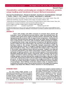
Size, Glycosaminoglycan Content, and Amino Acid Composition of Fetal Membrane Proteoglycan-To estimate the size of the fetal membrane proteoglycan on an analytical scale, a sample of the purified proteoglycan was radioiodinated and chromatographed on Sepharose CL-2B under associative conditions (Fig. L4). About 15% of the radiolabeled material was excluded from the column while 85% eluted as a broad peak with a mean K,, of 0.55. A similar pattern was observed when the proteoglycan was chromatographed on Sepharose CL-4B in 4 M guanidine HCl. Under the latter conditions, however, about 10% of the radioactivity was excluded fromthe column and 90% eluted with an approximate KaVof 0.4 (data not shown), suggesting that atleast part of the excluded material represented aggregated proteoglycan. The nature of the remaining void volume radioactivity was not investigated further because subsequent analyses showed the proteoglycan to be essentially homogeneous with regard to its core protein (see below). To estimate the size of the glycosaminoglycans,the proteoglycan was radiolabeled by &elimination in the presence of sodium borotritide, and chromatographed on a Sepharose CL6Bcolumn (Fig. 1B). Comparison withglycosaminoglycan
c ' 5! 0.5 P
0
-
Y
LI
n r
Vo
CS
HS
Vt
1
TABLE I Purification of proteoglycan from human fetalmembranes Purification step
Total
uronic
protein mg
1. Starting material (50 g, wet weight) 2. 8 M urea, 1 M NaCl, 0.5% deoxycholate extraction 3. DE52 cellulose (batchwise preparation) 4. Ethanol precipitation 5. Sepharose CL-4B column gel chromatography 6. DE52 cellulose chromatography 7 . CsCl gradient centrifugation Fraction Density
25.0 13.3 6.0 5.0 4.0 1.8
1.6
glml
1 (bottom) 2 3
1.50 1.46 1.43
1.30 (72)b 0.68 (50) 0.26 (14) 0.26 (19) 0.20 (15) 0.17 (9) 0.06 (3) 0.11 (8) 4 1.41 0.03 (1) 0.05 (4) 5 1.39 0.01 (1) 0.05 (4) 6 1.37 a Uronic acid content was measured by the carbazole method. In purification steps 1-4 the samples were first isolated in the void volume of a Sephadex G-50 column after digestion with papain (41). Per cent of total amount separated.
Fraction Number
FIG. 1. Elution profiles of purified proteoglycanon Sepharose CL-2B, and glycosaminoglycanson Sepharose CL-BB. A, purified '261-labeled proteoglycan from fetal membrane tissue (lo6 cpm) was applied to a Sepharose CL-2B column (1.5 X 115 cm) and eluted with 0.5 M sodium acetate, pH 7.0. Fractions of 3 ml were collected and assayed for radioactivity. B, purified proteoglycan was radiolabeled with [3H]sodiumborohydride and a sample (5 X lo' cpm) was chromatographed on Sepharose CL-GB. The column (1.5 X 115 cm) was eluted with 0.1% SDS, Tris-HC1, pH 8.0, and 3.4-ml fractions were collected and assayed for radioactivity. The K., of alkali-treated rat yolk sac tumor chondroitin sulfate proteoglycan glycosaminoglycan chains (CS), M,= 40,000 (41), and bovine liver heparan sulfate (HS), M,= 31,000 (40), are shown for comparison. Voand V, are the void volume and total volume of the columns.
Membrane Fetal
and Proteoglycan Fibroblast
standards indicated that thepolysaccharide side chains of the fetal membrane proteoglycan have an average M, = 55,000. The proteoglycan was analyzed by enzymatic degradation to determine the natureof its glycosaminoglycan. After treatment with chondroitinase AC-11, approximately 75% of the uronic acid-containing material elutedin theincluded volume on Sephadex G-50 while the remaining 25% came out in the 101 void volume (Fig. 2). About 94% of the material which eluted in the void volume was degraded upon further treatmentwith chondroitinase ABC. Based on the criterion of sensitivity to the chondroitinases, the polysaccharide chains of the proteoglycan appear to consist of 75% chondroitin sulfate and 23% dermatan sulfate. The amino acid and amino sugar compositions of the proteoglycan are shown inTable 11. In agreement with the glycosaminoglycan analysis, the main amino sugar detected was galactosamine. NHzterminul Amino Acid Sequence of Proteoglycan Core Protein-The polypeptide portion of the proteoglycan before and after CsCl purification was studied by subjecting the intact proteoglycan to NH2-terminal amino acid sequence analysis using a gas phase sequencer. The amountof protein applied was determined by protein assay (47). A single sequence was obtained in both cases (Fig. 3), suggesting the presence of only one core protein. Taking the average M,= 44,000 as the size of the core protein (see below), the initial yield of this sequence was calculated to be 93% and 76% in the two runs shown in Fig. 3. Electrophoretic and Immunoblotting Analysts of the Core Protein-The intact proteoglycan did not lend itself well to analysis by SDS-PAGE. No bands could be visualized by Coomassie Blue protein staining even when 30 pg of proteoglycan was applied to the gel. However, the core protein of the proteoglycan could be visualized on SDS-PAGEfollowing chondroitinase digestion by protein staining and by immunoblotting as shown below. When the proteoglycan was digested with chondroitinase AC-11 or ABC, two main bands a t M, = 45,000 and 43,000
13745
TABLEI1 Amino acid and amino sugar composition of fetal membrane proteoglycan Residues/1000
ASP Thr Ser Glu Pro GlY Ala CYS Val Met Ile Leu
108 60 77
72 103 62 11 66 14 46 99 TYf 17 Phe 34 His 34 LYs 62 Arg 34 GalNH2 188 GlcNH2 15 Approximate value for tyrosine was estimated from the hydrolysis for amino sugars because it was obscured by a large galactosamine peak in the runs of the 24-h hydrolysates for amino acids. The value was calculated by assuming that theratio of tyrosine to phenylalanine was the same in the 4- and 24-h hydrolysates. ’The numbers of hexosamine residues are expressed per 1000 amino acid residues witbout correction for destruction during hydrolysis.
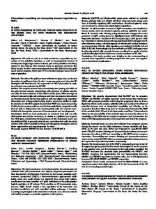
which were not present in the digestion buffer were seen in protein staining after SDS-PAGE (Fig. 4 A , lane 1).When the separated proteins were transferred onto nitrocellulose paper and stained with antibodies prepared against the intact proteoglycan as described under “Experimental Procedures,” the bands with M, = 45,000 and 43,000in the chondroitinase ABC-treatedproteoglycan sample were specificallyrecognized by the antibodies (Fig. 4B, lane I ) . The mobility of these bands was notsubstantially affected by reduction of the sample (result not shown), indicatingthat they are notdisulfide bonded. A Proteoglycan Immunologically Cross-reactive with the Fetal Membrane Proteoglycan in Tissues and Cultured Fibroblasts-Affinity-purified antibodiesagainst the fetal membrane proteoglycan were used to gain information on the cellular source of the proteoglycan. Tissue sections of human 0.4 fetal membranes and placenta analyzed by indirect immunofluorescence revealed strong staining in the compact layer E 0 and fibroblast layer of the amnion (Fig. 5a; see Ref. 39 for m ln descriptive histology). The brush border of the amniotic epiU thelial cells was also stained. Immunofluorescence was absent 0.2 in the chorionic membrane except for the staining of chorionic villi which penetrate the trophoblast layer. The connective tissue of placental villi and around blood vessels in placenta stained intensely with the anti-proteoglycan antibodies (Fig. 5b). Adult skin was also stained and found to be positive (result not shown). The lack of basement membrane tissue staining and the 10 30 50 strong stainingof the fibroblast layer of the amnion suggested Fraction Number FIG. 2. Analysis of glycosaminoglycans in fetal membrane that fibroblasts are likely to be the source of the proteoglycan proteoglycan by chondroitinase digestion. Purified fetal mem- in the fetal membrane. Moreover, human embryonic fibrobrane proteoglycan (0.3 mgof uronic acid) was alkali-treated and blasts have previously been shown to synthesize chondroitin/ digested with chondroitinase AC-I1 as described under “Experimental dermatan sulfate proteoglycans (13), and Vogel and Peterson Procedures.” The material was fractionated on acolumn of Sephadex (31) have partially characterized the proteoglycans syntheG-50 (0.5 X 50 cm) using 0.2 M ammonium bicarbonate and one-half of the 0.4-1111fractions collected were analyzed for uronic acid (0). sized by the IMR-90 human embryonic lung fibroblasts. We Fractions 18, 19, and 20 were pooled, lyophilized, degraded with therefore stained cultures of the IMR-90 fibroblasts with the chondroitinase ABC (O), and fractionated as described above. anti-fetal membrane proteoglycan antibodies. Staining was
13746
Membrane Fetal
12
DEA?GIGPEVPDDRDFEP?LGQ?V D ?
and Proteoglycan Fibroblast 1
A
B 2
1
2
%7:
4
E
12 -
8-
412rA
h
12y
4
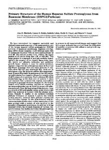
FIG.4. Immunological analysis of proteoglycan core proteins after separation on SDS-PAGE. A, Chondroitinase ABCtreated proteoglycan (lane I , 15 pg of protein) and molecular mass standards (lane 2) were separated by SDS-PAGE in the presence of 2-mercapt~ethanol and stained with Coomassie Blue. Arrows indicate two bands derived from the proteoglycan. B, material from the polyacrylamide gel was transferred to a nitrocellulose filter, reacted with the anti-proteoglycan antiserum and stained in an immunoassay as described under “ExperimentalProcedures.” Two major bands are visualized in the chondroitinase ABC-treated proteoglycan by the antiserum (lane I ) . The molecular mass markers are myosin (200,000), phosphorylase a (94,000), albumin (67,000), ovalbumin (43,000), and carbonic anhydrase (30,000).
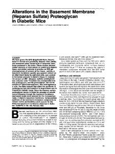
branes and that these cells could serve as a useful source of the proteoglycan, we characterized the antigen isolated from 12 the IMR-90 media. Properties of the Fibroblast Proteoglycan-The immunoprecipitated radioactivity from sulfate-labeled IMR-90 culture medium eluted on Sepharose CL-2B as one broad peak with 12 an average K,,, of 0.57 (Fig. 7A), indicating a size similar to that of the fetal membrane proteoglycan (Kay= 0.55). To estimate the size of the glycosaminoglycan side chains, an 2 4 6 8 1012 1416182022 aliquot of the immunoprecipitate was alkali-treated and chroResidue matographed on Sepharose CL-GB (Fig. 7 B ) . The 35S-radioFIG.3. Determination of NH*-terminal amino acid se- activity eluted as a single homogeneous peak with a K., of quence of fetal membrane proteoglycan. Yields of amino acids 0.42. Based on the elution pattern of standards, the average obtained in each cycle of two independent sequencing runs areshown. M,of the glycosaminoglycans can be estimated to be 40,000. Purified material prior to (60 pg of protein, O),and after CsCl density To determine the glycosaminoglycan composition, [35S]su1gradient centrifugation (30 pg of protein, O), in the form of intact fate-labeled immunoprecipitate was digested with papain and proteoglycan was sequenced. These amounts represent 1.4 nmol and further treated with chondroitinase AC-I1 or chondroitinase 700 pmol of polypeptide, respectively. ABC. Only 20% of this material was sensitive to digestion detected in a punctate distribution pattern in the pericellular with chondroitinase AC-I1 while 86% was sensitive to chonmatrix of these cells (Fig. 5c), identifying embryonic fibro- droitinase ABC degradation as determined by gel filtration on Sephadex G-50 (Fig. 8). This patternwas distinctly differblasts as a possible in vitro source of a similar proteoglycan. In order to study further theproteoglycan recognized in the ent from that obtained withthe fetal membraneproteoglycan IMR-90 fibroblast cultures by our antibodies, the cells were which had a much higher proportion of glycosaminoglycan metabolically labeled with [35S]sulfate.A maximum of about sensitive to chondroitinase AC-I1 (compare Figs. 2 and 8). In 50% of the macromolecule-associated sulfate released by these neither case were chondroitinase AC-I1 digestion products cells into the culture fluid was precipitated by the antiserum with intermediate molecular weights seen inthe Sephadex G(Fig. 6A). The fetal membrane proteoglycan inhibited, in a 50 profile, suggesting that the chondroitin sulfate and derdose-dependent fashion, the precipitation of sulfate-labeled matan sulfate exist in separate chains or as large blocks in material from conditioned medium by the antibodies (Fig. the same chains. SDS-PAGE analysis of the 35S-labeledIMR-90 media and 6B).Sincetheseresultsindicated that the IMR-90 cells synthesize a sulfated macromolecule(s) immunologically re- material immunoprecipitated from it showed that of the sevlated to theproteoglycan purified from the human fetal mem- eral sulfated components in the IMR-90 media (Fig. 9, lane
+’
‘W
Fetal Membrane and Fibroblast Proteoglycan
13747
A
, I
-1 J
I
2
6
1
I
I
I
60 200 600 Reciprocal Antiserum Dilution
20
B
L
FIG. 5. Immunofluorescent staining with anti-fetal membrane proteoglycan antibodies.Affinity-purified proteoglycan antibodies wereused to stain human fetal membranes ( a ) , human placenta ( b ) , and IMR-90 human embryonic lung fibroblasts (c) by immunofluorescence. Panels a and b X 270; panel c, X 435.
I ) the antiserum precipitated a single component which migrated as a diffuse band with an average M,= 120,000 (Fig. 9, lane 2). No significant 35S-radioactivity was precipitated when the antiserum was replaced with normal rabbit serum. Little decrease in the intensityof the M,= 120,000 band was seen following digestion with chondroitinase AC-I1 (Fig. 9, lane 3) while there was a complete loss of this band after treatment with chondroitinase ABC (Fig. 9, lane 4). Concomitantly a faint, but discrete radioactive band appeared at M, = 45,000. This apparently represents the core protein that has retained some sulfated sugars as has been suggested by others (51). To study further the specificity of the antibodies and to confirm the size of the core protein of the immunoreactive proteoglycan in the IMR-90 cells, the cells were metabolically labeled with [3H]leucine and themedium was immunoprecipitated and analyzed by SDS-PAGE (Fig. 10). As was seen in the [35S]sulfate experiments, the M, = 120,000 band was visualized in the immunoprecipitate from amino acid-labeled culture as theonly specifically precipitated band (Fig. 10, lane 2). This band did not appear in control immunoprecipitates obtained with normal rabbit IgG (Fig. 10, lane 3). The M,= 120,000 component inthe [3H]leucine-labeledimmunoprecipitate was mostly resistant to digestion with chondroitinase AC-I1 (not shown) but following chondroitinase ABC digestion, this band disappeared and a major band at M,= 45,000 appeared (Fig. 10, lane 4). In addition, this procedure revealed a minor core protein band a t M,= 43,000 which had not been visible in the sulfate-labeled samples, presumably because its
Concentration of Inhibitor (pg/ml) FIG. 6. Immunoprecipitation of [a6S]sulfate-labeled proteoglycans from fibroblast culture media by anti-fetal membrane antibodies and inhibition of binding by unlabeled tissue proteoglycan. A, antiserum raised against human fetal membrane proteoglycan was titrated against ["Sjsulfate-labeled conditioned media fromhuman IMR-90 fibroblasts and the bound radioactivity was determined as described under"ExperimentalProcedures."The maximal binding was 49%. E , inhibition of the binding of ["Slsulfatelabeled IMR-90 proteoglycans to antiserum against fetal membrane proteoglycan by the purified fetal membrane proteoglycan. The undiluted inhibitor had a concentration of 20 pg of uronic acid/ml.
amount was insufficient for it to be detectable. Incubation without the enzyme did not alter theappearance of the M,= 120,000 proteoglycan band (not shown). DISCUSSION
Since fetal membranesare a readily available human tissue rich in connective tissue (34, 35) and basement membrane components (36),we considered this tissue as a possible source of proteoglycans. Glycosaminoglycan analysis indicated that more than 85% of the uronic acid content of this tissue is chondroitin/dermatan sulfate. Heparansulfateapparently makes up less than 10% of the glycosaminoglycans even though this tissue contains anepithelial basementmembrane (34). Considering the abundance of chondroitin sulfate in the fetal membrane tissue,it was not unexpected that theproteoglycan we purified from this tissue was found to carry polysaccharide side chains of this type. These chains have an
Fetal Membrane and Fibroblast Proteoglycan
-
13748 A
1
2
3
4
-e,,
200K94K67K43K-
30K-
.
. .
FIG.9. SDS-PAGE analysis of [S"S]sulfate-labeledIMR-90 fibroblast-conditioned medium immunoprecipitated by antifetal membrane proteoglycan antibodies. Thesamples were: [3SS]sulfate-labeled IMR-90 culture medium ( l a n e 1 , 4 X IO' cpm); untreatedmaterialimmunoprecipitatedfromsuch medium using antibody against fetal membrane proteoglycan ( l a n e 2 , 6 X lo3 cprn); the immunoprecipitated materialdigested with chondroitinase AC-I1 ( l a n e 3 ) ;or chondroitinaseABC ( l a n e 4 ) . The arrow marks the topof the 10% separating gel. Molecular mass markers are as described in the legend to Fig. 4. The bandswere visualized by autoradiography.
200,000 core protein of the major cartilage proteoglycan and the M,= 25,000 core protein of a smaller chondroitin sulfate proteoglycan, which is present in cartilage and other tissues and which we have isolated from a rat yolk sac tumor (41), are each productsof different genes (7,23). Moreover, several lines of evidence suggest that the fetal membrane proteoglycan described here has yet another core protein, This core protein differs in size from the other two proteoglycans, and the small number of glycosaminoglycan chains ( 2 for a M , = 45,000 core protein) also distinguishes it from the other two which have 20-40 side chains (1, 41). Finally, NHz-terminal amino acid sequence of the yolk sac tumorproteoglycan shows no homology with the NII,-terminal sequence of the fetal membrane proteoglycan reported here.' Cartilage contains, in addition to the major cartilage proteoglycan and the smaller chondroitin sulfate proteoglycan, a third chondroitin sulfate proteoglycan which has a collagenous core protein (53). There appear, then, tobe at least four core proteins for chondroitin/ dermatan sulfate proteoglycans. The proteoglycan we describe here is similar to small chondroitin/dermatan sulfate proteoglycans identified in a number of tissue and fluids including aorta (16), uterine cervix (30), cornea (54), bone (52), skin (29, 55), serum (33), and culture media of human fibroblasts (31). The core proteins of two of these proteoglycans, one from bovine skin (55) and the other from bone tissue of various species (52), are of particular interest in this context. Both of these proteoglycans have a core protein with a size similar to that of the one described here. An NHp-terminalsequence was recently reported for the one from bovine skin (55). Eight of the firstnine amino acids in this sequence are identical to our sequence (Table 111). Position four in both proteoglycans fail to give an identifiable
Fetal Membrane and
13750
Fibroblast Proteoglycan
in the fibroblast layer of the amnion but does not appear to be a component of the epithelial basement membrane. The antibodies against this proteoglycan should enable us to determine what tissuesand cells synthesize this macromolecule (as demonstrated here for placenta and fibroblasts) and whether it might occur in various body fluids. Finally, the NHz-terminal sequence reported here will provide a basis for the molecular cloning of the cDNA for the core protein. Acknowledgments-We thank Dr. Minoru Fukuda and John Harper for help with the labeling of proteoglycans, Dr. Edward Miller for amino acid analyses and helpful discussions, Dr. Magnus Hook for comments on the manuscript, and Yvonne Ohgren and Khanh Nguyen for technical assistance. REFERENCES 1. Hascall, V.C., and Hascall, G. T. (1981) in Cell Biology of Extracellular Matrix (Hay, E. D., ed) pp, 39-63, Plenum Press, New York 2. Brennan, M. J., Oldberg, A., Hayman, E. G., and Ruoslahti, E. (1983) Cancer Res. 43,4302-4307 3. Culp, L.A., Murray, B. A., and Rollins, B. J. (1979) J. Supramol. Struct. 11,401-427 4. Kraemer, P. M., and Tobey, R.A. (1972) J. Cell Bwl. 5 5 , 713717 5. Poole, A. R., Pidoux, I., and Rosenberg, L. (1982) J. Cell Biol. 92,249-260 6. Kanwar, Y. S., Hascall, V. C., and Farquhar, M. G. (1981) J. Cell Biol. 90,527-532 7. Kimata, K.,Barrach, H.-J., Brown, K. S., and Pennypacker, J. P. (1981) J. Biol. Chum. 256,6961-6968 8. Schwartz, N. B., Ostrowski, V., Brown, K. S., and Pratt, R.M. (1978) Biochem. Bwphys. Res. Commun. 82,173-178 9. Toole, B. P., Okayama, M., Orkin, R. W., Yoshimura, M., Muto, M., and Kaji, A. (1977) in Cell and Tissue Interactwm (Lash, J. W., and Burger, M. M.,eds) pp. 139-154, Raven Press, New York 10. Thompson, H.A., and Spooner, B. S. (1983) J. CellBiol. 9 6 , 1443-1450 11. Dietrich, C. P., Sampaio, L. O., Toledo, 0. M. S., and Cassaro, C. M. F. (1977) Bwchem. Biophys. Res. Commun. 75,329-336 12. Iozzo, R. V., and Wight, T. N. (1982) J. Biol. Chem. 257,1113511144 13. Glimelius, B., Norling, B., Westermark, B., and Wasteson, A. (1979) J. Cell Physwl. 98,527-538 14. Muir, H., and Hardingham, T. E. (1983) in Biochemistry of Carbohydrates (Whelan, W. H., ed) pp. 153-222, Butterworths, London 15. Rosenberg, L. (1975) in Dynamics of Connective Tissue Macromolecules (Burleigh, P. M. C., and Poole, A. R., eds) pp. 105128, Elsevier/North-Holland Press, Amsterdam 16. Salisbury, B.G. J., and Wagner, W.D. (1981) J. Bwl. Chern. 256,8050-8057 17. Norling, B., Glimelius, B., Westermark, B., and Wasteson, A. (1978) Bwchem. Biophys. Res. Commun. 84,914-921 18. Wight, T. N., and Hascall, V. C. (1983) J. Cell Biol. 9 6 , 167-176 19. Chang, Y., Yanagishita, M., Hascall, V., and Wight, T. N. (1983) J. Bwl. Chem. 258,5679-5688 20. Sugahara, K., Ho, P.-L., and Dorfman, A. (1981) J. Cell Biol. 9 0 , 527-532 21. Swann, D.A., Powell, S., and Sotman, S. (1979) J. Biol. Chem. 254,945-954
22. Kimata, K., Oike, Y., Ito, K., Karasawa, K., and Suzuki, S. (1978) Biochem. Biophys. Res. CoFmun. 85, 1431-1439 23. Brennan, M. J., Oldberg, A., Ruoslahti, E., Brown, K., and Schwartz, N. (1983) Dev. Biol. 98,139-147 24. Oegema, T. R., Jr., Hascall, V.C., and Eisenstein, R. (1979) J. Biol. Chem. 2 5 4 , 1312-1318 25. Hassell, J. R., Newsome, D. A., Krachmer, J. H., and Rodrigues, M. M. (1980) Proc. Natl. Acad. Sei. U. S. A. 77,3705-3709 26. Gregory, J. D., Coster, L., and Damle, S. P. (1982) J. Bwl. Chem. 257,6965-6970 27. Yanagishita, M., Rodbard, D., and Hascall, V.C. (1979) J. Bwl. Chem. 254,~911-920 28. Fransson, L.-A., Coster, L., Malmstrom, A., and Sheehan, J. K. (1982) J. Biol. Chern. 257,6333-6338 29. Damle. S. P.. Coster.. L.,. and Gregory. J. D. (1982) J. Biol. Chem. 257,552315527 30. Uldbiere. N.. Malmstrom. A.. Ekman. G.. Sheehan. J.. Ulmsten. U.,-aid Wingerup, L. (1983) Biochek. 209,497-503 31. Vogel,K.G., and Peterson, D. W. (1981) J. Biol. Chem. 2 5 6 , 13235-13242 32. Huang, S. S., Huang, J. S., and Deuel, T. F. (1982) Dev. Bwl. 8 5 , 180-189 33. Silvestri, L., Baker, J. R., RodBn, L., and Stroud, R. M. (1981) J. Biol. Chem. 2 5 6 , 7383-7387 34. Alitalo, K., Kurkinen, M., Vaheri, A., Krieg, T., and Timpl, R. (1980) Cell 1 9 , 1053-1062 35. Burgeson, R. E., El Adli, F. A., Kaitila, I. I., and Hollister, D. W. (1976) Proc. Natl. Acad. Sci. U. S. A. 7 3 , 2579-2583 36. Hessle, H., Sakai, L. H., Hollister, D. W., Burgeson, R. E., and Engvall, E. (1984) Differentiation 26,49-54 37. Russo, R. C., Liotta, L. A., Thorgeirssen, U., Brundage, R., and Schiffmann, E. (1981) J. Cell Biol. 9 1 , 459-467 38. Liotta, L. A,, Goldfarb, R. H., Brundage, R., Siegal, G. P., Terranova, V., and Garbisa, S. (1981) Cancer Res. 4 1 , 4629-4636 39. Bourne, G. L. (1980) Am. J. Obstet. Gynecol. 7 9 , 1070-1073 40. KjellBn, L., Oldberg, A., and Hook, M. (1980) J. Biol. Chem. 2 5 5 , 10407-10413 41. Oldberg, A., Hayman, E. G., and Ruoslahti, E. (1981) J. Biol. Chem. 256,10847-10852 42. Engvall, E., and Ruoslahti, E. (1977) Znt. J . Cancer 2 0 , l - 5 43. Engvall, E., and Perlmann, P. (1972) J. Immunol. 109,129-135 44. Laemmli, U. K. (1970) Nature ( L o r d . ) 227,680-685 45. Bell, M. L.,and Engvall, E. (1982) Anal. Biochem. 123,329-335 46. Bitter, T., and Muir, H. M. (1962) Anal. Biochem. 4,330-334 47. Lowry, 0.H., Rosebrough, N. J., Farr, A. L., and Randall, R. J. (1951) J. Biol. Chem. 1 9 3 , 265-275 48. Saito, H., Yamagata, T., and Suzuki, S. (1968) J. Biol. Chem. 243, 1536-1542 49. Miller, E. J. (1972) Biochemistry 1 1 , 4309-4909 50. Hewick, R. M., Hunkapiller, M. W., Hood, L. E., and Dreyer, W. J.(1981) J . Biol. Chem. 256,7990-7997 51. Oike, Y., Kimata, K., Shinomura, T., Nazakawa, K., and Suzuki, S. (1980) Biochem. J. 191,193-207 52. Fisher, L. W., Termine, J. D., Dejter, S. W., Jr., Whitson, S. W., Yanagishita, M., Kimura, J. H., Hascall, V. C., Kleinman, H. K., Hassell, J. R., and Nilsson, B. (1983) J. Biol. Chem. 258, 6588-6594 53. Noro, A,, Kimata, K., Oike, Y., Shinomura, T., Maeda, N., Yano, S., Takahashi, N., and Suzuki, S. (1983) J. Biol. Chem. 258, 9323-9331 54. Hassell, J. R., Newsome, D. A., and Hascall, V. C. (1979) J. Biol. Chem. 254, 12346-12354 55. Pearson, C. H., Winterbottom, N., Fackre, D. S., Scott, P. G., and Carpenter, M. R. (1983) J. Bwl. Chem. 2 5 8 , 15101-15104 "
i.