Objetivos: El objetivo del presente estudio es analizar los factores que influyen en la progresión de la insuficiencia renal crónica en pacientes con enfermedad ...
http://www.revistanefrologia.com © 2012 Revista Nefrología. Official Publication of the Spanish Nephrology Society
originals
Chronic kidney disease progression in patients with autosomal dominant polycystic kidney disease Nayara Panizo1, Marian Goicoechea1, Soledad García de Vinuesa1, David Arroyo1, Claudia Yuste1, Abraham Rincón1, Úrsula Verdalles1, Caridad Ruiz-Caro2, Borja Quiroga1, José Luño1 1 2
Servicio de Nefrología. Hospital General Universitario Gregorio Marañón. Madrid (Spain) Clínica Dialcentro. Madrid. (Spain)
Nefrologia 2012;32(2):197-205 doi:10.3265/Nefrologia.pre2011.Dec.11177
ABSTRACT Objectives: The aim of this study was to analyse the factors influencing chronic kidney disease (CKD) progression in patients with autosomal dominant polycystic kidney disease (ADPKD). Material and Method: We studied 101 patients (mean age: 43±17.3 years, 43.56% male) followed during a median (interquartile range) follow-up time of 69 (35-128) months from 1997 to 2010. The primary end point was: time to a 50% decrease of estimated glomerular filtration rate (eGFR) (CKD-EPI) since the first-time visit and/or time to initiation of renal replacement therapy. The mean anual eGFR change was also analysed. Clinical and demographic data, blood pressure, concomitant medications, and analytical parameters were collected at each visit. Baseline kidney size was also recorded by ultrasound. Results: Thirty-one patients achieved the primary end point after a median (IQR) time of 102 (53-131) months. Those patients who achieved the primary end point had higher SBP and DBP (P=.017 and P=.001), higher LDL-cholesterol (P=0.011), higher creatinine (P=.006), higher uricemia (P=0.041), more severe proteinuria (P=.033) and greater kidney size (P=.05). The mean annual eGFR change was of –3.52±7.3ml/min/1.73m2. Forty-nine patients had a rapid decline in renal function: Group A (higher than 3.52ml/min/1.73m2) and 52 patients had a lower renal disease progression: Group B ( –3,52 ml/min/1,73 m2) y 52 pacientes tuvieron una progresión lenta de la insuficiencia renal: Grupo B (< –3,2 ml/min/1,73 m2). Por regresión de Cox, en un modelo ajustado, la PAS y la menor edad al diagnóstico son las variables que mantienen su poder predictivo de mal pronóstico renal (p = 0,026). Conclusiones: La función renal inicial, proteinuria, tamaño renal, hipercolesterolemia, hiperuricemia y PAS basal son factores que influyen en la progresión de la insuficiencia renal en la PQRAD, siendo la PAS y la menor edad los factores que mantienen su poder predictivo independiente en el análisis multivariante.
period that, although was retrospective in nature, has been surpassed by only a few studies published to date. In contrast to other studies, the importance of systolic (SBP) as opposed to diastolic (DBP) blood pressure stands out as a factor for the progression of renal failure in these patients. Additionally, we have underlined the prognostic importance of age at diagnosis as a possible variable suggestive of which type of genetic mutation is present. As such, our study provides a simple and practical focus for the evaluation of the risk of rapid progression towards ESRD. We have also evaluated the role of other factors of progression such as dyslipidemia and hyperuricemia, which have been poorly studied until now.
Palabras clave: Enfermedades renales poliquísticas.
We included a total of 101 patients that were monitored at our clinic from 1997 to the present. Patients had been diagnosed with ADPKD based on standardised radiological criteria.16 Patients with ADPKD with less than two visits to our practice were excluded, whether because the patient abandoned the follow-up protocol or when a renal event occurred within the first year after diagnosis.
INTRODUCTION Autosomal dominant polycystic kidney disease (ADPKD) is one of the most common hereditary diseases observed and the most frequent genetic cause of chronic kidney disease (CKD).1-3 This disease affects one out of every 800-1000 people in the global population, and constitutes the aetiology for end-stage renal disease (ESRD) in 5%-10% of patients on renal replacement therapy programmes.4 Despite the importance of this health issue, it was not been the subject of much research until recent years, when several studies were performed with the objective of identifying predictive factors for a rapid progression of renal disease.5-7 Some of these factors can be modified by using different therapeutic interventions that are currently under study, such as tolvaptan,8,9 m-TOR (mammalian target of rapamycin) inhibitors,10 somatostatin analogues,11 and even rosiglitazone,12 which have been used successfully in animal models. Among the factors that have been identified in the progression of ADPKD, the most important one is hypertension,8,14 but other possible factors for the progression of disease have been identified, such as African American race,15 male sex, haematuria, proteinuria, renal cyst volume, and polycystin 1 (PKD1) vs polycystin 2 (PKD2) mutations.1 The objective of our study was to perform a retrospective analysis of the factors that influence the progression of chronic renal failure in incident patients with ADPKD treated at our department throughout the last 13 years. Our study, which dealt with patients at a single centre and thus a small sample size, stands out for the long follow-up 198
MATERIAL AND METHOD
At each visit, we collected demographical variables, SBP and DBP values, concomitant medications, haemoglobin, creatinine (Cr), uric acid, total cholesterol, HDL-cholesterol, LDLcholesterol, triglycerides, calcium, phosphorous, parathyroid hormone (PTH), proteinuria, and haematuria. Biochemical variables were measured using standard methods. PTH was measured using IRMA (intact 10-60pg/ml molecule, Allegro Nichols). Albuminuria was measured in 24-hour urine samples using immunonephelometric methods. Haematuria was defined in urine sediments as the presence of more than three red blood cells per microscopic field in at least two of three correctly collected samples.17 Kidney size (cm) was measured using renal ultrasound. Although each patient underwent several ultrasounds throughout the follow-up period, only baseline values were taken into account for the analysis, both for the largest renal cyst and maximum kidney diameter in centimetres. Since this was a retrospective analysis, we did not have an initially standardised methodology for taking measurements, and multiple professionals performed the procedures. Over the 13 years of the study, different radiologists evaluated the ultrasounds (three different doctors evaluated the majority of images). Inter and intra-observer variability was not taken into account. Blood pressure (BP) was measured using an automatic sphygmanometer in accordance with the recommendations of the Spanish Society of Hypertension. 18 Patients were considered hypertensive when SBP and DBP were equal to or greater than 140mm Hg and 90mm Hg, respectively, or if they were receiving anti-hypertensive drugs. The treatment objective was set at 130/80mm Hg, in accordance with the recommendations of the seventh report of the JNC Nefrologia 2012;32(2):197-205
Nayara Panizo et al. Factors for progression in polycystic kidney disease
(Joint National Committee on Prevention, Detection, Evaluation, and Treatment of High Blood Pressure).19 Dyslipidemia was defined as total cholesterol greater than 200mg/dl and LDL-cholesterol greater than 130mg/dl, in accordance with the European guidelines for cardiovascular prevention,20 or if the patient was receiving lipid lowering treatment. Renal function was evaluated as estimated glomerular filtration rate (eGFR) using the CKD-EPI formula.21 We analysed the progression of renal dysfunction using the following two methods: 1) Based on the appearance of a renal event, defined as a 50% reduction in eGFR as measured by CKD-EPI from the initial values from the outpatient visit and/or entrance to dialysis. 2) Based on mean change in eGFR/year. According to the decrease in eGFR/year above or below the mean, we defined two different groups: group A: patients with a fast progression of CKD; and group B: patients with a normal progression of CKD. Patients diagnosed with ADPKD were included in the study from the moment of their first visit with a nephrologist, considering all measurements from this consultation as baseline values, until a renal event occurred. Data were compiled on an annual basis, with the number of visits ranging between 1 and 10. The longest follow-up period lasted 13 years. The median follow-up time was 69 (35-128) months. Values are expressed as mean ± SD (standard deviation) or median (interquartile range) if the variables did not follow a normal distribution. We used the Kolmogorov-Smirnov test to evaluate normal distribution for the different parameters assessed. For the analysis of differences between patients that suffered a renal event and those that did not, we used chi-square tests for qualitative variables and Student’s t-tests or Mann-Whitney U-tests for quantitative variables with a normal distribution or non-Gaussian distribution, respectively. We assumed that a difference was statistically significant when P.05). Group A patients had higher baseline SBP and proteinuria (Table 4). The logistic regression analysis revealed that the predictive variable for a rapid disease progression was still SBP, with the addition of a younger age at the first consultation, regardless of proteinuria, sex, initial renal function, and DBP (Table 5). 200
DISCUSSION The primary result of our study, stable patients with with ADPKD diagnosed an evaluated in nephrological consultations, was the confirmation that systolic arterial hypertension and a younger age at diagnosis are the most important factors that influence the long-term progression of renal failure. Many recent studies on ADPKD have focused on the importance of renal volume as a determining factor in the deterioration of renal function, which advances parallel to the increase in cyst sizes.22 There are also other studies that correlate clinical and biochemical parameters with kidney size and glomerular filtration rate.5 However, there are few examples in the literature that correlate a patient’s baseline clinical characteristics with long-term clinical evolution as our study has. Recently, a retrospective cohort study was published with a follow-up period of 22 years, including 194 patients with ADPKD, which analysed the influence of mutation type on clinical characteristics and the development of renal events.23 Cyst growth appears to be one of the most important factors in determining the progression of CKD in these patients. As such, in many clinical trials focusing on therapeutic interventions, cyst growth has been used as the reference marker for identifying patients that could benefit from treatment.5,24 However, one of the most reliable methods for measuring renal structure and volume, standard magnetic resonance,25 is an expensive method that is not available in all hospitals and requires a large amount of time from the radiologist. A similar pattern occurs in computed tomography, which has been employed in volumetric measurements, with good correlation with total renal volume, renal cyst volume, and decrease in eGFR.26,27 Some authors have indicated that the deterioration in renal function does not depend on cyst volume as much as on the amount of intermediate tissue in polycystic kidneys measured by computed tomography.28 Other methods of measurement such as ultrasound do not provide as good a correlation for renal function,27 and so we need other variables from our clinical practice that aid in identifying patients that would benefit from early treatment. In our study, we evaluated kidney size using ultrasound, which was an imprecise method, taking into account that the measurements were taken by various radiologists. Although the univariate analysis revealed a difference in kidney size between patients that had a renal event and those that did not, this variable did not retain its predictive value when other parameters were adjusted for. As such, in routine clinical practice, it would be very beneficial to have access to markers, aside from cyst growth, that would identify patients who, starting at an early age, will suffer a rapid progression in their kidney disease, and as such will be candidates for more aggressive therapeutic options, when sufficient evidence exists in the literature to support their use. Nefrologia 2012;32(2):197-205
Nayara Panizo et al. Factors for progression in polycystic kidney disease
originals
Linear R2 = 0,078
Linear R2 = 0,027
Linear R2 = 0,012
CKD-EPI (ml/min/1.73 m2)
120.00
100.00
80.00
60.00
40.00
20.00
.00
2.00
4.00
6.00
8.00
10.00
12.00 .00
2.00
Visit number
Total
4,.0
6.00
8.00 10.00 12.00 .00
2.00
4.00
6.00
8.00 10.00 12.00
Visit number
Visit number
Group A
Group B
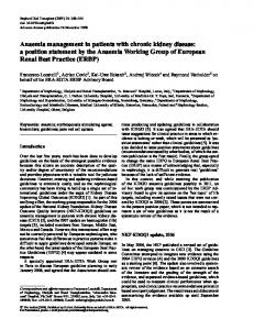
Figure 1. Decrease in glomerular filtration rate in all patients and in the two different groups of disease progression. Mean decrease in glomerular filtration rate for all patients: -3.52±7.3ml/min/1.73m2. Group A (rapid progression): decrease was greater than the mean. Group B (normal progression): decrease was less than or equal to the mean.
Arterial hypertension is a common clinical manifestation in patients with ADPKD. A recent demographic study performed in Turkey with 1139 patients with ADPKD identified this as the most common clinical manifestation in these patients, as it was present in 76% of cases.29 It commonly occurs early on, before the appearance of DBP (mmHg)
SBP (mmHg)
180 160 140 120 100 80 60 40 1
2
3
4
5
6
7
8
9
10
Visit number n
101
95
83
63
49
37
27
17
8
2
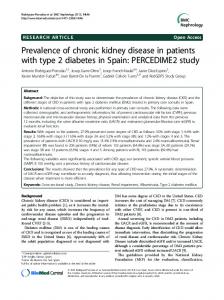
Figure 2. Evolution over time of SBP and DBP values during follow-up. Mean (SD). SD: standard deviation; DBP: diastolic blood pressure; SBP: systolic blood pressure. Nefrologia 2012;32(2):197-205
deteriorated renal function in 60% of cases.30 Several studies have related hypertension with the speed of progression in renal disease. One retrospective study with 94 patients compared rapid progression with slow progression patients, finding that those who progressed faster had a higher incidence of hypertension, haematuria, proteinuria, and urinary infections.31 Another study points out that hypertensive patients with ADPKD have worse renal function than normotensive patients, with an added increase in cardiovascular risk.32 There have also been studies showing a lower mortality rate33 and a progressive delay in the age at which these patients reach ESRD, relating this to a progressively better control of blood pressure levels in recent years,1,34 especially from the use of ACE inhibitors and ARB, although no differences have been found between the two drugs in controlling BP and Cr values in these patients.35 In a recently published study in Spain, kidney size was correlated with a pathological profile involving BP as measured in outpatient monitoring programmes, highlighting a greater variability in BP, especially diastolic, and lower pulse pressure with greater kidney size, as measured using ultrasound, even in a stage of pre-hypertension.36 Our study corroborated these findings, with the novel result that it is SBP, not DBP, that maintains its independent predictive value for the rapid progression of kidney disease, which is in agreement with the results from other studies, such as the report by Cadnapaphornchai et al.,37 which analysed the association between renal volume, SBP and DBP, and left ventricular mass index in patients with ADPKD. As in our case, they detected an association between both BP variables and renal volume in the univariate analysis, but only SBP 201
Nayara Panizo et al. Factors for progression in polycystic kidney disease
originals
Table 3. Predictive factors for renal events. Univariate analysis Event yesa
Event no
(No.=31)
(No.=70)
P
SBP (mmHg)
143.5±20
132.5±22.8
.02
DBP (mmHg)
88.3±9.8
81.8±13.7
.01
Uric acid (mg/dl)
6.5±1.2
5.7±2
.03
Cholesterol (mg/dl)
237±45
202±40
.00
146.5±22.4
120.3±35
.01
1.4±0.5
1.1±0.5
.01
102 (131.5-53.7)b
16.9 (33.8-0)b
.03
15.6±2.6
14.3±2.9
.05
LDL-cholesterol (mg/dl) Baseline creatinine (mg/dl) Proteinuria (mg/24 h) Kidney size (cm)
Event: 50% decrease in CKD-EPI eGFR from the values recorded at the initial visit and/or if the patient started a haemodialysis programme. Values expressed as median (interquartile range). Baseline creatinine: serum creatinine on the initial visit; DBP: diastolic blood pressure; SBP: systolic blood pressure. a
b
remained after adjusting for age and sex in the multivariate analysis. The percentage of patients that were already receiving treatment with renin-angiotensin-aldosterone system (RAAS) blockers at the start of treatment was 41.4%; 32.6% started treatment with these drugs during the follow-up period. In total, 74% received treatment with RAAS blockers over the study period. We observed no differences between patients receiving this treatment and those that did not, whether in terms of the appearance of renal events or the speed of progression. However, it is well known that control of BP levels, especially systolic, is a fundamental factor for slowing the progression of kidney disease, not only in ADPKD, but in CKD in general, as demonstrated by several studies.38-40 In the specific case of ADPKD, the HALT study is currently under way, with the goal of elucidating the potential benefit of strict BP control using RAAS blockers in slowing the progression of ESRD.41 In the univariate analysis, we found that patients with a renal event or rapid disease progression had higher initial proteinuria values than those with a slow disease progression/absence of
renal events. However, this variable did not maintain its predictive value in the Cox regression analysis, despite the fact that proteinuria is a known factor for the progression of kidney disease.42 This fact was probably due to the low values of proteinuria displayed by our patients (median: 19.75; IQR: 058.5), as proteinuria is an uncharacteristic manifestation of this disease. Our second notable finding was that younger patients had a higher rate of decrease in eGFR. These results concur with those from Torres et al., who showed that a younger age at diagnosis is associated with a greater increase in total renal volume, and this, in turn, is correlated with a greater rate of decrease in glomerular filtration rate.5 Although we did not perform a genetic study, we can put this finding in the context of the higher probability that these patients have a mutation on the PKD1 gene, which tends to produce a greater pathological repercussion and earlier deterioration in renal function.43,44 These results are supported by those from the genetic study by Torra et al., which found that, in patients with ADPKD that had not reached ESRD by the age of 63 years, the prevalence of PKD2 mutations was three times higher than the estimated proportion for the general
Table 4. Predictive factors for a rapid progression in kidney disease. Univariate analysis (Student’s t-test) Rapid progressiona (No.=49)
Normal progression (No.=52)
P
SBP (mmHg)
145±21.6
127.3±19.9
.001
DBP (mmHg)
87.2±12.7
80.7±12.5
Microalbuminuria (mg/day)
36.7 (111-0)
b
.010
15 (130.2-34.5)
b
.010
Rapid progression: annual decrease in glomerular filtration rate higher than the mean for the population studied. Values expressed as median (interquartile range). DBP: diastolic blood pressure; SBP: systolic blood pressure. a
b
202
Nefrologia 2012;32(2):197-205
Nayara Panizo et al. Factors for progression in polycystic kidney disease
originals
Table 5. Predictive variables for a rapid progression in kidney disease. Logistic regression OR
95% CI
P
SBP (mmHg)
1.058
(1.020-1.068)
.001
Age at diagnosis
0.961
(0.931-0.991)
.010
Model adjusted for proteinuria, sex, initial renal function, and diastolic blood pressure. CI: confidence interval; OR: odds ratio; SBP: systolic blood pressure.
population with ADPKD.45 Other studies also coincide on the finding that, in families diagnosed with the disease in which at least one member reached ESRD at an age younger than 55, there is a high probability that the mutation is on the PKD1 gene.46 Our results also demonstrate the importance of controlling other cardiovascular risk factors, such as cholesterol and uric acid, both for the progression of CKD in patients with ADPKD and in other renal pathologies. Although these parameters do not independently influence the progression of renal dysfunction, they do appear to be important and controllable variables in the appearance of renal events in the univariate analysis. Several studies have suggested that ADPKD produces an increase in uric acid levels associated with decreased renal function,47 but there are no data regarding the influence of uric acid levels on the progression of the kidney disease in this particular case. However, in recent years, the relationship between hyperuricemia and the progression of CKD has been investigated,48,49 and recently the use of uric acid lowering drugs (allopurinol) has been shown to reduce disease progression.50 As regards lipid control, several studies have indicated that lower HDLcholesterol levels are correlated with increased deterioration of renal function,5 although we have not found data in the literature that show the influence of LDL-cholesterol levels on renal function in patients with ADPKD. Even so, the fact that patients with worse cholesterol levels have more renal events supports the hypothesis held by some trials that have attempted to show that renal function in these patients could be improved by using statins, through an increase in renal plasma flow derived from improved endothelial function.51 Our study is not without its limitations. The primary limitation in our case was that, while the study design was a prospective, longitudinal type, the data collection was retrospective, with the inherent drawbacks from the point of view of data analysis, and so we can only show evidence of an association between systolic hypertension and increased disease progression. Additionally, the sample was not sufficiently large due to the fact that our data were derived from one single centre. However, despite the small sample size, the long follow-up period has been surpassed by only a Nefrologia 2012;32(2):197-205
few isolated studies available in the medical literature,1,33,34 which bolsters the validity of our results. We should also point out the limitations of the absence of a genetic analysis, along with the use of renal ultrasound instead of a computed tomography or nuclear magnetic resonance for the evaluation of initial renal volume. We can conclude that a younger age at diagnosis and higher SBP values are the two factors associated with a greater progression of renal disease in our patients with ADPKD. These two variables, which are easily measured, allow us to detect patients that have a high probability of a faster progression towards ESRD in centres that do not have easy access to genetic analyses or magnetic resonance. This in turn provides the possibility of selecting these patients as candidates for possible future treatments that show long-term advantages in slowing the progression towards the need for renal replacement therapy. Interventional studies are needed to establish the potential benefit of controlling SBP values, cholesterol, and uric acid in slowing the progression towards ESRD in these patients.
Conflicts of Interest The authors have no potential conflicts of interest related to the content of this article to declare.
REFERENCES 1. Schrier RW, McFann KK, Johnson AM. Epidemiological study of kidney survival in autosomal dominant polycystic kidney disease. Kidney Int 2003;63(2):678-85. 2. Igarashi P, Somlo S. Polycystic kidney disease. J Am Soc Nephrol 2007;18(5):1371-3. 3. Cadnapaphornchai MA, George DM, Masoumi A, McFann K, Strain JD, Schrier RW. Effect of statin therapy on disease progression in pediatric ADPKD: design and baseline characteristics of participants. Contemp Clin Trials 2011;32(3):437-45. 4. Bleyer AJ, Hart TC. Polycystic kidney disease. N Engl J Med 2004;350(25):2622. 5. Torres VE, Grantham JJ, Chapman AB, Mrug M, Bae KT, King BF, et al. Potentially modifiable factors affecting the progression of autosomal 203
originals
Nayara Panizo et al. Factors for progression in polycystic kidney disease
dominant polycystic kidney disease. Clin J Am Soc Nephrol 2011;6(3):640-7. 6. Meijer E, Rook M, Tent H, Navis G, van der Jagt EJ, de Jong P, et al. Early renal abnormalities in autosomal dominant polycystic kidney disease. Clin J Am Soc Nephrol 2010;5(6):1091-8. 7. Reiterova J, Obeidova H, Lenicek M, Stekrova J, Merta M, Maixnerova D, et al. Influence of VEGF polymorphism on progression of autosomal dominant polycystic kidney disease. Kidney Blood Press Res 2008;31(6):398-403. 8. Torres VE, Meijer E, Bae KT, Chapman AB, Devuyst O, Gansevoort RT, et al. Rationale and design of the TEMPO (Tolvaptan Efficacy and Safety in Management of Autosomal Dominant Polycystic Kidney Disease and its Outcomes) 3-4 Study. Am J Kidney Dis 2011;57(5):692-9. 9. Irazabal MV, Torres VE, Hogan MC, Glockner J, King BF, Ofstie TG, et al. Short-term effects of tolvaptan on renal function and volume in patients with autosomal dominant polycystic kidney disease. Kidney Int 2011;80(3):295-301. 10. Grantham JJ, Bennett WM, Perrone RD. mTOR inhibitors and autosomal dominant polycystic kidney disease. N Engl J Med 2011;364(3):286-7. Author reply 287-9. 11. Ruggenenti P, Remuzzi A, Ondei P, Fasolini G, Antiga L, Ene-Iordache B, et al. Safety and efficacy of long-acting somatostatin treatment in autosomal-dominant polycystic kidney disease. Kidney Int 2005;68(1):206-16. 12. Dai B, Liu Y, Mei C, Fu L, Xiong X, Zhang Y, et al. Rosiglitazone attenuates development of polycystic kidney disease and prolongs survival in Han: SPRD rats. Clin Sci (Lond) 2010;119(8):323-33. 13. Schrier RW. Hypertension and autosomal dominant polycystic kidney disease. Am J Kidney Dis 2011;57(6):811-3. 14. Schrier RW, Johnson AM, McFann K, Chapman AB. The role of parental hypertension in the frequency and age of diagnosis of hypertension in offspring with autosomal-dominant polycystic kidney disease. Kidney Int 2003;64(5):1792-9. 15. Yium J, Gabow P, Johnson A, Kimberling W, Martinez-Maldonado M. Autosomal dominant polycystic kidney disease in blacks: clinical course and effects of sickle-cell hemoglobin. J Am Soc Nephrol 1994;4(9):1670-4. 16. Belibi FA, Edelstein CL. Unified ultrasonographic diagnostic criteria for polycystic kidney disease. J Am Soc Nephrol 2009;20(1):6-8. 17. Grossfeld GD, Wolf JS Jr, Litwan MS, Hricak H, Shuler CL, Agerter DC, et al. Asymptomatic microscopic hematuria in adults: summary of the AUA best practice policy recommendations. Am Fam Physician 2001;63(6):1145-54. 18. Marín R, de la Sierra A, Armario P, Campo C, Banegas JR, Gorostidi M. 2005 Spanish guidelines in diagnosis and treatment of arterial hypertension. Med Clin (Barc) 2005;125(1):24-34. 19. Jones DW, Hall JE. Seventh report of the Joint National Committee on Prevention, Detection, Evaluation, and Treatment of High Blood Pressure and evidence from new hypertension trials. Hypertension 2004;43(1):1-3. 20. Lobos JM, Royo-Bordonada MA, Brotons C, Álvarez-Sala L, Armario P, Maiques A, et al. European Guidelines on Cardiovascular Disease Prevention in Clinical Practice. CEIPC 2008 Spanish adaptation. Rev Clin Esp 2009;209(6):279-302. 21. Levey AS, Stevens LA, Schmid CH, Zhang YL, Castro AF 3rd, Feldman 204
HI, et al. A new equation to estimate glomerular filtration rate. Ann Intern Med 2009;150(9):604-12. 22. Tokiwa S, Muto S, China T, Horie S. The relationship between renal volume and renal function in autosomal dominant polycystic kidney disease. Clin Exp Nephrol 2011;15(4):539-45. 23. Dicks E, Ravani P, Langman D, Davidson WS, Pei Y, Parfrey PS. Incident renal events and risk factors in autosomal dominant polycystic kidney disease: a population and family-based cohort followed for 22 years. Clin J Am Soc Nephrol 2006;1(4):710-7. 24. Chapman AB. Approaches to testing new treatments in autosomal dominant polycystic kidney disease: insights from the CRISP and HALTPKD studies. Clin J Am Soc Nephrol 2008;3(4):1197-204. 25. Chapman AB, Guay-Woodford LM, Grantham JJ, Torres VE, Bae KT, Baumgarten DA, et al. Renal structure in early autosomal-dominant polycystic kidney disease (ADPKD): The Consortium for Radiologic Imaging Studies of Polycystic Kidney Disease (CRISP) cohort. Kidney Int 2003;64(3):1035-45. 26. King BF, Reed JE, Bergstralh EJ, Sheedy PF, Torres VE. Quantification and longitudinal trends of kidney, renal cysts and renal parenchyma volumes in Autosomal dominant polycystic kidney disease. J Am Soc Nephrol 2000;11(8):1505-11. 27. Chapman, AB, Wei W. Imaging approaches to patients with polycystic kidney disease. Semin Nephrol 2011;31(3):237-44. 28. Antiga L, Piccinelli M, Fasolini G, Ene-Iordache B, Ondei P, Bruno S, et al. Computed tomography evaluation of autosomal dominant polycystic kidney disease progression: a progress report. Clin J Am Soc Nephrol 2006;1(4):754-60. 29. Kazancioglu R, Ecder T, Altintepe L, Altiparmak MR, Tuglular S, Uyanik A, et al. Demographic and clinical characteristics of patients with autosomal dominant polycystic kidney disease: a multicenter experience. Nephron Clin Pract 2011;117(3):c270-5. 30. Ecder T, Schrier RW. Hypertension in autosomal-dominant polycystic kidney disease: early occurrence and unique aspects. J Am Soc Nephrol 2001;12(1):194-200. 31. Ahmed ER, Tashkandi MA, Nahrir S, Maulana A. Retrospective analysis of factors affecting the progression of chronic renal failure in adult polycystic kidney disease. Saudi J Kidney Dis Transpl 2006;17(4):511-5. 32. Idrizi A, Barbullushi M, Strakosha A, Kodra S, Thereska N, Zaimi E, et al. The relation of hypertension, renal function and cardiovascular events in autosomal dominant polycystic kidney disease. G Ital Nefrol 2007;24(6):595-9. 33. Patch C, Charlton J, Roderick PJ, Gulliford MC. Use of antihypertensive medications and mortality of patients with autosomal dominant polycystic kidney disease: a population-based study. Am J Kidney Dis 2011;57(6):856-62. 34. Orskov B, Romming Sorensen V, Feldt-Rasmussen B, Strandgaard S. Improved prognosis in patients with autosomal dominant polycystic kidney disease in Denmark. Clin J Am Soc Nephrol 2010;5(11):2034-9. 35. Nakamura T, Sato E, Fujiwara N, Kawagoe Y, Yamada S, Ueda Y, et al. Changes in Urinary Albumin Excretion, Inflammatory and Oxidative Stress Markers in ADPKD Patients with Hypertension. Am J Med Sci 2012;343(1):46-51. 36. Sans L, Roca-Cuchars A, Torra R, Calero F, Arias P, Ballarin J, et al. Relationship between kidney size and blood pressure profile in paNefrologia 2012;32(2):197-205
Nayara Panizo et al. Factors for progression in polycystic kidney disease
37.
38.
39. 40. 41.
42.
43.
tients with autosomal dominant polycystic kidney disease without renal failure. Nefrologia 2010;30(5):567-72. Cadnapaphornchai MA, McFann K, Strain JD, Masoumi A, Schrier RW. Increased left ventricular mass in children with autosomal dominant polycystic kidney disease and borderline hypertension. Kidney Int 2008;74(9):1192-6. Agarwal R. Blood pressure components and the risk for end-stage renal disease and death in chronic kidney disease. Clin J Am Soc Nephrol 2009;4(4):830-7. Wuhl E, Schaefer F. Managing kidney disease with blood-pressure control. Nat Rev Nephrol 2011;7(8):434-44. Wenzel RR. Renal protection in hypertensive patients: selection of antihypertensive therapy. Drugs 2005;65 Suppl 2:29-39. Chapman AB, Torres VE, Perrone RD, Steinman TI, Bae KT, Miller JP, et al. The HALT polycystic kidney disease trials: design and implementation. Clin J Am Soc Nephrol 2010;5(1):102-9. Sarnak MJ, Astor BC. Implications of proteinuria: CKD progression and cardiovascular outcomes. Adv Chronic Kidney Dis 2011;18(4):258-66. Hateboer N, van Dijk MA, Bogdanova N, Coto E, Saggar-Malik AK, San Millan JL, et al. Comparison of phenotypes of polycystic kidney disease types 1 and 2. European PKD1-PKD2 Study Group. Lancet 1999;353(9147):103-7.
originals
44. Grantham JJ. Clinical practice. Autosomal dominant polycystic kidney disease. N Engl J Med 2008;359(14):1477-85. 45. Torra R, Badenas C, Pérez-Oller L, Luis J, Millán S, Nicolau C. Increased prevalence of polycystic kidney disease type 2 among elderly polycystic patients. Am J Kidney Dis 2000;36(4):728-34. 46. Barua M, Cil O, Paterson AD, Wang K, He N, Dicks E, et al. Family history of renal disease severity predicts the mutated gene in ADPKD. J Am Soc Nephrol 2009;20(8):1833-8. 47. Hosoya T, Ichida K, Tabe A, Sakai O. A study of uric acid metabolism and gouty arthritis in patients with polycystic kidney. Nippon Jinzo Gakkai Shi 1993;35(1):43-8. 48. Kuo CF, Luo SF, See LC, Ko YS, Chen YM, Hwang JS, et al. Hyperuricaemia and accelerated reduction in renal function. Scand J Rheumatol 2011;40(2):116-21. 49. Feig DI. Uric acid: a novel mediator and marker of risk in chronic kidney disease? Curr Opin Nephrol Hypertens 2009;18(6):526-30. 50. Goicoechea M, de Vinuesa SG, Verdalles U, Ruiz-Caro C, Ampuero J, Rincon A, et al. Effect of allopurinol in chronic kidney disease progression and cardiovascular risk. Clin J Am Soc Nephrol 2010;5(8):1388-93. 51. van Dijk MA, Kamper AM, van Veen S, Souverijn JH, Blauw GJ. Effect of simvastatin on renal function in autosomal dominant polycystic kidney disease. Nephrol Dial Transplant 2001;16(11):2152-7.
Sent to review: 1 Oct. 2011 | Accepted: 6 Dec. 2011 Nefrologia 2012;32(2):197-205
205