Chronobiology International, 27(5): 934–944, (2010) Copyright © Informa UK Ltd. ISSN 0742-0528 print/1525-6073 online DOI: 10.3109/07420528.2010.488981
CHRONIC SLEEP DEFICIT AND PERFORMANCE OF A SUSTAINED ATTENTION TASK—AN ELECTROOCULOGRAPHY STUDY
Magdalena Fafrowicz,1 Halszka Oginska,2 Justyna Mojsa-Kaja,1 Tadeusz Marek,1 Krystyna Golonka,1 and Kinga Tucholska1 1
Department of Neuroergonomics, Institute of Applied Psychology, Jagiellonian University, Krakow, Poland 2 Department of Ergonomics and Exercise Physiology, Collegium Medicum, Jagiellonian University, Krakow, Poland
Electrooculography (EOG) was used to explore performance differences in a sustained attention task during rested wakefulness (RW) and after 7 days of partial sleep deprivation (SD). The RW condition was based on obtaining regular sleep, and the SD condition involved sleep restriction of 3 h/night for a week resulting in a total sleep debt of 21 h. The study used a counterbalanced design with a 2-wk gap between the conditions. Participants performed a sustained attention task for 45 min on four occasions: 10:00–11:00, 14:00–15:00, 18:00–19:00, and 22:00–23:00 h. The task required moving gaze and attention as fast as possible from a fixation point to a target. In each session, 120 congruent and 34 incongruent stimuli were presented, totaling 1232 observations/participant. Correct responses plus errors of omission (lapses) and commission (false responses) were recorded, and the effect of time-of-day on sustained attention following SD was investigated. The analysis of variance (ANOVA) model showed that SD affected performance on a sustained attention task and manifested itself in a higher number of omission errors: congruent stimuli (F(1,64) = 13.3, p < .001) and incongruent stimuli (F(1,64) = 14.0, p < .001). Reaction times for saccadic eye movements did not differ significantly between experimental conditions or by time-of-day. Commission errors, however, exhibited a decreasing trend during the day. The visible prevalence of omissions in SD versus RW was observed during the mid-afternoon hours (the so-called post-lunch dip) for both congruent and incongruent stimuli (F(1,16) = 5.3, p = .04 and F(1,16) = 5.6, p = .03, respectively), and at 18:00 h for incongruent stimuli (F(1,13) = 5.7, p = .03). (Author correspondence:
[email protected]) Keywords Chronic sleep deficit; Omission and commission errors; Post-lunch dip; Saccadic eye movements; Sustained attention
This paper was presented at the 19th International Symposium on Shiftwork and Working Time, August 2–6, 2009, San Servolo Island, Venice, Italy Address correspondence to Magdalena Fafrowicz, Department of Neuroergonomics, Institute of Applied Psychology, Jagiellonian University, 4 Lojasiewicza Street, 30-348 Krakow, Poland. E-mail:
[email protected]
934
An Electrooculography Study
935
INTRODUCTION Attention, defined as the mental ability to select stimuli, responses, memories, or thoughts that are behaviorally relevant (Corbetta & Shulman, 2002), governs all human cognitive processes, from perception to decision and execution (e.g., Fafrowicz & Marek, 2007). According to current theories, attention can be divided into two broad domains, representing intensity and selection aspects (e.g., Sturm & Willmes, 2001). Whereas the intensity aspects are related to alertness and sustained-attention processes, selection aspects are linked to orienting and executive attention. Intensity aspects appear more fundamental and constitute prerequisite conditions for more complex and capacity-demanding attention selectivity (e.g., Fan et al., 2003). Over the last two decades, sleep-deprivation studies have recognized alertness and sustained-attention as two components of cognition that are drastically affected by periods without sleep (Lim & Dinges, 2008). Total sleep-deprivation leads to an extreme form of sleep deficit (Casagrande et al., 2006; Chee et al., 2006). However, chronic sleep-restriction (partial sleep-deprivation) is pervasive in modern society due to work demands and social/lifestyle factors. Both acute sleep-deprivation (≥24 h) and short-term, chronic sleep-restriction (6.5 h/each night the month before the study. Participants completed a survey that provided demographic data, chronotype, and sleepiness. Subjective phase and amplitude were derived from the Chronotype Questionnaire (Oginska et. al, 2000), and the Epworth Sleepiness Scale (Johns, 1991) was used to assess daytime sleepiness level. These measures were used to exclude participants who were extreme circadian types and who experienced excessive/ pathological sleepiness. A laboratory experiment was conducted under two conditions: (1) rested wakefulness (RW) where participants had their usual sleep and (2) after undergoing sleep deprivation (SD) of 3 h/night for 7 nights (amounting to 21 h of sleep debt). Three participants were excluded from the final analyses due to technical errors or an excessive number of lapses (>25% of total trials) during the RW session. Participants were briefed on the study requirements, and they signed a written consent form. They were remunerated for their participation. The study protocol met the ethical standards established by the journal for the conduct of human research (Portaluppi et al., 2008). Procedure The study was conducted in a laboratory environment that provided dim light, low noise level, and controlled room temperature. Participants visited the laboratory three times. The first session consisted of a briefing on the experimental procedure, and those that volunteered undertook some practice on the attention task. The second and third visits involved participating in the EOG experiment. The first experimental session took place 1 day after the initial visit. The order of the two experimental sessions (RW, SD) was counterbalanced across all participants. The sessions were separated by at least 2 wk to minimize the residual effects of sleep deficit on performance of a sustained-attention task. During experimental days, participants were allowed to engage in nonstrenuous activities (e.g., reading, conversing, and watching videos). Research assistants observed the participants and were instructed to prevent them from napping by verbal reminders. Subjects were not allowed to drink or eat substances containing alcohol or caffeine (e.g., tea, chocolate) either during the day of the experiment or during the previous 48 h. Before attempting each of the performance tasks, participants completed the Activation-Deactivation Adjective Check List (Thayer, 1989) to reflect their energy, tiredness, tension, and calmness. Participants also recorded their sleepiness using the Karolinska Sleepiness Scale (Åkerstedt & Gillberg, 1990).
An Electrooculography Study
937
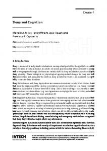
Eye Tracking The saccadometer system (Ober-Consulting, Poland) was used to record saccadic responses to visual stimuli. Horizontal saccadic eye movements were registered using a noninvasive, infrared reflection recording technique with a spatial resolution of 0.1° and time resolution of 1 ms. This system allowed data to be recorded at a sample rate of 250 Hz. Stimuli generated by red and green laser diodes were presented on a screen integrated with the EOG system. The system was attached to the subject’s head with an adjustable headband (Figure 1), and the screen was located approximately 3 cm from the participant’s eyes. Participants completed the sustained-attention task in an isolated room, with low levels of artificial light and assumed a recumbent position. Experimental Task The sustained attention task consisted of two unpredictable conditions: one with a cue congruent to a target and the other in which the cue and target were incongruent. The spatial cuing paradigm, designed for the classic work of Posner (e.g., Posner, 1980; Vecera & Rizzo, 2003), was modified for the experiment with the central cue (engaging covert orienting attention) used to voluntarily shift the spatial attention and eye movement to the target location (employing overt orienting attention). Each experimental trial began with the presentation of a fixation point (green laser diode) in the center of the screen. After 3000 or 6000 ms, a cue (red diode) appeared at 1° to the right or left of the fixation point for 100, 250, or 500 ms. After a further 300–500 ms, a target
FIGURE 1 Eye-tracking system used in the study.
938
M. Fafrowicz et al.
FIGURE 2 Schematic description of congruent task.
FIGURE 3 Schematic description of incongruent task.
stimulus (red diode) was flashed for 100, 250, or 500 ms at 10° to the right or left of the fixation point, with stimulus-onset asynchronies (SOAs) randomly distributed (Figures 2 and 3).
An Electrooculography Study
939
Each participant was instructed to direct her gaze and attention straight ahead towards the fixation point. When the target appeared to the left or right side, the participant was asked to execute a saccadic eye movement, that is, to move gaze and attention from the fixation point to the target and back to the fixation point as fast as possible. Execution of an eye movement for a given cue was not allowed to strengthen top-down mechanisms involved in voluntary shift of attention. The participants performed the sustained-attention tasks four times during the day: at about 10:00–11.00, 14:00–15.00, 18:00–19:00, and 22:00–23:00 h. In each 45-min session, 120 congruent (78%) and 34 incongruent (22%) stimuli were presented. RESULTS The mean length of the self-reported sleep in the RW condition was 7 h 52 min (SD ± 45 min), and in the SD condition sleep duration was 4 h 57 min (SD ± 33 min). The number of correct responses and two types of erroneous reactions (omissions and commissions) occurring in the four sessions for the two conditions (normal sleep versus sleep debt) were analyzed. There were three kinds of commission errors: error of direction, error of place, and error of both place and direction). Omissions were defined as noreaction within 1 s from target appearance. Each participant provided a maximum of 1232 measurements. Statistical analyses were performed via analysis of variance (ANOVA) using Statistica (V. 8.0) software. The results show that chronic partial sleep-deprivation produces considerable performance decrements on a sustained-attention task. As Table 1 shows, sleep deficit resulted in significantly more errors of omission. Significant differences in reaction time of saccadic eye movements were not found, but a tendency for slower reactions was observed. TABLE 1 Parameters of performance of sustained attention task in state of rested wakefulness (RW) and chronic partial sleep deficit (SD; total sleep debt of 21 h)
Congruent cue % of correct responses % of omissions % of commissions Mean RT in correct response (ms) Incongruent cue % of correct responses % of omissions % of commissions Mean RT in correct response (ms)
RW (n = 34)
SD (n = 32)
ANOVA F(1, 64) p
70.6 ± 29.7 4.8 ± 6.4 24.5 ± 26.7 194 ± 37
64.0 ± 24.2 16.2 ± 16.9 19.8 ± 19.1 204 ± 44
1.321 13.344 0.685 0.987
.255 .001 .411 .324
68.7 ± 29.9 2.0 ± 4.0 23.7 ± 28.9 217 ± 47
58.1 ± 23.3 13.6 ± 17.6 20.4 ± 20.7 235 ± 72
2.577 14.011 0.287 3.371
.113 < .001 .594 .071
940
M. Fafrowicz et al.
FIGURE 4 Average percentage of omissions in a congruent sustained-attention task performed in rested wakefulness (RW) and sleep deficit (SD) conditions (n = 9, 120 trials/session).
For both congruent (Figure 4) and incongruent stimuli (not shown), chronic partial sleep-deprivation effect on performance was particularly evident at 14:00 h (F(1,16) = 5.31, p = .04; F(1,16) = 5.63, p = .03, respectively) and at 18:00 h for incongruent stimuli (F(1,13) = 5.73, p = .03). In contrast, the average percentage of commission errors actually decreased during the day, and this tendency appeared especially strong under SD conditions (Figure 5). DISCUSSION The effects of sleep loss and circadian rhythmicity have a detrimental impact on the cognitive functioning of individuals. Sleep deprivation induces performance decline modulated by the interaction of two agents: a homeostatic effect related to the increased effort of maintaining wakefulness and an endogenous circadian effect (e.g., Durmer & Dinges, 2005; Chee et al., 2006). The findings from this study are not consistent with studies that have employed the Psychomotor Vigilance Test (PVT). The PVT has been widely used for the last two decades, and these findings confirm that sleep deprivation results in more errors of omission and commission (Lim & Dinges, 2008). Omission errors, defined as lapsing or failing to respond in a timely fashion to a presented stimulus are related to microsleeps. Errors of commission or false alarms are considered to be compensatory responses to drowsiness (Lim & Dinges, 2008; Miccoli et al. 2008; Rogers et al., 2003). In the present study using EOG, chronic
An Electrooculography Study
941
sleep deficit was found to significantly stimulate only the appearance of errors of omission. Dorrian et al. (2007) observed the effects of self-rated fatigue in train drivers using a rail simulator. The results indicated a different pattern of errors depending on the level of fatigue. Under high-fatigue conditions, errors of omission increased, whereas errors of commission decreased, a phenomenon that Dorrian et al. explained as cognitive disengagement. Although the present study did not involve any workload causing fatigue, and followed very basic visual attention processes, an analogous effect (of increased omissions and decreased commissions) was observed, especially under sleep deficit conditions, during the daytime. It may be hypothesised that sleep deficit affects the individual’s interaction with the environment in a similar way as fatigue. The results described above are not congruent with the classical effects listed among various cognitive performance consequences of sleep deprivation studied using the PVT. This observation is also inconsistent with the current concept of state instability (Doran et al., 2001), which assumes lapses of performance and compensatory increases in effort, especially in highly motivated individuals. Thus, people exhibiting poor performance increase their effort and may produce both correct responses and errors of commission. It may be that participants in the present study, although highly motivated, failed the challenge of sustained-attention in subsequent sessions and disengaged from the task. The results also found a significant prevalence of omissions during the mid-afternoon for both the congruent and incongruent stimuli in SD compared to RW subjects. This period—also referred to as the post-lunch dip in performance—has attracted much attention (e.g., Campbell, 1992; Carskadon & Dement, 1992; Wever, 1985). Based on a series of laboratory tests, Lavie (1986) confirmed the occurrence of a two-phase cycle of sleepiness in which the first strongly experienced phase of sleepiness occurred between 01:00 and 06:00 h, whereas the second, less intense phase occurred in the mid-afternoon. Strogatz et al. (1987) conducted laboratory studies using a procedure of free-running methodology and likewise found that the sleepiness experience had a bi-phase character. In this study, participants experienced sleepiness most sharply when body temperature dropped significantly: the very early morning hours. Other research (Owens et al., 2000) suggested that afternoon sleepiness was felt most intensely between the 8th and 9th h following the moment of waking. Recent EOG and functional magnetic resonance imaging (fMRI) studies (Fafrowicz, 2006, 2008) investigating the effects of time-of-day on attention disengagement also support the ‘post-lunch dip’ hypothesis and suggested that a period of decreased alertness occurs 8–9 h after usual wakening time. Based on our finding that the dip in performance of a sustained attention task during the mid-afternoon hours, we suggest this
942
M. Fafrowicz et al.
may be attributed to the effect of circadian factor and exacerbated by homeostatic mechanisms (sleep debt). In the present study, 7 days of partial sleep deprivation was shown to impair performance on a sustained-attention task, manifesting itself in a growing number of omission errors. A significantly higher level of omissions, associated with deficits in top-down attention mechanisms, was observed at 14:00 h. This finding suggests there is a critical time-of-day when stronger attention performance decrease occurs than at other times. This performance decrement in sustained-attention task during the midafternoon hours has potentially serious implications for work safety. The experimental protocol used in the present study represents a relatively new approach to the investigation of sustained attention. Performance based on saccadic eye movements requires voluntary control of elementary cognitive processes. Although it is widely known that no single task covers a single cognitive process, tasks are frequently considered as if they were, in themselves, pure cognitive processes. The challenge in identifying all cognitive processes associated with a given experimental task is that all of them are ‘dirty’ (Firth et al., 2004), that is, they do not examine a single process. Our protocol addresses this issue by focussing on visual attention only and, thus, it provides a more precise methodological approach. Another promising approach in studying vigilant attention is linked with the development of neuroimaging techniques. According to Chee and coworkers (2008), performance lapses during rested wakefulness and in sleep-deprivation conditions are similar from a behavioral perspective, but exhibit different neural activation
FIGURE 5 Average percentage of commission errors during performance of a congruent sustained attention task in rested wakefulness (RW) and sleep deficit (SD) conditions (n = 9, 120 trials/session).
An Electrooculography Study
943
patterns. Therefore, further research should focus on applying neuroimaging data while continuing to analyze changes in cognitive processes.
ACKNOWLEDGMENTS The research work detailed in this study was supported by a grant from the Polish Ministry of Science and Higher Education N N106 283935 (2008–2011). Declaration of Interest: The authors report no conflicts of interest. The authors alone are responsible for the content and writing of the paper.
REFERENCES Åkerstedt T, Gillberg M. (1990). Subjective and objective sleepiness in the active individual. J. Neurosci. 52:29–37. Åkerstedt T, Ingre M, Kecklund G, Folkard S, Axelsson J. (2008). Accounting for partial sleep deprivation and cumulative sleepiness in the three-process model of alertness regulation. Chronobiol. Int. 25:309–319. Axelsson J, Kecklund G, Åkerstedt T, Donofrio P, Lekander M, Ingre M. (2008). Sleepiness and performance in response to repeated sleep restriction and subsequent recovery during semilaboratory conditions. Chronobiol. Int. 25:297–308. Campbell SS. (1992). The timing and structure of spontaneous naps. In Stampi C. (ed.). Why we nap: evolution, chronobiology, and function of polyphasic and ultrashort sleep. Boston: Birkhauser, pp. 71–81. Carskadon MA, Dement WC. (1992). Multiple sleep latency tests during the constant routine. Sleep 15:396–399. Casagrande M, Martella D, Di Pace E, Pirri F, Guadalupi F. (2006). Orienting and alerting: effect of 24 h prolonged wakefulness. Exp. Brain Res. 171:184–193. Chee MWL, Chuach LYM, Venkatraman V, Zen Chan W, Philip P, Dinges DF. (2006). Functional imaging of working memory following normal sleep and after 24 and 35 h of sleep deprivation: correlations of fronto-parietal activation with performance. NeuroImage 31:419–428. Chee MWL, Chow Tan J, Zeng H, Parimal S, Weissman DH, Zagorodnov V, Dinges DF. (2008). Lapsing during sleep deprivation is associated with distributed changes in brain activation. J. Neurosci. 28:5519–5528. Corbetta M, Shulman GL. (2002). Control of goal-directed and stimulus driven attention in the brain. Nat. Rev. 3:201–215. Doran SM, Van Dongen HPA, Dinges DF. (2001). Sustained attention performance during sleep deprivation: evidence of state instability. Arch. Ital. Biol. 139:253–267. Dorrian J, Rogers NL, Dinges DF. (2005). Psychomotor vigilance performance: neurocognitive assay sensitive to sleep loss. In Kushida CA. (ed.). Sleep Deprivation. Clinical issues, pharmacology, and sleep loss effects. New York: Marcel Dekker, pp. 39–70. Dorrian J, Roach GD, Fletcher A, Dawson D. (2007). Simulated train driving: fatigue, self-awareness and cognitive disengagement. Appl. Ergon. 38:155–166. Durmer JS, Dinges DF. (2005). Neurocognitive consequences of sleep deprivation. Semin. Neurol. 25:117–129. Fafrowicz M. (2006). Operation of attention disengagement and its diurnal variability. Ergonomia IJEH. 28:13–32. Fafrowicz M, Marek T. (2007). Neuronal attention networks, task demands, and human error. Occup. Ergon. 7:73–81.
944
M. Fafrowicz et al.
Fafrowicz M, Golonka K, Marek T, Mojsa-Kaja J, Tucholska K, Oginska H, Urbanik A, Orzechowski T. (2008). Diurnal variability of attention disengagement process—EOG and fMRI studies. In Karwowski W, Salvendy G. (eds.). Conference Proceedings. 2008 Applied Human Factors and Ergonomics Conference (AHFE). USA Publishing, pp. 1–9. Fan J, Raz A, Posner MI. (2003). Attentional Mechanisms. In Aminoff MJ, Daroff RB. (eds.). Encyclopedia of neurological sciences. New York: Elsevier Science, pp. 292–299. Firth C, Gallagher H, Maguire E. (2004). Mechanism of control. In Frackowiak SJ, Friston KJ, Frith CD, Dolan RJ, Price CJ, Zeki S, Ashburner J, Penny W. (eds.). Human brain function. New York: Elsevier Science, pp. 329–362. Johns MW. (1991). A new method for measuring daytime sleepiness: the Epworth Sleepiness Scale. Sleep 14:540–545. Lavie P. (1986). Ultrashort sleep—waking schedule. “Gates” and “forbidden zones” for sleep. Electroenceph. Clin. Neurophysiol. 63:934–944. Lim J, Dinges DF. (2008). Sleep deprivation and vigilant attention. Ann. N. Y. Acad. Sci. 1129:305–322. Miccoli L, Versace F, Koterle S, Cavallero C. (2008). Comparing sleep-loss and sleep inertia: lapses make the difference. Chronobiol. Int. 25:725–744. Oginska H, Pokorski J, Oginski A, Pietsch E. (2000). Predicting individual shiftwork tolerance – practical aspects. In Marek T, Oginska H, Pokorski J, Costa G, Folkard S. (eds.). Shiftwork 2000—implications for science, practice, and business. Krakow: Jagiellonian University, pp. 267–292. Owens DS, Macdonald I, Tucker P, Sytnik N, Totterdell P, Minors D, Waterhouse J, Folkard S. (2000). Diurnal variations in the mood and performance of highly practiced young women living under strictly controlled conditions. Br. J. Psychol. 91:41–60. Portaluppi F, Touitou Y, Smolensky MH. (2008). Ethical and methodological standards for laboratory and medical biological rhythm research. Chronobiol. Int. 25:999–1016. Posner MI. (1980). Orienting of attention. Q. J. Exp. Psychol. 41A:19–45. Rogers NL, Dorrian J, Dinges DF. (2003). Sleep, waking and neurobehavioural performance. Frontiers Biosci. 8:1056–1067. Strogatz SH, Kronauer RE, Czeisler CA. (1987). Circadian pacemaker interferes with sleep onset at specific times of day: role of insomnia. Am. J. Physiol. 253:R172–R178. Sturm W, Willmes K. (2001). On the functional neuroanatomy of intrinsic and phasic alertness. NeuroImage 14:S76–S84. Thayer RE. (1989). The biopsychology of mood and arousal. New York: Oxford University Press, Appendix I. Van Dongen HPA, Maislin G, Mullington JM, Dinges DF. (2003). The cumulative cost of additional wakefulness: dose-response effect on neurobehavioral functions and sleep physiology from chronic sleep restriction and total sleep deprivation. Sleep 26:117–126. Vecera SP, Rizzo M. (2003). Spatial attention: normal processes and their breakdown. Neurol. Clin. N. Am. 21:575–607. Wever RA. (1985). Men in temporal isolation: basic principles of the circadian system. In Folkard S, Monk TH. (eds). Hours of work: temporal factors in work scheduling. Chichester: J. Wiley & Sons, pp. 15–28.
Copyright of Chronobiology International: The Journal of Biological & Medical Rhythm Research is the property of Taylor & Francis Ltd and its content may not be copied or emailed to multiple sites or posted to a listserv without the copyright holder's express written permission. However, users may print, download, or email articles for individual use.