18. Smolensky MH, D'Alonzo GE: Nocturnal asthma: circadian compo- nents and implications for chronotherapeutic strategies. In: Hekkens, WTJM, Kerkhof GA, ...
CLINICAL CLINICAL INVESTIGATION ARTICLE
Circadian heart rate variability in asthma Barbara Zahorska-Markiewicz1, Ewaryst Tkacz3, Stefan Kossmann2, Bogus³aw Konieczny2, Jolanta Hefczyc2, Micha³ Nitka2 1
Department of Pathophysiology, Silesian Medical University, Katowice, Poland Department of Internal Medicine, Silesian Medical University, Katowice, Poland 3 Biomedical Electronics Department, Silesian Technical University, Gliwice, Poland 2
key words: circadian, heart rate variability, asthma, fenoterol
SUMMARY The 24-h heart rate variability (HRV), as a means of assessing the modulation of the autonomic nervous function, was examined. The study was performed in asthmatic patients who used no drugs or received fenoterol four times daily, and in control subjects. HRV was assessed by calculation of indices based on statistical operations and power spectrum analysis of R-R intervals from continuous 5 min electrocardiograms. Fifteen patients receiving fenoterol were tested in the morning and in the afternoon before and after inhalation. The electrocardiograms of the remaining 16 asthmatic subjects and 10 controls were recorded 10 times during a 24-h span. The asthmatics not receiving the drug had an increased parasympathetic drive during the 24-h recording, especially at night, as compared to control groups, whereas asthmatic patients receiving fenoterol showed an increased adrenergic drive. Med Sci Monit, 1997; 3(1): 52-56
Introduction A worsening of the symptoms of asthma is very common during the night and in the early morning. Several mechanisms have been proposed to explain nocturnal wheezing [1]. A diurnal variation in peak expiratory flow rate (PEF) occurs in normal and asthmatic adults with a peak at about 4 p.m. and a trough at about 4 a.m. [2,3]. The mean amplitude of the diurnal variation of PEF was higher in adults with asthma than in normals. Airways tone is strongly regulated by autonomic nervous function [4]; ß-adrenoceptor agonists are potent bronchodilators, a-adrenergic and cholinergic stimulation causes bronchoconstriction [5]. a-adrenergic [6] and cholinergic hyperresponsiveness together with ß-adrenergic hyperresponsiveness Received: 96.11.03 Accepted: 97.01.20 Correspondence address: Prof. Barbara Zahorska-Markiewicz MD PhD, Department of Pathophysiology, Silesian Medical University, ul. Medyków 18, 40-752 Katowice, Poland, tel. (48-32) 1526-091
52
have been reported in asthma [7]. But there is no direct information on whether this symptom varies over the 24-h [5]. The night time dominance of the cholinergic tone and the nocturnal decline of airways patency suggest that parasympathetic mechanisms may play an important role in the nocturnal exacerbation of asthma. Changes in heart rate variability (HRV) reflect the action of the sympathetic and parasympathetic nervous systems [8]. Analysis of the beat-to-beat interval variations in a continuous electrocardiogram provides a non-invasive means of assessing the modulation of the autonomic nervous function [9,10]. ß2-adrenoceptor agonists are the most effective bronchodilators in current use, but recently their safety has been questioned. The aim of the present study was to evaluate the effect to the ß2 adrenomimetic fenoterol on HRV and to compare the 24-h variations of the HRV between asthmatic
Zahorska-Markiewicz B et al. – Circardian heart rate…
patients receiving or non-receiving fenoterol and normal subjects. Material and methods The study was carried out in 31 patients, aged 42±7 years, with moderate asthma. Twenty-five of them received fenoterol (Berotec) inhalation 0.2 mg given by a metered-dose inhaler four times daily at 06.00, 12.00, 18.00 and 24.00; 6 patients did not receive adrenergic or cholinolytic drugs. Ten healthy subjects, aged 37±5 years, were used as a control group. All of them were admitted to hospital.
0.15 Hz) and the respiratory frequency range (FR=0.15-0.40 Hz). The magnitudes of these components respectively provide indices of vagal cardiac control (an increase in R-R intervals, SD of R-R and power spectrum in the respiratory frequency range and a decrease in the load index) and of sympathetic cardiac control with vagal modulation (LF and the ration of LF to RF powers). Two-way analysis of variance (ANOVA) and Student’s t-test were used for statistical analysis. Results
Fifteen patients receiving fenoterol were tested twice a day in the morning and in the afternoon. The ECG was recorded during 5 min periods in the supine position immediately before, and 5 and 15 min after the inhalation of fenoterol. Simultaneously, the bronchodilatory effect was analysed by peak expiratory flow (PEF) measurement before and 10 min after inhalation.
The asthmatic patients did not suffer from asthma symptoms during the 24-h registration.
The 5 min ECGs of the remaining 10 patients who received fenoterol, of the 6 patients without drugs and of the 10 controls were recorded 10 times during a 24 hours span: at 01.00, 02.00, 03.00, 04.00, 05.00, 06.00, 12.00, 18.00, 22.00 and 24.00. During the recording, the subjects were in the supine position, breathing spontaneously and were not awakened during the night. PEF was assessed at 06.00, 12.00, 18.00 and 24.00 (the best from three measurements of PEF was used in the analysis). The results are expressed as a percentage of the predicted values [11].
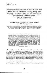
In the course of 24 hrs the average R-R intervals were longer at night than during the day (Fig. 1); they were the longest in the asthmatic patients without drugs (group A) and the shortest in the asthma patients who received fenoterol (group B). The differences between all groups were significant at night (i.e. at 18.00, 22.00, 24.00, 01.00 and 02.00).
Indices of HRV of the 15 patients tested in the morning and in the afternoon before and after inhalation of fenoterol were similar (Table 1). Mean PEF value was significantly higher after fenoterol inhalation.
In group A of asthma patients without the drug, the mean 24 h values of R-R intervals and of the SD of
Heart rate variability (HRV) was estimated from the beat-to-beat R-R interval variations. The R-R variability was extracted from the sampled ECG and analysed on line using a personal computer (IBM XT). The HRV was assessed by calculation of indices based on: 1) statistical operations on R-R intervals (time domain): - average R-R intervals - standard deviations of R-R intervals - load index [12] calculated as % of R-R intervals equal modal value divided by (2 x amplitude of modal value x R-R variability range) 2) spectral analysis (frequency domain) - the amplitudes and powers of the spectral density computed with a program for the autoregressive model [13,14] in the low frequency range (LF=0.04-
Figure 1. Average R-R intervals. asthmatic without drugs, asthmatics with fenorol controls Significant p