Unexpectedly, MRI showed a mucocele of the ethmoid sinus with compression of the right optic nerve (Figure 3A and B). The patient was immediately referred to ...
Therapeutic Advances in Neurological Disorders
Clinical approach to optic neuritis: pitfalls, red flags and differential diagnosis Elke Voss, Peter Raab, Corinna Trebst and Martin Stangel
Review
Ther Adv Neurol Disord (2011) 4(2) 123134 DOI: 10.1177/ 1756285611398702 ! The Author(s), 2011. Reprints and permissions: http://www.sagepub.co.uk/ journalsPermissions.nav
Abstract: Demyelinating optic neuritis (ON) is the most common cause of optic neuropathy typically presenting with a subacute painful visual loss. In 20% of patients with multiple sclerosis (MS), ON is the presenting symptom and half of the patients with isolated ON develop MS within 15 years. The diagnosis of ON plays an important role in neurological practice. A correct and early diagnosis is necessary to ensure optimal further investigations and treatment. Other causes of optic neuropathies such as connective tissue disorders, infectious diseases, tumours or ischaemic neuropathies are less frequent but clinical and therapeutic management can differ dramatically. We present five patients admitted to our hospital with suspected demyelinating ON, but the clinical work up revealed different causes of optic neuropathy. We discuss the differential diagnosis of ON and clinical red flags that require careful diagnostic assessment of other diseases. A workflow for the diagnosis of optic neuropathies is presented. Keywords: differential diagnosis, multiple sclerosis, optic neuritis
Introduction Optic neuritis (ON) is the most common cause of acute unilateral visual loss in young adults having an incidence of 15 in 100,000 per year. Women are more often affected than men with a peak manifestation between the ages of 15 and 49 [Rodriguez et al. 1995; Optic Neuritis Study Group, 1991]. Patients with ON typically present with the triad of subacute unilateral loss of vision, periocular pain and impaired colour vision. The symptoms usually worsen over the course of a few days to 2 weeks but spontaneously recover in >90% of cases after 23 weeks regardless of steroid treatment. In most patients a relative afferent pupillary defect (RAPD) is obvious, although this is not specific to ON but is present in most optic neuropathies [Balcer, 2006; Optic Neuritis Study Group, 1991]. An atypical clinical presentation, e.g. missing pain, a marked visual loss, bilateral visual loss or absence of spontaneous recovery after 23 weeks should always give reason to careful diagnostic search for other differential diagnoses. In most individuals ON is caused by idiopathic inflammatory demyelination. This may occur as
http://tan.sagepub.com
an isolated syndrome or in association with multiple sclerosis (MS). Approximately 50% of patients with isolated ON develop definite MS within 15 years [Beck et al. 2004; Rodriguez et al. 1995]. The most predictive factor for the development of MS after ON is the presence of asymptomatic demyelinating lesions in the central nervous system (CNS). In the Optic Neuritis Treatment Trial (ONTT) the 5-year risk for definite MS was 52% in those patients with one or more asymptomatic white matter lesions on brain MRI compared with a 5-year risk of only 16% in patients with normal brain MRI [Optic Neuritis Study Group, 1997].
Correspondence to: Martin Stangel Hannover Medical School, Carl-Neuberg-Strasse 1, Hannover 30625, Germany stangel.martin@ mh-hannover.de Elke Voss Peter Raab Corinna Trebst Hannover Medical School, Hannover, Germany
Nonetheless, inflammatory ON may also be caused by other autoimmune diseases such as sarcoidosis, systemic lupus erythematosus (SLE), Sjo¨gren’s syndrome (SS) or Behc¸et’s disease that require different immunosuppressive long-term therapies compared with MS [Cikes et al. 2008]. Although infectious causes of ON such as herpes Zoster, Lyme disease, syphilis, tuberculosis or toxoplasmosis are rare, they should be kept in mind because a specific treatment regime is required rapidly. Many other conditions such as tumours or ischaemic diseases can also cause an optic neuropathy and clinically
123
Therapeutic Advances in Neurological Disorders 4 (2) mimic idiopathic inflammatory ON [Hickman et al. 2002]. In those cases in particular, in which the patient history reveals atypical clinical aspects for ON, a careful clinical work up is required in order to establish the correct treatment regimen. Furthermore, in those patients with ON with a high risk of developing MS, an early and accurate diagnostic assessment to rule out other causes is necessary to initiate a diseasemodifying treatment [Kappos et al. 2009; Comi et al. 2001]. Our case series of patients admitted for suspicious ON demonstrates the spectrum of underlying conditions for acute visual loss. We discuss the differential diagnosis of ON and compile a clinical work up for the assessment of ON. Patients We present the history and clinical course of five patients who were admitted to our hospital with suspected ON between 2007 and 2010 (two females, three males). All patients suffered from subacute visual loss. The age range was 2651 years. All patients underwent general examination, neurological examination and ophthalmological evaluation including measurement of visual acuity, colour vision with an Ishihara pseudoisochromatic plate, visual field, fundoscopy and visual evoked potentials (VEPs). Routine blood tests in patients included serum electrolytes, renal function tests, C-reactive protein (CRP), erythrocyte sedimentation rate (ESR), liver enzymes, creatine kinase (CK), glucose, complete blood count (CBC), partial thromboplastin time (PTT), and international normalized ratio (INR). In all patients except case 3, additional blood tests were performed including antinuclear antibodies (ANA), anti-double-stranded DNA antibodies (anti-dsDNA-Ab), protoplasmic/cytoplasmic antineutrophil cytoplasmic antibodies (p/c ANCA), extractable nuclear antigens (ENA), angiotensin converting enzyme (ACE), interleukin-2 receptor (IL-2R), antiphospholipid Ab, rheumatoid factor (RF), calcium and vitamin B12. Lumbar puncture was performed in three patients. Analysis of cerebrospinal fluid (CSF) included determination of total protein, albumin, IgG, IgA, IgM, glucose, lactate, cell count, microbiological and virological analysis and oligoclonal bands. In the first two cases a chest X-ray and CT scan of the chest were added for assessment of typical findings for sarcoidosis. Orbital and
124
brain MRI with gadolinium was performed in all patients. MRI examinations were conducted on a 1.5-Tesla system (Signa LX NV/I). Case presentations Case 1 A 26-year-old female public employee was admitted with a relapse of visual loss on the right eye to 25% visual acuity with severe eye pain inhibiting any eye movement. An ON on the same eye was diagnosed 1 and 2 years prior to this admittance. In both cases she suffered from identical symptoms and visual acuity partially recovered after high-dose intravenous (i.v.) methylprednisolone. The clinical work up after the first episode 2 years before included MRI scan of the brain, VEP, CSF analysis and routine blood tests. Aside from a gadolinium enhancement of the right optic nerve, no other lesions suspicious for MS or other chronic inflammatory CNS diseases were present on MRI scans. Routine blood tests and CSF analysis were normal, oligoclonal bands were negative. The VEP showed a prolonged latency on the right eye. After the second relapse 1 year later the clinical work up was repeated and showed identical results. Furthermore MRI scan of the spine and additional laboratory tests including ANA, p/cANCA, anti-dsDNA-Ab, antiphospholipid Ab, RF, ACE, HbA1c, vitamin B12, folic acid, HIV, hepatitis B and C, borrelia, and lues in serum and CSF as well as virology in the CSF were performed without any pathological findings. The MRI performed at the third episode was suggestive for a meningioma or glioma rather than a typical ON (Figure 1A and B). Therefore, a biopsy including decompression of the optical canal was performed. Surprisingly, the histopathological analysis revealed a granulomatous inflammation typical for sarcoidosis. Further investigations showed no systemic manifestation of sarcoidosis (ACE, calcium, IL-2R, CT scan of the chest) therefore suggesting an isolated neurosarcoidosis. The patient was again treated with steroids and later switched to azathioprine. For the last 2 years, visual acuity remained stable on the right eye and no other symptoms occurred. Case 2 A 42-year-old mechanical engineer with severe pain on eye movement on both eyes for 2 weeks complained of a progressive reduction of visual acuity. In addition, he suffered from painful joints and neck as well as headache for several years.
http://tan.sagepub.com
E Voss, P Raab et al.
Figure 1. Optic nerve sheath diseases (cases 1 and 3). (A) and (B) demonstrate imaging findings of case 1, showing intense contrast enhancement of the optic nerve extending through the optic canal almost reaching the chiasm (B) due to neurosarcoidosis. There is an asymmetrical thickening of the optic nerve compared with the symmetric ‘tram track’-like enhancement in (C) and (D), representing the optic nerve sheath meningioma found in case 3. Neurosarcoidosis can mimic a nerve sheath meningioma, the two entities can be difficult to discern especially in cases of primary optic manifestation of sarcoidosis. Axial T1-weighted sequences with contrast enhancement and fat suppression pulse.
A diagnosis of rheumatoid arthritis (RA) 4 years ago was based on an increased RF and typical joint alterations. Under treatment with prednisolone and methotrexate the joint pain recovered completely, but whenever prednisolone was reduced below 30 mg per day the headache and neck pain worsened. When admitted to the hospital, visual acuity was reduced to 80% on both eyes. Severely painful eye movement and neck pain were the most prominent findings during neurological examination. On brain MRI scan before lumbar puncture, a pachymeningeal gadolinium enhancement and thickening of the meninges was evident (Figure 2A and B). CSF analysis excluded infectious meningitis and meningeal carcinomatosis, but oligoclonal bands were positive. Immunological parameters were negative except for RF. X-ray and CT scan of the
http://tan.sagepub.com
chest and laboratory tests including calcium, ACE and IL-2R were not suggestive for sarcoidosis. The symptoms improved after increasing prednisolone therapy from 20 mg per day to 50 mg per day. An advised meningeal biopsy was refused by the patient. Five months later the patient suffered from a relapse after the prednisolone has been reduced monthly by 5 mg to 25 mg per day. Brain MRI scan again showed the meningeal enhancement and the patient now agreed to a meningeal biopsy. The histopathological analysis showed perivascular infiltrates with lymphocytes, plasma cells and epithelioid histiocytes but no neoplasia, vasculitis, granulomas or infectious agents. The pathological findings were consistent with an idiopathic hypertrophic pachymeningitis that can be associated with RA [Kupersmith et al. 2004].
125
Therapeutic Advances in Neurological Disorders 4 (2)
Figure 2. Pachymeningitis (case 2). Both images clearly show the linear dural thickening and enhancement found in case 2. The differential diagnosis includes carcinomatous meningitis, intracranial hypotension, sarcoidosis, histiocytosis, idiopathic hypertrophic cranial pachymeningitis and dural sinus thrombosis. (A) Coronal T2-weighted sequence. (B) Coronal T1-weighted sequence with contrast enhancement.
Despite treatments with methotrexate, azathioprine, infliximab, adalimumab or abatacept it was not possible to reduce prednisolone without reoccurrence of symptoms. At present a treatment with rituximab has been initiated. Case 3 A 45-year-old male electrician was admitted due to a painless substantial visual loss on the right eye for 4 weeks. He reported that he had noticed a slightly progressive reduction of visual acuity on the right eye around 1.5 years ago. At the time of examination visual acuity was 10% on the right eye without any further signs or symptoms in the neurological examination. Fundoscopy and routine blood tests were normal. Because of the atypical history of slowly progressive painless visual loss over 1.5 years, an orbital and brain MRI scan was performed that revealed an optic nerve sheath meningioma (Figure 1C and D). Diagnosis was confirmed by optic nerve biopsy. A fractionated stereotactic radiotherapy was performed and resulted in an improvement of visual acuity to 20% that remained stable over the next 2 years. Case 4 A local ophthalmologist referred a 32-year-old technician to our emergency unit with suspected ON. She suffered from a progressive visual loss on the right eye with painful eye movement for 1 day. She had a history of squint amblyopia on the right eye with a reduced visual acuity to 50%, but had no previous other neurological symptoms. The ophthalmologist found a reduced visual
126
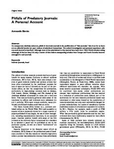
acuity to 15% with decreased colour vision and a diffuse visual field defect. Fundoscopy, routine blood testing as well as further neurological examination were normal. We suggested an ON and arranged a MRI scan to determine the risk of developing MS. Unexpectedly, MRI showed a mucocele of the ethmoid sinus with compression of the right optic nerve (Figure 3A and B). The patient was immediately referred to the ear, nose and throat (ENT) department and a surgical decompression of the orbit was performed. Case 5 A 51-year-old male civil servant was admitted by an ophthalmologist with a sudden visual loss on the right eye on the previous day. Because of a swollen disc on fundoscopy the ophthalmologist suggested an ON. The patient had a history of arterial hypertension, obesity and hypercholesterolemia. Further neurological examination and routine blood tests were normal. No pathological parameters were found in the additional blood tests and CSF. MRI scan showed some white matter lesions suggestive of microangiopathy but no typical lesions for MS or other inflammatory diseases. Morphology of the optic nerves appeared normal on MRI and no pathological contrast enhancement was detected. ECG showed a sinus rhythm and echocardiography was normal. The intima-media thickness (IMT) of the carotid arteries was increased on ultrasound but no stenosis was obvious. Owing to the sudden onset of visual loss without pain, no evidence of inflammation, typical changes of microangiopathy on brain MRI and the presence
http://tan.sagepub.com
E Voss, P Raab et al.
Figure 3. Ethmoidal mucocele. In (A) an abnormal enhancement of the intraconal space surrounding the optic nerve can be seen. This indicates intraorbital and intraconal inflammation caused by the expanding mucocele. (B) shows an enlarged ethmoid cell and a bony defect between this nasal sinus and the tip of the orbit due to the enlarging sinus. (A) Coronal T1-weighted sequence with fat suppression. (B) Axial CT scan.
of cardiovascular risk factors we diagnosed an anterior ischaemic optic neuropathy (AION). Discussion Here we have presented five cases of optic neuropathies that were all referred with suspicion of demyelinating ON. In four of the five cases the patient history or the clinical signs revealed aspects that were not typical for ON. The first two cases demonstrate different causes of noninfectious inflammatory optic neuropathy. An important differential diagnosis of ON are granulomatous optic neuropathies such as sarcoidosis or Wegener’s granulomatosis [Montagnac et al. 2009; Graham et al. 1986]. Connective tissue disorders such as SLE, RA, SS or systemic vasculitis can all be associated with optic neuropathy [Lin et al. 2009; Chen et al. 2008; Kansu and Kadayifcilar, 2006]. In most patients with connective tissue disorders, sarcoidosis or vasculitis a history of other symptoms of a multisystem disorder can be found, although the inflammatory activity can be, at least initially, restricted to the optic nerve. Typically, these optic neuropathies come along with severe eye pain and progressive visual loss as reported in case 1. The bilateral presentation, absence of spontaneous improvement and prompt recurrence of symptoms after steroid treatment was withdrawn in the second patient are further alerting symptoms atypical for demyelinating ON but more suggestive of other corticosteroid-responsive optic neuropathies (Tables 1 and 2). Immunological laboratory
http://tan.sagepub.com
tests (e.g. ANA, ENA, anti-dsDNA-Ab, p/c ANCA, RF, ACE, antiphospholipid Ab) may lead to the correct differential diagnosis [Cikes et al. 2008; Sastre-Garriga et al. 2001]. However, in some cases these parameters were negative not excluding a primary CNS manifestation of connective tissue disorders, sarcoidosis or vasculitis and only a biopsy can reveal the correct diagnosis. On the other hand, some patients present with a comorbidity of both a systemic autoimmune disease together with a demyelinating CNS disease such as MS and neuromyelitis optica (NMO) [Kang et al. 2010; Cooper et al. 2009; Pittock et al. 2008]. In these patients the differentiation between two independent autoimmune disorders or one systemic autoimmune disease with CNS manifestation is sometimes complicated and individual decisions on treatment management have to be considered carefully. ON also typically occurs in Devic’s disease or NMO, a demyelinating autoimmune disease that in contrast to MS primarily affects the optic nerve and spinal cord [Wingerchuk et al. 2006]. The clinical presentation of ON in NMO is variable but often occurs bilaterally and recurrently, and visual loss is more severe with less improvement when compared with ON seen in MS. Characteristically, oligoclonal bands are negative and brain MRI is normal in NMO, but spinal MRI shows transverse myelitis often spanning three or more spinal cord segments [Wingerchuk et al. 2007, 2006]. Serum NMO immunoglobulin G Ab (NMO-IgG), that bind to the water-channel protein aquaporin-4,
127
Therapeutic Advances in Neurological Disorders 4 (2) Table 1. Typical and atypical presentations of optic neuritis. Typical signs and symptoms of optic neuritis
Red flags: atypical signs and symptoms of optic neuritis
# # # #
Young adult patient 50 or 2 weeks Severe visual loss to no perceptions of light
#
Periocular pain and painful eye movement
# #
Previous history of ON or MS Neurological signs and symptoms suggestive of MS
# # # # #
# # #
Normal or swollen optic disc Normal macula and peripheral retina Uveitis or retinal periphlebitis possible
# #
Spontaneous improvement after 23 weeks No deterioration after withdrawal of steroids
Bilateral visual loss No pain Severe or persistent pain >2 weeks Previous history of neoplasia Clinical symptoms suggestive of other diseases than MS (NMO, connective tissue disorders, sarcoidosis, vasculitis) Severe optic disc oedema Optic disc haemorrhage Marked uveitis or retinal periphlebitis Optic atrophy without history of ON or MS Absence of recovery >3 weeks after onset Deterioration after withdrawal of steroids
# # # # # #
ON, optic neuritis; MS, multiple sclerosis; NMO, neuromyelitis optica.
has been recently established as a significant biomarker for NMO. These antibodies are highly specific (>90%) with a sensitivity of approximately 6070% [Wingerchuk et al. 2006; Lennon et al. 2004]. The differentiation of NMO from MS is of considerable importance for the long-term treatment, as the established disease-modifying drugs (DMDs) for MS are usually ineffective in NMO. Actually, even a worsening of NMO has been described under the treatment with DMD [Palace et al. 2010; Shimizu et al. 2010]. Controlled clinical trials for the management of NMO are missing but according to the EFNS guidelines on diagnosis and management of NMO an immunosuppressive treatment regime is recommended, e.g. with azathioprine or rituximab as a first-line or with mitoxantrone, cyclophosphamide or mycophenolate mofetil as a second-line therapy [Sellner et al. 2010]. Autoimmune mediated demyelinating ON may also appear as an isolated optic nerve disorder with one single episode (single isolated ON [SION]) or with relapsing ON (relapsing isolated ON [RION]). In patients with SION neither the MRI nor the CSF provide evidence for MS. For these patients the risk of developing MS is estimated to be low if the MRI remains negative for demyelinating white matter lesions over time [Nilsson et al. 2005; Ghezzi et al. 2000; Optic Neuritis Study Group, 1997]. The frequency of
128
NMO-IgG in patients with RION differs from 6% to 25%, suggesting that some of these patients have a higher risk to develop NMO and therefore may profit from early immunosuppressive treatment [de Seze et al. 2008; Matiello et al. 2008; Petzold, 2008]. In chronic relapsing inflammatory optic neuropathy (CRION), patients typically suffer from severe, painful and often sequentially bilateral visual loss that is ameliorated by corticosteroids but often relapses after withdrawal of steroids. Comparable to NMO these patients usually require long-term immunosuppression [Kidd et al. 2003]. Infections such as syphilis, Lyme disease, tuberculosis, cat-scratch disease, toxoplasmosis or viruses (e.g. herpes, hepatitis A virus or enteroviruses) may also cause optic neuropathies that are characterized with a progressive visual loss and severe optic disc oedema on fundoscopy [Bodaghi and LeHoang, 2000; Karma et al. 1995]. In children postinfectious or postvaccination optic neuropathies and neuroretinitis presenting with swollen optic disc and macular star should also be considered as these conditions usually have a very good prognosis [Ray and Gragoudas, 2001]. Different kinds of tumours can cause clinical symptoms suggestive of ON due to their compression of the optic nerve. Patients with compressive optic neuropathies caused by metastases
http://tan.sagepub.com
E Voss, P Raab et al. Table 2. Aetiology of optic neuropathies and clinical presentation in comparison to typical optic neuritis (ON). Diagnosis
Clinical presentation
Inflammatory optic neuropathies Autoimmune optic neuritis Single and relapsing isolated ON (SION, RION) [Petzold, 2008] Chronic relapsing inflammatory optic neuropathy (CRION) [Kidd et al. 2003] Neuromyelitis optica (NMO) [Wingerchuk et al. 2007] Acute disseminated encephalomyelitis (ADEM) [Tenembaum et al. 2007] Connective tissue disorders and vasculitis [Theodoridou and Settas, 2006; Cikes et al. 2008] Sarcoidosis Systemic lupus erythematosus (SLE) Sjo ¨gren’s syndrome Antiphospholipid antibody syndrome Behc¸et’s disease Wegener’s granulomatosis Giant cell arteritis (GCA) [Carroll et al. 2006] Other inflammatory optic neuropathies Postinfectious and postvaccination Neuroretinitis [Ray and Gragoudas, 2001] TolosaHunt syndrome [La Mantia et al. 2006] Infectious optic neuropathies Lyme disease [Karma et al. 1995] Syphilis Tuberculosis [Bodaghi and LeHoang, 2000] Viral optic neuritis Compressive optic neuropathies Primary tumours (meningiomas, gliomas, and pituitary tumours) [Eddleman and Liu, 2007] Metastases Thyroid ophthalmopathy [Vardizer et al. 2010]
Ischemic optic neuropathies [Fontal et al. 2007] Anterior ischaemic optic neuropathy (AION) Posterior ischaemic optic neuropathy (PION) Diabetic papillopathy Toxic and nutritional optic neuropathies [Orssaud et al. 2007] Vitamin B12 deficiency Tobaccoalcohol amblyopia Methanol intoxication Drug induced [Li et al. 2008] Inherited optic neuropathies [Milea et al. 2010] Leber’s hereditary optic neuropathy Kjer autosomal-dominant optic atrophy Ocular causes Posterior scleritis Big blind spot syndrome and acute zonal occult outer retinopathy Maculopathies and retinopathies
http://tan.sagepub.com
Progressive or relapsing severe visual loss, often very painful Rare, often unilateral, sometimes associated with transverse myelitis Often bilateral, severe visual loss Rare, often unilateral, sometimes associated with transverse myelitis Papillitis, uveitis, chorioretinitis, and retinal vasculitis Papillitis, scleritis, conjunctivitis, uveitis, retinal vasculitis Sudden visual loss (AION, PION), headache, muscle pain, age >50 years, jaw claudication Bilateral, often in childhood, good prognosis Swollen optic disc and macular star, spontaneous recovery Painful ophthalmoplegia Progressive visual loss with exposure to infectious agent Rare, more often occurring at later stages of disease Also manifestation as uveitis, retinitis Rare, more often presenting as choroiditis or uveitis Most frequently associated with herpes Zoster infection Painless and progressive visual loss Optic atrophy
Arterial aneurysms Sinus mucoceles
or primary tumours complain of a slowly progressive visual loss without pain and often an atrophy of the optic nerve is evident, comparable to patient 3 with optic nerve sheath meningioma
Steroid responsive ON without signs for other demyelinating CNS disease Often bilateral, severe and painful visual loss, relapse after withdrawal of steroids ON and transverse myelitis Usually monophasic, triggered by infections and vaccination, encephalomyelitis, can be bilateral Worsening of symptoms after withdrawal of steroids
History of or evidence for primary tumour Protrusion of one or both eyes, dry eyes, systemic signs for hyperthyroidism Painful progressive visual loss, general headache History of sinusitis, may be painful and with subacute visual loss Sudden onset of painless visual loss, age >50 years Swollen optic disc Optic disc atrophy History of diabetes, diabetic retinopathy Painless, bilateral, and symmetrical visual loss Associated with sensory ataxia, pernicious anaemia History of alcohol and tobacco abuse History of intoxication with contaminated alcohol Medication history, e.g. ethambutol, linezolid, amiodarone Painless, progressive, sequential bilateral visual loss Family history Family history, manifestation in childhood Severe pain, less visual symptoms Visual field loss and photopsiasis, normal fundus and colour vision Painless, metamorphosia, normal colour vision
[Eddleman and Liu, 2007] (Table 2). However, as in case 4, a painful eye movement is sometimes reported in patients with mucoceles [Avery et al. 1983; Rothstein et al. 1984] or arterial aneurysms
129
Therapeutic Advances in Neurological Disorders 4 (2) [Raps et al. 1993]. Moreover, a thyroid eye disease should be kept in mind as a cause of compressive optic neuropathy [Vardizer et al. 2010]. Conventional and gadolinium-enhanced orbital MRI is the most important diagnostic tool to ascertain an expanding orbital or retroorbital lesion. Nonetheless, as illustrated in case 1, a differentiation between primary tumours and other inflammatory diseases on MRI is not always possible (Figure 1) and a biopsy may be required to make the correct diagnosis. Patients with ischaemic optic neuropathy typically present with of a sudden onset of visual loss without pain. These patients are usually of older age and have a history of cardiovascular risk factors [Kerr et al. 2009]. As reported in case 5, in some patients with AION a swollen optic disc can be assessed (Table 2), whereas posterior ischaemic optic neuropathy (PION) is characterized by sudden vision loss without disc oedema but subsequent optic disc atrophy [Fontal et al. 2007]. In elderly patients, especially when visual loss is associated with eye pain, giant cell arteritis (GCA) should always be considered as differential diagnosis and routine blood testing should include ESR. Ischaemic optic complications appear in approximately 25% of patients suffering from GCA, most frequently presenting as AION [Hernandez-Rodriguez et al. 2007; Salvarani et al. 2005]. In patients with a history of diabetes a diabetic papillopathy may also be a cause of painless visual loss associated with sequential transient disc swelling [Barbera et al. 1996]. In most patients additionally a diabetic retinopathy can be assessed on fundoscopy at presentation. Other maculopathies and retinopathies could also mimic ON and cause a subacute visual loss. However, these patients typically complain about a slowly progressive, painless visual loss and alterations of the macula or retina are in most cases evident in ophthalmological examination. Posterior scleritis is often also associated with visual loss but in contrast to ON eye pain is the most prominent symptom [Maggioni et al. 2007]. The big blind spot syndrome and acute zonal occult outer retinopathy describe other rare ophthalmologic differential diagnosis. Patients typically present with an acute onset of a scotoma associated with photopsia and enlargement of the blind spot [Monson and Smith, 2011].
130
In most cases an ophthalmologic examination including detection of visual acuity, colour vision, visual field and fundoscopy can exclude an ocular cause of visual loss. VEP can also help to differentiate between a retinal disease and an optic nerve dysfunction. Even though VEP are not useful to distinguish between different causes of optic neuropathy in the acute phase, they can facilitate to evaluate recovery in followup examination or to identify subclinical optic nerve dysfunctions. Optical coherence tomography (OCT) is a novel method that has been predominantly used in ophthalmologic diseases to evaluate the thickness of the retinal nerve fibre layer (RNFL). Recent data suggest that the RNFL can aid to estimate neurodegeneration in MS and to differentiate between ON in MS and ON in NMO [Jindahra et al. 2010]. Conclusions Whenever a patient with suspected ON presents with atypical findings (Table 1), further investigations should be conducted urgently to rule out other aetiologies such as tumours or other corticosteroid responsive optic neuropathies (Table 2) that require a distinct further management. A clinical workflow for the diagnosis of ON is summarized in Figure 4. For patients with atypical clinical presentation an orbital and brain MRI with gadolinium is mandatory. If a MRI scan is not possible, a CT scan with contrast should be performed. Additional laboratory tests (Table 3) and CSF analysis can help to reveal infectious causes or optic neuropathies due to connective tissue disorders, granulomatous inflammation or vasculitis. A careful ophthalmologic examination is also recommended to rule out other differential diagnosis and the measurement of VEP can verify an optic nerve dysfunction In three of our cases only a biopsy revealed the correct diagnosis. Therefore, a biopsy should be considered if additional tests are not capable of revealing a correct diagnosis that may have substantial consequences for further therapeutic management, e.g. with regard to inflammatory causes or tumours. In those patients with typical clinical presentation of ON, a MRI scan with gadolinium should also be performed to assess the risk for development of MS. According to the McDonald criteria in these patients other diseases have to be ruled out by appropriate laboratory tests (Table 3) and the diagnosis can only be made when there
http://tan.sagepub.com
E Voss, P Raab et al.
Suspected optic neuritis
Atypical symptoms
Typical symptoms
Obligatory blood tests* Consider: MS/CIS SION
Consider:
MRI of brain: Suspicious of MS? yes
Inflammatory optic neuropathy Infectious optic neuropathy Ischemic optic neuropathy Compressive optic neuropathy Toxic or nutritional optic neuropathy Inherited optic neuropathy Ocular causes
no Further diagnostics as appropriate CSF
MRI of brain and orbitae
CSF
Additional blood tests*
High risk to develop MS
Low risk to develop MS
i.v. corticosteroids
i.v. corticosteroids
If applicable: biopsy
Discuss early treatment with DMD
Discuss follow-up examination and MRI
Specific diagnosis/ treatment
Figure 4. Clinical workflow for patients with diagnosis of suspected optic neuritis. *Recommended obligatory and additional blood tests: see Table 3.
Table 3. Recommended blood tests for the diagnosis of optic neuropathies. Obligatory blood tests
Additional blood tests
CRP CBC Blood chemistry Glucose ANA Vitamin B12 Serological testing for Lyme borreliosis
RF ACE Antiphospholipid Ab Anti-dsDNA Ab ENA p/c ANCA Methylmalonic acid Serological testing for syphilis, HIV Virology AQP-4 Ab Tuberculosis (if applicable) Genetic testing (if applicable)
CRP, C-reactive protein; CBC, complete blood count; ANA, antinuclear antibody; RF, rheumatoid factor; ACE, angiotensin converting enzyme; Anti-dsDNA Ab, anti-double-stranded DNA antibody; ENA, extractable nuclear antigens; ANCA, protoplasmic/cytoplasmic antineutrophil cytoplasmic antibody; AQP-4 Ab, antibodies against aquaporin 4; Ab, antibody.
is no better explanation [McDonald et al. 2001]. In patients with ON a treatment with intravenous methylprednisolone (1 g per day for 35 days) can be considered to hasten recovery of visual
http://tan.sagepub.com
acuity, however, this does not affect the longterm visual outcome [Brusaferri and Candelise, 2000; Kaufman et al. 2000; Beck et al. 1992]. A second steroid pulse (2 g per day) can be
131
Therapeutic Advances in Neurological Disorders 4 (2) applied when the symptoms persist 1014 days after the first therapy. In those patients with no or incomplete recovery after steroid treatment a plasma exchange therapy (PE) may facilitate an improvement of visual function [Trebst et al. 2009; Tumani, 2008]. The initiation of a treatment with DMDs should be discussed in patients with a high risk of developing MS. Funding This research received no specific grant from any funding agency in the public, commercial, or notfor-profit sectors. Conflict of interest statement The authors declare that there are no conflicts of interest.
References
Cooper, G.S., Bynum, M.L. and Somers, E.C. (2009) Recent insights in the epidemiology of autoimmune diseases: improved prevalence estimates and understanding of clustering of diseases. J Autoimmun 33: 197207. de Seze, J., Arndt, C., Jeanjean, L., Zephir, H., Blanc, F., Labauge, P. et al. (2008) Relapsing inflammatory optic neuritis: is it neuromyelitis optica? Neurology 70: 20752076. Eddleman, C.S. and Liu, J.K. (2007) Optic nerve sheath meningioma: current diagnosis and treatment. Neurosurg Focus 23: E4. Fontal, M.R., Kerrison, J.B., Garcia, R. and Oria, V. (2007) Ischemic optic neuropathy. Semin Neurol 27: 221232. Ghezzi, A., Martinelli, V., Rodegher, M., Zaffaroni, M. and Comi, G. (2000) The prognosis of idiopathic optic neuritis. Neurol Sci 21: S865S869.
Avery, G., Tang, R.A. and Close, L.G. (1983) Ophthalmic manifestations of mucoceles. Ann Ophthalmol 15: 734737.
Graham, E.M., Ellis, C.J., Sanders, M.D. and McDonald, W.I. (1986) Optic neuropathy in sarcoidosis. J Neurol Neurosurg Psychiatry 49: 756763.
Balcer, L.J. (2006) Clinical practice. Optic neuritis. N Engl J Med 354: 12731280.
Hernandez-Rodriguez, J., Font, C., Garcia-Martinez, A., Espigol-Frigole, G., Sanmarti, R., Canete, J.D. et al. (2007) Development of ischemic complications in patients with giant cell arteritis presenting with apparently isolated polymyalgia rheumatica: study of a series of 100 patients. Medicine (Baltimore) 86: 233241.
Barbera, L.G., Weiss, M.J. and Hofeldt, A.J. (1996) Diabetic retinopathy and diabetic papillopathy. Semin Neurol 16: 179185. Beck, R.W., Cleary, P.A., Anderson Jr, M.M., Keltner, J.L., Shults, W.T., Kaufman, D.I. et al. (1992) A randomized, controlled trial of corticosteroids in the treatment of acute optic neuritis. The Optic Neuritis Study Group. N Engl J Med 326: 581588. Beck, R.W., Gal, R.L., Bhatti, M.T., Brodsky, M.C., Buckley, E.G., Chrousos, G.A. et al. (2004) Visual function more than 10 years after optic neuritis: experience of the optic neuritis treatment trial. Am J Ophthalmol 137: 7783. Bodaghi, B. and LeHoang, P. (2000) Ocular tuberculosis. Curr Opin Ophthalmol 11: 443448. Brusaferri, F. and Candelise, L. (2000) Steroids for multiple sclerosis and optic neuritis: a meta-analysis of randomized controlled clinical trials. J Neurol 247: 435442. Carroll, S.C., Gaskin, B.J. and Danesh-Meyer, H.V. (2006) Giant cell arteritis. Clin Experiment Ophthalmol 34: 159173. Chen, Y.H., Wang, A.G., Lin, Y.C. and Yen, M.Y. (2008) Optic neuritis as the first manifestation of rheumatoid arthritis. J Neuroophthalmol 28: 237238. Cikes, N., Bosnic, D. and Sentic, M. (2008) Non-MS autoimmune demyelination. Clin Neurol Neurosurg 110: 905912. Comi, G., Filippi, M., Barkhof, F., Durelli, L., Edan, G., Fernandez, O. et al. (2001) Effect of early
132
interferon treatment on conversion to definite multiple sclerosis: a randomised study. Lancet 357: 15761582.
Hickman, S.J., Dalton, C.M., Miller, D.H. and Plant, G.T. (2002) Management of acute optic neuritis. Lancet 360: 19531962. Jindahra, P., Hedges, T.R., Mendoza-Santiesteban, C.E. and Plant, G.T. (2010) Optical coherence tomography of the retina: applications in neurology. Curr Opin Neurol 23: 1623. Kang, J.H., Chen, Y.H. and Lin, H.C. (2010) Comorbidities amongst patients with multiple sclerosis: a population-based controlled study. Eur J Neurol 17: 12151219. Kansu, T. and Kadayifcilar, S. (2006) Visual aspects of Behcet’s disease. Curr Treat Options Neurol 8: 8188. Kappos, L., Freedman, M.S., Polman, C.H., Edan, G., Hartung, H.P., Miller, D.H. et al. (2009) Long-term effect of early treatment with interferon beta-1b after a first clinical event suggestive of multiple sclerosis: 5-year active treatment extension of the phase 3 BENEFIT trial. Lancet Neurol 8: 987997. Karma, A., Seppala, I., Mikkila, H., Kaakkola, S., Viljanen, M. and Tarkkanen, A. (1995) Diagnosis and clinical characteristics of ocular Lyme borreliosis. Am J Ophthalmol 119: 127135. Kaufman, D.I., Trobe, J.D., Eggenberger, E.R. and Whitaker, J.N. (2000) Practice parameter: the role of corticosteroids in the management of acute
http://tan.sagepub.com
E Voss, P Raab et al. monosymptomatic optic neuritis. Report of the quality standards subcommittee of the American Academy of Neurology. Am J Ophthalmol 130: 541. Kerr, N.M., Chew, S.S. and Danesh-Meyer, H.V. (2009) Non-arteritic anterior ischaemic optic neuropathy: a review and update. J Clin Neurosci 16: 9941000. Kidd, D., Burton, B., Plant, G.T. and Graham, E.M. (2003) Chronic relapsing inflammatory optic neuropathy (CRION). Brain 126: 276284. Kupersmith, M.J., Martin, V., Heller, G., Shah, A. and Mitnick, H.J. (2004) Idiopathic hypertrophic pachymeningitis. Neurology 62: 686694. La Mantia, L., Curone, M., Rapoport, A.M. and Bussone, G. (2006) TolosaHunt syndrome: critical literature review based on IHS 2004 criteria. Cephalalgia 26: 772781. Lennon, V.A., Wingerchuk, D.M., Kryzer, T.J., Pittock, S.J., Lucchinetti, C.F., Fujihara, K. et al. (2004) A serum autoantibody marker of neuromyelitis optica: distinction from multiple sclerosis. Lancet 364: 21062112. Li, J., Tripathi, R.C. and Tripathi, B.J. (2008) Druginduced ocular disorders. Drug Saf 31: 127141. Lin, Y.C., Wang, A.G. and Yen, M.Y. (2009) Systemic lupus erythematosus-associated optic neuritis: clinical experience and literature review. Acta Ophthalmol 87: 204210. Matiello, M., Lennon, V.A., Jacob, A., Pittock, S.J., Lucchinetti, C.F., Wingerchuk, D.M. et al. (2008) NMO-IgG predicts the outcome of recurrent optic neuritis. Neurology 70: 21972200. McDonald, W.I., Compston, A., Edan, G., Goodkin, D., Hartung, H.P., Lublin, F.D. et al. (2001) Recommended diagnostic criteria for multiple sclerosis: guidelines from the International Panel on the diagnosis of multiple sclerosis. Ann Neurol 50: 121127. Milea, D., Amati-Bonneau, P., Reynier, P. and Bonneau, D. (2010) Genetically determined optic neuropathies. Curr Opin Neurol 23: 2428. Maggioni, F., Ruffatti, S., Viaro, F., Mainardi, F., Lisotto, C. and Zanchin, G. (2007) A case of posterior scleritis: differential diagnosis of ocular pain. J Headache Pain 8: 123126. Monson, D.M. and Smith, J.R. (2011) Acute zonal occult outer retinopathy. Surv Ophthalmol 56: 2335. Montagnac, R., Nyandwi, J., Loiselet, G., Pradel, J. and Schillinger, F. (2009) [Ophthalmic manifestations in Wegener’s granulomatosis. Review of literature about an observation]. Nephrol Ther 5: 603613. Nilsson, P., Larsson, E.M., Maly-Sundgren, P., Perfekt, R. and Sandberg-Wollheim, M. (2005) Predicting the outcome of optic neuritis: evaluation of
http://tan.sagepub.com
risk factors after 30 years of follow-up. J Neurol 252: 396402. Optic Neuritis Study Group (1991) The clinical profile of optic neuritis. Experience of the Optic Neuritis Treatment Trial. Arch Ophthalmol 109: 16731678. Optic Neuritis Study Group (1997) The 5-year risk of MS after optic neuritis. Experience of the optic neuritis treatment trial. Neurology 49: 14041413. Orssaud, C., Roche, O. and Dufier, J.L. (2007) Nutritional optic neuropathies. J Neurol Sci 262: 158164. Palace, J., Leite, M.I., Nairne, A. and Vincent, A. (2010) Interferon Beta treatment in neuromyelitis optica: increase in relapses and aquaporin 4 antibody titers. Arch Neurol 67: 10161017. Petzold, A. (2008) Isolated, relapsing and progressive demyelinating diseases of the central nervous system. J Neurol 255(Suppl. 6): 6976. Pittock, S.J., Lennon, V.A., de Seze, J., Vermersch, P., Homburger, H.A., Wingerchuk, D.M. et al. (2008) Neuromyelitis optica and non organ-specific autoimmunity. Arch Neurol 65: 7883. Raps, E.C., Rogers, J.D., Galetta, S.L., Solomon, R.A., Lennihan, L., Klebanoff, L.M. et al. (1993) The clinical spectrum of unruptured intracranial aneurysms. Arch Neurol 50: 265268. Ray, S. and Gragoudas, E. (2001) Neuroretinitis. Int Ophthalmol Clin 41: 83102. Rodriguez, M., Siva, A., Cross, S.A., O’Brien, P.C. and Kurland, L.T. (1995) Optic neuritis: a populationbased study in Olmsted County, Minnesota. Neurology 45: 244250. Rothstein, J., Maisel, R.H., Berlinger, N.T. and Wirtschafter, J.D. (1984) Relationship of optic neuritis to disease of the paranasal sinuses. Laryngoscope 94: 15011508. Salvarani, C., Cimino, L., Macchioni, P., Consonni, D., Cantini, F., Bajocchi, G. et al. (2005) Risk factors for visual loss in an Italian population-based cohort of patients with giant cell arteritis. Arthritis Rheum 53: 293297. Sastre-Garriga, J., Reverter, J.C., Font, J., Tintore´, M., Espinosa, G. and Montalban, X. (2001) Anticardiolipin antibodies are not a useful screening tool in a nonselected large group of patients with multiple sclerosis. Ann Neurol 49: 408411. Sellner, J., Boggild, M., Clanet, M., Hintzen, R.Q., Illes, Z., Montalban, X. et al. (2010) EFNS guidelines on diagnosis and management of neuromyelitis optica. Eur J Neurol 17: 10191032. Shimizu, J., Hatanaka, Y., Hasegawa, M., Iwata, A., Sugimoto, I., Date, H. et al. (2010) IFNb-1b may severely exacerbate Japanese optic-spinal MS in neuromyelitis optica spectrum. Neurology 75: 14231427.
133
Therapeutic Advances in Neurological Disorders 4 (2) Tenembaum, S., Chitnis, T., Ness, J. and Hahn, J.S. (2007) Acute disseminated encephalomyelitis. Neurology 68: S23S36.
Vardizer, Y., Tomkins, O. and Briscoe, D. (2010) Clinical assessment of thyroid related orbitopathy: a review. Pediatr Endocrinol Rev 7(Suppl. 2): 186192.
Theodoridou, A. and Settas, L. (2006) Demyelination in rheumatic diseases. J Neurol Neurosurg Psychiatry 77: 290295.
Wingerchuk, D.M., Lennon, V.A., Lucchinetti, C.F., Pittock, S.J. and Weinshenker, B.G. (2007) The spectrum of neuromyelitis optica. Lancet Neurol 6: 805815.
Trebst, C., Reising, A., Kielstein, J.T., Hafer, C. and Stangel, M. (2009) Plasma exchange therapy in steroid-unresponsive relapses in patients with multiple sclerosis. Blood Purif 28: 108115. Visit SAGE journals online http://tan.sagepub.com
134
Tumani, H. (2008) Corticosteroids and plasma exchange in multiple sclerosis. J Neurol 255(Suppl. 6): 3642.
Wingerchuk, D.M., Lennon, V.A., Pittock, S.J., Lucchinetti, C.F. and Weinshenker, B.G. (2006) Revised diagnostic criteria for neuromyelitis optica. Neurology 66: 14851489.
http://tan.sagepub.com