BEAU J. ANDERSON,1 MARGARET M. BILLS,2 JOHN R. EGERTON,1 AND JOHN S. MATTICK2*. Department of Veterinary Clinical Studies, The University ...
Vol. 160, No. 2
JOURNAL OF BACTERIOLOGY, Nov. 1984, p. 748-754 0021-9193/84/110748-07$02.00/0 Copyright © 1984, American Society for Microbiology
Cloning and Expression in Escherichia coli of the Gene Encoding the Structural Subunit of Bacteroides nodosus Fimbriae BEAU J. ANDERSON,1 MARGARET M. BILLS,2 JOHN R. EGERTON,1 AND JOHN S. MATTICK2* Department of Veterinary Clinical Studies, The University of Sydney, Sydney, New South Wales, Australia 2006,1 and Division of Molecular Biology, Commonwealth Scientific and Industrial Research Organization, North Ryde, New South Wales, Australia 21132 Received 11 June 1984/Accepted 15 August 1984 Bacteroides nodosus is the primary causative agent of ovine foot rot. Virulent isolates of this bacterium
cohtain fimbriae which appear to play a major role in both infectivity and protective immunity. This paper presents the cloning and expression in Escherichia coli of the gene encoding the structural subunit of the fimbriae of B. nodosus. Total DNA was isolated from B. nodosus VCS 1001 (serogroup A), digested with HindIlI, and inserted into the positive-selection vector pTR262. Recoimbinant E. coli clones were screened directly with anti-fimbrial antiserum by using a colony immunoassay. Several positive colonies were identified, each of which con'tained the same 5.5-kilobase Hindlll insert. The prototype has been designated pBA101. Some clones also contained additional flanking sequences from the B. nodosus genome. Western transfer analyses verified that the positive clones were producing the B. nodosus fimbrial structural subunit, molecuilar weight ca. 17,500. The level of expression of the antigen in E. eoli was comparable to that in B. nodqsus itself and was unaffected by the insertion site or orientation of the cloned fragment, indicating that synthesis was being directed from an internal promoter. Restriction mapping and deletion analyses localized the fimbrial subunit gene to the vicinity of a PvuII site near the central region of the originai HindIII insert. The expressed antigen was located in the membrane-cell wall fraction and may be exposed on the surface of the recombinant E. coli cells.
weight of about 17,500 (10, 17, 19); in other serotypes the size appears to vary (15, 17). Isolated fimbrial preparations also contain another significant component, a polypeptide of ca. 80,000 molecular weight, which seems to represent the basal protein linking the fimbriae to the cell wall (17). Both the basal and structural subunit components of the fimbriae are prominent antigens in vivo and in vitro (17, 22). This paper presents the molecular cloning of the gene sequences encoding the fimbrial subunit of B. nodosus as a prelude to investigating the genetic organization and control of fimbrial biosynthesis in this organism as well as the structural basis for the serological differences between strains. The cloned gene is expressed in the recombinant Escherichia coli host.
Foot rot is a major disease affecting sheep in wet, temperate regions. The organism essentially responsible for this condition is Bacteroides nodosus (1, 9), an anaerobic bacterium whose virulent isolates are characterized (28, 29, 30, 38) by the presence of surface filaments termed fimbriae (23). In other bacteria, fimbriae have been shown to possess adhesive properties (see reference 23), and although their exact function in B. nodosus has not been clearly defined, it seems likely that they are involved in attachment to, and colonization of, the epithelial tissues in hooves. Over the past decade, a great deal of evidence has accumulated to suggest that the fimbriae play a central role in both the pathogenicity of the bacteria and the immunological response of the host (11, 29, 32, 38). The fimbriae are the principal antigenic determinants involved in the K-agglutination reaction (10, 31, 38), which has been used to classify B. nodosus isolates into at least eight major serogroups and a number of subtypes (4, 7, 15, 26). These groupings are reflected in the patterns of cross-immunity between strains; the range of effective immunity conferred by vaccination with purified fimbriae, as with whole cells, appears to be restricted to the serogroup involved (8, 32; P. D. Claxton, Ph.D. thesis, University of Sydney, 1981). Consequently, the currently available foot rot vaccines consist of a mixture of cells from each of the representative B. nodosus serogroups. Fimbriation is an unstable characteristic in B. nodosus. The cells undergo a spontaneous and apparently irreversible conversion to nonfimbriate types, especially in liquid culture media (29, 35). Such types are relatively benign and appear to provide only weak immunity (29, 32, 36). B. nodosus fimbrial strands consist of a single repeated polypeptide subunit, which in serotype Al has a molecular *
MATERIALS AND METHODS Bacterial culture. B. nodosus 198 (ATCC 25549) was used for this study. This strain is the prototype of B. nodosus serogroup A (subtype 1) and has been reclassified by this scheme as VCS 1001 (4). Cells were cultured on hoof-agar plates (34) in an atmosphere of 90% H2 and 10% CO2 at 370C as described previously (17). E. coli RR1 was cultured on LB broth (16), in the presence of antibiotic selection where
appropriate.
DNA isolation and analysis. Total DNA was isolated from washed B. nodosus cells (suspended in 20 mM Tris, 20 mM EDTA [pH 8.0]) after the cells were exposed to lysozyme (4 mg/ml at 40C for 20 min), followed by the addition of 1% sodium dodecyl sulfate (SDS) and treatment with proteinase K (200 p.g/ml) at 450C for 4 h. The lysate was thenl extracted with phenol-chloroform, and nucleic acids were recovered from the supernatant by ethanol precipitation (16). The pellet was dissolved gently overnight in TE (10 mM Tris, 1 mM EDTA [pH 8.0]), treated with RNase A (100 ,ug/ml) at 37°C for 30 min, and then digested with proteinase K (100
Corresponding author. 748
VOL. 160, 1984
CLONING OF B. NODOSUS FIMBRIAL SUBUNIT GENE
,g/ml) in the presence of 0.5% SDS for 1 h at 50°C. After another round of phenol extraction and ethanol precipitation, the purified DNA was dissolved and dialyzed against TE. Large-scale preparations of plasmid DNA were obtained from chloramphenicol-induced E. coli cultures (16) by a lysozyme-EDTA-Triton X-100 lysis method. The cells obtained from 750 ml of culture were suspended in 7.75 ml of a 25% sucrose-50 mM Tris-hydrochloride (pH 8.0) solution and treated with lysozyme (2 to 4 mg/ml) at 4°C for 5 min; 1.25 ml of 0.5 M disodium EDTA (pH 8.0) was then added, and the incubation continued at 4°C for a further 5 min. The cells were lysed by the dropwise addition of the suspension into 10 ml of 0.1% Triton X-100-62.5 mM disodium EDTA-50 mM Tris-hydrochloride (pH 8.0) and mixing on ice for 15 min. The lysate was cleared by centrifugation in a Sorvall SS34 rotor for 90 min at 19,000 rpm. Plasmid DNA was then purified from the lysate by cesium chloride-ethidium bromide isopycnic ultracentrifugation. Minipreparations of recombinant plasmids were obtained by the alkaline-SDS lysis procedure (2, 16). Restriction endonuclease digestions were carried out under standard conditions and analyzed on 0.4 or 0.6% agarose gels in a Tris-borate-EDTA buffer system (16) containing 0.5 ,ug of ethidium bromide per ml. DNA cloning. B. nodosus DNA was digested with HindIII and ligated into the corresponding site in the positive-selection plasmid vector pTR262. This procedure causes the insertional inactivation of the Cl repressor gene, thereby permitting expression of tetracycline resistance (TetD directed from a promoter (25). Ligation was carried out with T4 DNA ligase under standard conditions (16) at vector and insert DNA concentrations of 7.5 and 10 jig/ml, respectively (6). The ligation mixture was used to transform E. coli RR1 directly, and recombinant transformants were selected on tetracycline plates. Recloning of DNA sequences in pBR322 was carried out by analagous procedures. Immunological screening. Recombinant E. coli clones were replica plated onto nitrocellulose filters (BA85; Schleicher & Schuell Co.) and grown on plates overnight, and then the colonies were lysed in situ by exposure to SDS and chloroform vapor (14). The filters were placed in a blocking solution, containing 2.4% bovine serum albumin and 1% bovine gelatin in Tris-saline buffer (12 mM Tris-hydrochloride, 120 mM NaCl [pH 7.5]), for 1 h at 37°C. The filters were then incubated for 4 h at room temperature in antiserum diluted 1:1,000 with Tris-saline buffer containing 2.4% bovine serum albumin and 0.2% gelatin. After thorough washing with Tris-saline, the filters were incubated with '25I-labeled protein A (0.1 ,uCi/ml) in Tris-saline-2.4% bovine serum albumin-0.2% gelatin buffer at room temperature for 2 h. The filters were washed again with Tris-saline, then dried, and exposed to X-ray film. Antisera. Rabbit antiserum against isolated B. nodosus fimbriae was raised as described previously (17). The antiserum recognizes both the structural subunit (Mr, 17,500) and the basal protein (Mr, 80,000) associated with the fimbrial structure (17). This antiserum also reacts with lipopolysaccharides from B. nodosus, which appear as laddered formations in Western transfer analyses of whole cells (see reference 17). 'Western transfer. Samples of cells, or fractions thereof, were solubilized in a 1.6% SDS-0.5 M urea buffer (17) and displayed on linear 6 to 15% gradient polyacrylamide gels, containing 0.15% SDS and 0.5 M urea, by the modification of the Laemmli system detailed previously (18). Standard pro-
749
tein mixtures (Bethesda Research Laboratories) or 14C-labeled protein standards (Amersham Corp.) or both were included in peripheral gel lanes for the calibration of molecular weights. The gel displays were then electrophoretically transferred to nitrocellulose paper by the method of Towbin et al. (37). The sheets were then treated with the blocking solution and exposed to antiserum (diluted 1:1,000) and I251-labeled protein A, under similar conditions to those described above and described in detail elsewhere (17). The transfers were then developed by autoradiography. When appropriate, gels were stained with Coomassie blue R250 to visualize the complete spectrum of polypeptide components. Cell fractionation. Washed E. coli cells were lysed by the lysozyme-EDTA-Triton X-100 method used for DNA isolations. This mixture was then sonicated at maximum power for a total of 1 min with an MSE sonicator. The soluble fraction was separated from insoluble material by centrifugation at 40,000 x g for 1 h. RESULTS Construction of B. nodosus genomic library. Total DNA was isolated from B. nodosus VCS 1001 (serogroup A) and digested with the restriction endonuclease HindIII to yield a population of fragments of average sizes of ca. 5 to 10 kilobases (kb). The fragments were then ligated into the HindIII site in the X repressor gene of the positive-selection vector pTR262 (25) and transformed into E. coli. A library of some 2,000 recombinant (Tetr) clones was obtained, which corresponds to ca. 2 to 4 genomic equivalents of B. nodosus DNA (27). Immunological screening of cloned library. The recombinant E. coli library was screened for the presence of B. nodosus fimbrial genes using a solid phase radioimmunoassay (14). Colonies were replicaplated and grown on nitrocellulose filters, lysed in situ and probed with rabbit antiserum against isolated B. nodosus fimbriae. Several immunoreactive colonies were identified (Fig. 1A) and were rescreened in triplicate arrays interspersed with known negatives. This test yielded a total of eight cloqs which exhibited a clear and reproducible signal in colony immunoassay (Fig. iB). Characterization of expressed antigen. The antiserum used in colony screening recognizes both the structural subunit and the basal protein antigens associated with B. nodosus fimbriae (17). Therefore, in order to establish the exact identity of the antigen being expressed, each of the positive clones was subjected to Western transfer analysis, along with a range of controls which included negative clones, B. nodosus cells, and purified fimbriae (Fig. 2). Samples were solubilized in SDS buffer, displayed by electrophoresis, then transferred to nitrocellulose paper and challenged with antifimbrial antiserum. All eight of the positive clones were found to be producing an antigen which comigrated with the B. nodosus fimbrial subunit, molecular weight ca. 17,500. As judged from the signal intensity, the level of expression of the fimbrial subunit in the recombinant cells appeared to be comparable within an order of magnitude to that in B. nodosus itself (Fig. 2). There was no indication that any clone was expressing the other major antigen, the 80,000dalton (d) basal polypeptide of the fimbrial strand. Characterization of cloned gene sequences. The DNA inserts in each of the positive clones were analyzed by HindIll digestion of small-scale plasmid preparations (Fig. 3). Each clone was found to contain a common 5.5-kb HindlIl frag-
750
ANDERSON ET AL.
lll.'-
"". $ 07:1
J. BACTERIOL.
,.?
.4
A
..
£*Sff 61 -
*
*@@ji . ... +
..
41
*
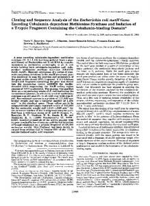
B FIG. 1. Colony immunoassay. Recombinant E. coli (Tetr) colonies containing HindIII fragments of B. nodosus DNA in the plasmid vector pTR262 were grown on nitrocellulose filters, lysed in situ, and exposed to rabbit anti-fimbrial antiserum. Colonies binding antibody were identified by autoradiography after incubation with "25I-labeled protein A. (A) shows a representative filter from the initial screening; the dark spots boxed at the top are reference controls of 0.1 and 1 ,ug of purified B. nodosus fimbriae. The arrows point to colonies considered putative positives. (B) shows the rescreening of putative positive colonies, replicated in triplicate and interspersed with known negatives in a zig-zag array.
ment of B. nodosus DNA, which therefore included the gene encoding the fimbrial structural subunit. Four of the eight clones contained additional inserts in the recombinant plasmids; two contained the 5.5-kb fragment plus a 1.2-kb HindIII fragment; one contained these 5.5- and 1.2-kb fragments as well as a 0.7-kb fragment (Fig. 3), and the other clone contained the 5.5-kb fragment plus HindIII segm.ents of about 8 and 3 kb (pBA104; not shown). The last clone grew very poorly and was difficult to maintain. The extended inserts in these plasmids probably represent the products of partial HindIII digestion of B. nodosus DNA, rather than any fortuitous cocloning of otherwise unrelated fragments. The original digestion was, in fact, incomplete. This conclusion is supported by the finding of apparently identical fragment extensions in three out of four clones (Fig. 3) as well as the fact that although we obtained both orientations of the inserted sequences in- pTR262, the relationship between the (5.5- and 1.2-kb) fragments was the same in each case (see below). It was therefore possible to construct a HindIII map of the fimbrial gene region, at least as it extended in the (arbitrary) rightward direction (Fig. 4). Although there was only the one example, it is likely that the other plasmid (pBA104), which contained a different set of additional HindIII fragments, probably represents the leftward extension of the same region. If this were so, the eight positive Qlones collectively represent sequences totaling ca. 18 kb which span the fimbrial subunit gene in B. nodosus, 1.9
kb to the right and possibly 11 kb to the left of the central 5.5-kb segment (Fig. 4). Restriction mapping and localization of the fimbrial subunit gene. The 5.5-kb HindIII fragment containing the fimbrial structural subunit gene was excised from its pTR262 vector (pBA101) and recloned into the HindIII site of pEIR322 to facilitate the restriction mapping of this segment (Fig. 4). Both orientations of the insert were obtained, designated pBA121 and pBA122, respectively, and again both were found to express high levels of the 17,500-d subunit (Fig. 5). The internal restriction map of the segment was aligned with the HindlIl map of the overall region (Fig. 4) by reference to the extended inserts in the other clones, such as those in pBA102 (Fig. 3). The location of the sequences encoding the fimbrial subunit was more closely defined by selective deletion, using restriction site pairs within the insert and vector. The strategy is outlined in Fig. 5. Both pBA121 and pBA122 were digested with EcoRI, NdeI, or PvuII and religated at low concentration. Ampicillin-resistant (Ap) subclones containing the corresponding deletions were then screened by colony immunoassay for expression of the fimbrial subunit (Fig. 5). These experiments showed that pBA122:AEcoRI and pBA121:ANdeI were positive for fimbrial expression, whereas the complementary clones containing pBA121: AEcoRI and pBA122:ANdeI were not. In the case of PvuII,
A
B
C
D
E
F
G
H
I
4
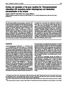
FIG. 2. Western transfer analysis of positive clones. Samnples of cells (ca. 101) or puirified fimbriae ('20 ±Lg) were electrophoresed on SDS-linear 6 to 15% gradient polyacrylamide gels. The resulting displays were transferred to nitrocellulose paper, incubated with rabbit anti-fimbrial antiserum and then "II1-labeled protein A, and autoradiographed. Lane contents are as follows: lane A, B. nodosus cells; lane B, purified fimbriae from B. nodosus; lanes C, F, and I, negative controls consisting of E. coli RR1 host cells (lane. C) or recombinant cells (lanes F and I) containing pTR262 with irrelevant inserts of B. nodosus DNA (cf. Fig. 3); lanes D, E, G, and H, independently isolated recombinant clones exhibiting a positive signal in colony immunoassays (cf. Fig. 1). The open arrowhead on the right marks the position of a nonspecific cross-reacting' species of abouit 60,000 molecular weight, present in the E. coli cells. The closed arrowhead indicates the position of the fimbrial structural subunit, molecular weight 17,500. The large amount of immu'noreactive material in lane, A is primarily due to the heterogeneous lipopolysaccharide antigens present in B. nodosus cells (17).
CLONING OF B. NODOSUS FIMBRIAL SUBUNIT GENE
VOL. 160, 1984
751
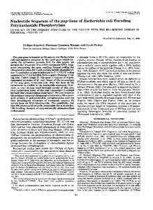
FIG. 3. Analysis of recombinant plasmids in positive clones. Minipreparations of plasmid DNA were digested with HindIll and displayed by electrophoresis in 0.6% agarose gels as described in the text. Lanes A to F contain DNA from six independent E. coli clones expressing the fimbrial subunit antigen: lanes A to C (pBA101) show a single 5.5-kb insert, lanes D and E (pBA102) show double inserts, and lane F (pBA103) shows a triple insert (see text). Lanes G and H contain DNA from two negative clones (cf. Fig. 2), lane I contains pTR262 vector DNA alone, and lane J contains HindIII-digested X phage DNA standards. The numbers on the right refer to the sizes, in kb, of the X standards. The arrowhead on the left points to the pTR262 vector DNA band.
neither subclone (pBA121:APvuII or pBA122:APvuII) was found to express the fimbrial subunit, indicating that this site 1.2 0.7 occurred within the functional gene. Another subclone span-11 5.5 ning this region, from the PvuI-PstI sites, ca. 1.25 kb to the ' '