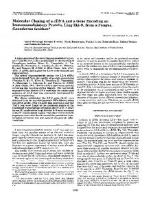
INFECTION AND IMMUNITY, Aug. 1996, p. 3134–3141 0019-9567/96/$04.0010 Copyright q 1996, American Society for Microbiology
Vol. 64, No. 8
Cloning of a DNA Fragment Encoding a Heme-Repressible Hemoglobin-Binding Outer Membrane Protein from Haemophilus influenzae HONGFAN JIN,1,2 ZHEN REN,1,2 JUDITH M. POZSGAY,3 CHRISTOPHER ELKINS,4 PAUL W. WHITBY,1 DANIEL J. MORTON,1 AND TERRENCE L. STULL1,2* Departments of Pediatrics1 and Microbiology and Immunology,2 University of Oklahoma Health Sciences Center, Oklahoma City, Oklahoma 73104; Departments of Pediatrics and Microbiology and Immunology, Medical College of Pennsylvania and Hahnemann University, Philadelphia, Pennsylvania 191293; and Department of Medicine, School of Medicine, University of North Carolina, Chapel Hill, North Carolina 275994 Received 15 March 1996/Returned for modification 30 April 1996/Accepted 27 May 1996
Haemophilus influenzae is able to use hemoglobin as a sole source of heme, and heme-repressible hemoglobin binding to the cell surface has been demonstrated. Using an affinity purification methodology, a hemoglobinbinding protein of approximately 120 kDa was isolated from H. influenzae type b strain HI689 grown in heme-restricted but not in heme-replete conditions. The isolated protein was subjected to N-terminal amino acid sequencing, and the derived amino acid sequence was used to design corresponding oligonucleotides. The oligonucleotides were used to probe a Southern blot of EcoRI-digested HI689 genomic DNA. A hybridizing band of approximately 4.2 kb was successfully cloned into pUC19. Using a 1.9-kb internal BglII fragment of the 4.2-kb clone as a probe, hybridization was seen in both typeable and nontypeable H. influenzae but not in other bacterial species tested. Following partial nucleotide sequencing of the 4.2-kb insert, a putative open reading frame was subcloned into an expression vector. The host Escherichia coli strain in which the cloned fragment was expressed bound biotinylated human hemoglobin, whereas binding of hemoglobin was not detected in E. coli with the vector alone. In conclusion, we hypothesize that the DNA fragment encoding an approximately 120-kDa heme-repressible hemoglobin-binding protein mediates one step in the acquisition of hemoglobin by H. influenzae in vivo. the circulatory system by hepatocytes (3). Hemoglobin and the hemoglobin-haptoglobin, heme-hemopexin, and heme-albumin complexes can all be utilized by H. influenzae as heme sources (48). We have recently shown that H. influenzae binds hemoglobin directly at the cell surface, possibly as an initial step in the utilization of hemoglobin-associated heme (14). We have also demonstrated that both hemoglobin binding and the binding of transferrin, an iron-binding glycoprotein which H. influenzae uses as an iron source, are repressible by heme but not by elemental iron alone (14, 34). In addition, we have noted heme-repressible hemolytic activity expressed by H. influenzae (unpublished data), which may represent an important first step in acquiring hemoglobin in vivo. The objectives of the current study were to clone and to characterize the gene(s) encoding the protein(s) mediating the binding of hemoglobin by H. influenzae.
Haemophilus influenzae is responsible for many human infections, including otitis media, meningitis, epiglottitis, and pneumonia (52). The incidence of invasive disease caused by strains with the type b capsule has been radically reduced following the introduction of vaccines based on the type b capsular polysaccharide (32, 37, 38, 43). However, vaccine failures occur, and although adults presumably have natural immunity, two thirds of H. influenzae blood isolates from adults are type b (10, 24, 46). Since currently available vaccines are based on the type b capsule, they provide no protection against disease caused by unencapsulated strains of H. influenzae, and such strains are a significant cause of otitis media in childhood, of neonatal sepsis, and of pneumonia in adults (12, 51, 53, 54). Viable vaccine candidates to provide protection against all H. influenzae disease would include surface exposed proteins which are widely distributed across the species and expressed during disease. H. influenzae has an absolute growth requirement for an exogenous source of protoporphyrin IX, which is the immediate precursor of heme (11). In vivo, all heme is intracellular, in the form of hemoglobin or heme-containing enzymes, and thus unavailable to invading microorganisms (3, 18, 26). Hemoglobin released by erythrocytes is avidly bound by the serum protein haptoglobin, and the hemoglobin-haptoglobin complex is rapidly cleared by hepatocytes (3, 40). Free heme, principally derived from the degradation of methemoglobin, is bound by the serum proteins hemopexin and albumin and cleared from
MATERIALS AND METHODS Bacterial strains and growth conditions. H. influenzae type b and serologically nontypeable strains (Table 1) were kindly provided by J. Musser, Baylor University, Houston, Tex., and have been described previously (34, 39). Strains of H. influenzae serotypes a to f were obtained from the American Type Culture Collection (ATCC). Strains of Haemophilus aphrophilus, Haemophilus parainfluenzae, Haemophilus haemolyticus, and Haemophilus parahaemolyticus were kindly provided by J. Mortensen, St. Christopher’s Hospital for Children, Philadelphia, Pa. Strains of Haemophilus segnis, Actinobacillus pleuropneumoniae, Gardnerella vaginalis, and Branhamella catarrhalis were obtained from the ATCC. Strains of Haemophilus, Actinobacillus, Gardnerella, and Branhamella spp. were routinely maintained on brain heart infusion (BHI) agar (Difco, Detroit, Mich.) supplemented with 10 mg of both hemin and b-NAD per ml. For long-term storage, strains were stored at 2708C in skim milk. For experiments in heme-replete medium, H. influenzae was grown at 378C in BHI broth (Difco) supplemented with 10 mg of b-NAD per ml and 10 mg of heme per ml (supplemented BHI; sBHI). Heme-restricted growth of H. influenzae was performed in BHI supplemented with 10 mg of b-NAD per ml and 0.1 mg of hemin per ml (hemin-
* Corresponding author. Mailing address: Department of Pediatrics, CHO 2B300, 940 N. E. 13th St., Oklahoma City, OK 73104. Phone: (405) 271-4401. Fax: (405) 271-8710. Electronic mail address:
[email protected]. 3134
HEMOGLOBIN BINDING BY HAEMOPHILUS INFLUENZAE
VOL. 64, 1996 TABLE 1. Strains and plasmids Strain or plasmid(s)
Strains H. influenzae 689 701 1371 1373 1375 1378 1384 1389 1392 1397 1403 1412 1423 1429 9006 10211 9007 9008 8143 9833 Rd E. coli DH5a BL21(DE3)pLysS JM109 Plasmids pRSETA
pHFJ1, pHFJ2 pXHGP pHFJ.19
Relevant characteristic(s)a
Reference or source
Type b, ET 22 Type b, ET 90 NT, ET 11 NT, ET 13 NT, ET 27 NT, ET 31 NT, ET 39 NT, ET 44 NT, ET 50 NT, ET 57 NT, ET 63 NT, ET 72 NT, ET 83 NT, ET 93 Type a Type b Type c Type d Type e Type f Capsule-deficient type d
39 39 39 39 39 39 39 39 39 39 39 39 39 39 ATCC ATCC ATCC ATCC ATCC ATCC
D(lacZYA-argF)U169 F2, ompT, (DE3)pLysS, Cmr
BRLb Novagen
Ampr, carrying T7 promoter, metal binding domain polylinker, F1 origin pUC19, carrying a 4.2-kb EcoRI fragment from H. influenzae pRSETA, carrying 3.2-kb PCR product derived from pHFJ2. pUC19, carrying a 1.9-kb BglII fragment from pHFJ2
Invitrogen
This work This work This work
a NT, nontypeable; ET, multilocus enzyme electrophoretic type; F2, F episome negative; Ampr, ampicillin resistance (50 mg/ml); Cmr, chloramphenicol resistance (50 mg/ml). b BRL, Bethesda Research Laboratories.
restricted BHI; hrBHI). Table 1 also shows the Escherichia coli strains which were maintained on Luria-Bertani (LB) medium and supplemented with antibiotics, as appropriate, at the indicated concentrations. Plasmids used in these studies and their relevant properties are also listed in Table 1. Biotinylation of hemoglobin. Human hemoglobin (Sigma, St. Louis, Mo.) was biotinylated as described previously (14). Hemoglobin (1 mg/ml) was dissolved in phosphate-buffered saline (PBS) (pH 7.4), and NHS-LC [sulfosuccinimidyl-6(biotinamido) hexanoate] biotin (Pierce, Rockford, Ill.) was dissolved to a concentration of 1 mg/ml in water at 508C. To 5 ml of the hemoglobin solution was added 430 ml of the biotin solution, and following incubation for 2 h at room temperature, unbound biotin was removed by passage through a Sephadex G-15 column (Pharmacia, Piscataway, N.J.). Preparation of outer membrane proteins. Outer membrane proteins were isolated by selective solubilization with Triton X-100 essentially as previously described (49). H. influenzae HI689 was grown to late logarithmic phase in sBHI or hrBHI and harvested by centrifugation at 6,000 3 g for 10 min. The cell pellets were resuspended in distilled water to an optical density at 605 nm of 0.6, and 10 ml of the suspension was sonicated (Model CL4 with microtip, set to microtip limit; Heat Systems, Farmingdale, N.Y.). Sonication was carried out in an iceNaCl bath for a total time of 2 min in 10-s bursts with 50 s between bursts. Triton X-100 was added to the sonicate at a final concentration of 2% (vol/vol), and following incubation for 5 min on ice, the mixture was sonicated as above or until the solution was clear. The sonicate was centrifuged at 35,000 3 g for 1 h at 48C, and the pellet was resuspended in 2 ml of 10 mM Tris-HCl (pH 7.4). Affinity chromatography purification of H. influenzae hemoglobin-binding protein (Hgp). Resuspended outer membranes, as described above, were divided equally into two microcentrifuge tubes and 100 ml of biotinylated hemoglobin (1 mg/ml) was added. Following incubation for 1 h with gentle agitation, mixtures
3135
were centrifuged at 13,000 3 g for 10 min. The pellet was resuspended in 1 ml of buffer I (0.75% [wt/vol] Sarkosyl, 100 mM NaCl, 100 mM EDTA, 50 mM Tris-HCl [pH 8.0]). The resuspended pellet was incubated for 1 h with 100 ml of streptavidin-agarose (diluted 1:1 in sterile distilled water; Sigma). Samples were centrifuged at 1,000 3 g for 30 s and the supernatant was carefully removed. One ml of buffer II (0.5% [wt/vol] Sarkosyl, 100 mM NaCl, 100 mM EDTA, 50 mM Tris-HCl, [pH 8.0]) was added to the beads, and following incubation for 5 min with gentle agitation, the mixture was centrifuged at 1,000 3 g and the supernatant was removed. The beads were washed twice more in buffer II and once in buffer III (100 mM NaCl, 50 mM Tris-HCl [pH 8.0]) as described above. All incubations and washes were performed at room temperature. Following the final wash the beads were resuspended in 20 ml of sodium dodecyl sulfatepolyacrylamide gel electrophoresis (SDS-PAGE) sample buffer (50 mM TrisHCl [pH 6.8], 2% [vol/vol] 2-mercaptoethanol, 10% [vol/vol] glycerol, 1% [wt/ vol] SDS, 0.01% [wt/vol] bromophenol blue) and heated in a 1008C water bath for 5 min to elute bound proteins. Eluted proteins were separated by SDS-PAGE on 6.5% acrylamide gels by using the discontinuous buffer system of Laemmli (25). Approximately 30 ml of protein preparation was loaded per lane. N-terminal amino acid sequencing. Affinity chromatography-purified proteins from heme-restricted H. influenzae HI689 were separated by SDS-PAGE on 7.5% acrylamide gels and transferred to membranes for N-terminal amino acid sequencing as described by Moos (33). Thioglycolic acid (sodium salt) (0.066% [wt/vol]) was added to the upper buffer reservoir during SDS-PAGE to remove the by-products of acrylamide polymerization (33). Proteins were transferred in a solution containing 10 mM 3-cyclohexylamino-1-propanesulfonic acid (CAPS) (pH 10.5), 1% (vol/vol) methanol, and 0.05% (wt/vol) dithiothreitol to a polyvinyldene difluoride membrane (Millipore, Bedford, Mass.) and visualized by staining with 0.1% Coomassie blue in 50% (vol/vol) methanol. The entire membrane was submitted to the UCLA Medical School Protein Microsequencing Facility, where the N-terminal amino acid sequence of the 120-kDa protein was determined. DNA isolation. Bacterial genomic DNA was isolated by standard techniques as previously described (44), or by using the DNA Now reagent (Biogentex, Seabrook, Tex.) as directed by the manufacturer. Plasmid DNA was isolated by the use of Qiagen plasmid kits (Qiagen, Chatsworth, Calif.) as directed by the manufacturer. DNA concentrations were assessed spectrophotometrically with a Shimadzu UV-1201S Spectrophotometer with a DNA/Protein program pack (Shimadzu, Kyoto, Japan). Southern blot and DNA hybridization. DNA was digested with restriction enzymes as directed by the manufacturers, separated on agarose gels (0.8% [wt/vol] agarose) in TBE buffer (0.045 M Tris-borate, 0.001 M EDTA), and transferred to Magnagraph nylon membranes (MSI, Westbrook, Mass.) by the method of Southern as described by Sambrook et al. (44). For screening of partial H. influenzae libraries in E. coli, bacterial colonies were transferred to a Magnagraph nylon membrane and prepared for hybridization as described by Sambrook et al. (44). The enhanced chemiluminescence (ECL) 39-oligolabeling system (Amersham Life Science, Arlington Heights, Ill.) was used as directed by the manufacturer to label the 39 end of the oligonucleotides. The ECL random prime labeling kit (Amersham) was used as directed by the manufacturer to label DNA probes. Labeled oligonucleotides or DNA were used to probe Southern blots or colony blots. For oligonucleotide probes, hybridization at 448C was followed by stringency washes as follows: 53 SSC (13 SSC is 8.8 g of NaCl per liter plus 4.4 g of sodium citrate per liter [pH 7.0])–0.1% (wt/vol) SDS twice for 5 min at room temperature and 13 SSC–0.1% (wt/vol) SDS twice for 15 min at 448C. For DNA probes the hybridization temperature was 608C and stringency washes were 13 SSC–0.1% (wt/vol) SDS for 15 min and 0.53 SSC–0.1% (wt/vol) SDS for 15 min both at 608C. Hybridization was detected by using ECL nucleic acid detection reagents (Amersham) as directed by the manufacturer. Blots were subsequently exposed to X-ray film (Fuji Photo Film Co., Tokyo, Japan). Cloning of the gene encoding Hgp. H. influenzae HI689 chromosomal DNA was digested with EcoRI and separated on a 0.8% (wt/vol) agarose gel. Bands in the region 4 to 6 kb were excised and purified from the gel by using the Prep-A-Gene DNA purification kit (Bio-Rad, Hercules, Calif.) as directed by the manufacturer. The isolated DNA fragments were ligated by standard techniques to dephosphorylated EcoRI-digested pUC19 (44). The ligation mixture was transformed into competent E. coli DH5a, and transformants were selected on LB plates containing 50 mg of ampicillin per ml. Ampicillin-resistant colonies were subcultured on selective media and colonies were probed with the Nterminal oligonucleotides to identify positive clones. Putative positive plasmids were isolated and confirmed by Southern blot and hybridization with the oligonucleotide probes. Partial automated sequencing (ABI model 373A; Recombinant DNA/Protein Resource Facility, Oklahoma State University, Stillwater) of a positive clone allowed us to identify a putative leader sequence and start codon and also a presumed C-terminal sequence showing significant homology with other bacterial iron- and heme-related proteins at the amino acid level. The length of DNA flanked by the proposed start codon and C-terminal sequence would account for a protein of approximately 120 kDa. Primers were designed for use in PCR to allow for cloning of the putative coding region into a controlled expression vector. Primers were synthesized (Molecular Biology Resource Facility, University of Oklahoma Health Sciences Center, Oklahoma City) with the sequences
3136
JIN ET AL.
INFECT. IMMUN.
59-GACCAGGGATCCATGACCAATTTTAG-39 and 59-GGAAGGGGTACC CTAGAATTCAAACTG-39. A BamHI site upstream of the start codon and a KpnI site downstream of the stop codon were included in the primers to allow for directional cloning of the PCR product in the vector pRSETA in the correct reading frame. PCR was performed in 50-ml reactions with 30 ng of pHFJ2, linearized by digestion with HindIII, as template. PCRs contained 2 mM MgCl2, 200 mM each deoxynucleoside triphosphate, 100 ng of each primer and 2 U of Taq DNA polymerase. PCR was carried out for 30 cycles with each cycle consisting of denaturation at 958C for 1 min, annealing at 558C for 1 min, and primer extension at 728C for 3.5 min, with a final extension time of 10 min. Amplicons of the expected size (3.2 kb) were gel purified and mapped by digestion with BglII. The PCR products were digested with BamHI and KpnI, and ligated to gel-purified BamHI- and KpnI-digested pRSETA. The ligation mixture was transformed into E. coli BL21(DE3)pLysS and recombinants were selected on LB agar containing 50 mg of ampicillin per ml. Plasmids were isolated from ampicillin-resistant colonies and mapped by restriction enzyme digestion to identify clones containing the expected product. A positive clone was identified and designated pXHGP. Expression of Hgp in E. coli. A hemoglobin-binding dot blot assay was used to determine whether E. coli containing pXHGP bound hemoglobin. Recombinant E. coli BL21(DE3)pLysS was grown to mid-logarithmic phase in LB medium with appropriate antibiotic supplementation. After induction with 1 mM isopropyl b-D-thiogalactopyranoside (IPTG) for 4 h, cells were harvested and resuspended in PBS to a concentration of 108 CFU/ml. In a dot blot manifold (Bio-Rad), 100 ml of cell suspension (107 CFU) was filtered onto nitrocellulose membranes. Membranes were air dried for 15 min and then incubated in 10% (wt/vol) skim milk in PBS for 1 h. Membranes were incubated in 500 ng of biotinylated human hemoglobin per ml in PBS for 1 h, and then washed three times for 10 min each in PBS. A second blocking step in 10% (wt/vol) skim milk in PBS was performed for 1 h; this was followed by incubation in 25 ng of streptavidin-horseradish peroxidase conjugate (Jackson Immunoresearch, West Grove, Pa.) per ml in PBS for 1 h. Membranes were washed three times for 10 min each in PBS and developed with ECL Western blot (immunoblot) detection reagents (Amersham) as directed by the manufacturer. All steps were performed at room temperature. In some experiments bacteria were harvested and resuspended to 108 CFU/ml and 5-ml samples were sonicated (Model CL4 with microtip, set to microtip limit; Heat Systems). Sonication was carried out in an ice-NaCl bath for a total time of 2 min in 10-s bursts with 50 s between bursts. Sonicates (100 ml) were filtered onto nitrocellulose membranes in a dot blot manifold and the membranes were probed and developed as above. Western immunoblot analysis. The 100-kDa hemoglobin-binding protein of Haemophilus ducreyi 35000 was affinity purified as previously described (9). Recombinant protein from IPTG-induced BL21(DE3)pLysS harboring pXHGP was purified by using the Xpress protein purification system (Invitrogen) as directed by the manufacturer. These proteins and the affinity-purified 120-kDa protein from H. influenzae HI689 were separated by SDS-PAGE on 7.5% acrylamide gels and electrophoretically transferred to nitrocellulose membranes. Membranes were blocked in 1% (wt/vol) skim milk in PBS for 1 h and then probed with a 1:1,000 dilution in PBS containing 1% (wt/vol) skim milk of an antibody raised against the 100-kDa HgbA of H. ducreyi in rabbits (9). Following four washes in PBS for 15 min each, membranes were probed with 1:10,000 donkey anti-rabbit immunoglobulin-horseradish peroxidase conjugate (Amersham) in PBS with 1% (wt/vol) skim milk. Membranes were washed four times for 15 min each in PBS and developed by using ECL detection reagents as directed by the manufacturer.
RESULTS Affinity purification of Hgp. H. influenzae is heme dependent and utilizes hemoglobin as a heme source in vitro (48). Using a whole cell dot blot assay, we have previously shown that hemoglobin binds to the H. influenzae cell surface in a manner indicative of a specific receptor, and we have demonstrated that binding is induced by limiting heme levels in the growth medium (14). To further investigate the possibility that hemoglobin binding to H. influenzae is mediated through an outer membrane protein regulated by heme levels, we subjected resuspended outer membrane proteins of strain HI689 to affinity purification with biotinylated hemoglobin. A 120-kDa protein was isolated from outer membranes derived from strain HI689 grown under heme-restricted conditions (hrBHI) (Fig. 1, lane C) but not from outer membrane proteins derived from HI689 grown under heme-replete conditions (sBHI) (Fig. 1, lane B). N-terminal amino acid sequencing. An N-terminal amino acid sequence of the affinity-purified Hgp was obtained from affinity-purified outer membranes of H. influenzae HI689. The
FIG. 1. Identification of the 120-kDa Hgp in H. influenzae. An SDS-PAGE (6.5% acrylamide) gel stained with Coomassie blue is shown. Lane A, molecular mass marker; lane B, affinity-purified outer membrane protein from H. influenzae type b grown in sBHI; lane C, affinity-purified outer membrane protein from H. influenzae type b grown in hrBHI. Numbers at left are molecular masses.
determined N-terminal amino acid sequence was AQPTNQPT NQ. The synthetic oligonucleotides 59-CAACCAACTAATCA ACCAAC-39 and 59-CAGCCTACAAATCAACCAAC-39, synthesized by Ransom Hill Bioscience, Inc., Ramona, Calif., were designed on the basis of the N-terminal amino acid sequence of the 120-kDa Hgp after consulting the H. influenzae codon preference data of Gilsdorf et al. (15). These two oligonucleotides were used in a mixture to probe Southern blots of H. influenzae HI689 genomic DNA digests. The oligonucleotides hybridized to an approximately 4.2-kb EcoRI fragment (Fig. 2, lane B) which was deemed of appropriate size for cloning. No hybridization of the oligonucleotides to either E. coli chromosomal DNA (data not shown) or pUC19 (Fig. 2, lane C) was detected, suggesting that the oligonucleotides could be used to identify recombinant clones containing the structural gene encoding the 120-kDa protein. Cloning of the DNA fragment encoding Hgp. A limited genomic library of H. influenzae HI689 was constructed in the vector pUC19 as described in Materials and Methods. Two recombinant clones were isolated from this library by using the N-terminal amino acid sequence-derived oligonucleotides as probes. Both clones contained inserts of approximately 4.2 kb which hybridized in Southern analyses with the oligonucleotide probes (Fig. 2, lanes D and F). These plasmids were designated pHFJ1 and pHFJ2. Partial mapping of the two clones revealed them to contain the same fragment in reverse orientation (Fig. 3). From preliminary sequencing analysis, the N-terminal nucleotide sequence and a putative C-terminal sequence flanking a region of the correct size to encode a 120-kDa protein were identified. The N-terminal and C-terminal regions showed significant homology at the amino acid level with other bacterial iron- and heme-related proteins (Fig. 4 and 5), and a predicted leader peptide of 23 amino acids was identified (Fig. 4). On the basis of the sequence data, primers were designed to amplify the entire putative coding region by PCR. Restriction enzyme sites were included in the primers to permit directional cloning in the expression vector pRSETA. By using pHFJ2 as the template in the PCR, an approximately 3.2-kb product was
VOL. 64, 1996
HEMOGLOBIN BINDING BY HAEMOPHILUS INFLUENZAE
3137
FIG. 4. Peptide alignments between the N-terminal region of the putative Hgp and the N-terminal regions of the following iron- and heme-related proteins: HgbA, a hemoglobin-binding protein of H. ducreyi (9); HmbR, a hemoglobin-binding protein of N. meningitidis (47); Tbp1-Hi, a transferrin-binding protein of H. influenzae (16); and Tbp1-Nm, a transferrin-binding protein of N. meningitidis (29). The 2 indicates the proposed leader peptide cleavage site of Hgp.
FIG. 2. Southern blot probed with ECL-labeled oligonucleotide specific for the 120-kDa Hgp N terminus and developed with ECL detection reagents as directed by the manufacturer (Amersham). Lane A, labeled l HindIII fragments; lane B, H. influenzae HI689 chromosomal DNA EcoRI digest; lane C, EcoRIdigested pUC19; lane D, EcoRI-digested pHFJ1; lane E, undigested pHFJ1; lane F, EcoRI-digested pHFJ2; lane G, undigested pHFJ2. Arrow indicates 4.2-kb hybridizing bands.
amplified; restriction analysis of the amplicon revealed it to be the expected product. The gel-purified PCR product was successfully cloned into pRSETA, as demonstrated by restriction mapping (Fig. 3), and the resulting plasmid was designated pXHGP. Expression of Hgp in E. coli. A hemoglobin-binding dot blot assay was used to investigate whether the cloned hemoglobinbinding protein gene in E. coli resulted in expression of a hemoglobin-binding phenotype. Sonicated E. coli BL21(DE3)pLysS harboring pXHGP bound biotinylated human hemoglobin following induction with IPTG (Fig. 6, row D). Whole cells of IPTG-induced E. coli BL21(DE3)pLysS harboring pXHGP did not bind hemoglobin (Fig. 6, row B), probably because the recombinant protein is not expressed at the E. coli cell surface. E. coli BL21(DE3)pLysS harboring pRSETA alone and induced with IPTG did not bind hemoglobin, whether or not the cells were lysed by sonication (Fig. 6, rows A and C), and all tested strains were unable to bind hemoglobin when uninduced (data not shown). These data demonstrate that pXHGP contained a fragment of DNA from H. influenzae HI689 which encodes the protein Hgp, which is expressed in E. coli.
FIG. 3. Partial restriction enzyme maps of the 4.2-kb insert of pHFJ2, the predicted ORF encoding Hgp, and pHFJ.19. The boxed area represents the putative ORF encoding Hgp and the insert of pXHGP. Numbers are sizes in kilobase pairs.
Distribution of hgp. Samples of genomic DNA (1 mg) from a number of H. influenzae strains and other bacterial species were digested with EcoRI, separated on agarose gels, blotted to nylon membranes, and probed with a labeled 1.9-kb BglII fragment internal to the putative hgb gene (Fig. 3). The labeled insert hybridized strongly with a band of approximately 4.2 kb in H. influenzae strains of types a, b, and c, and in seven nontypeable strains (Table 2). The labeled insert also hybridized with H. influenzae strains of types d, e, and f and also with five nontypeable strains, although the hybridization pattern was variable (Table 2). No hybridization was seen with either H. influenzae Rd or any other tested species (Table 2). These data indicate that the 120-kDa Hgp may be widely distributed across the species H. influenzae. Lack of cross-reactivity between HgbA of H. ducreyi and Hgp of H. influenzae. A conserved 100-kDa hemoglobin-binding protein from H. ducreyi, HgbA, has been recently identified by Elkins (8, 9). To determine if antibodies generated against HgbA would recognize the 120-kDa Hgp described in this paper, Western blots were probed with antibodies raised against the 100-kDa hemoglobin-binding protein of H. ducreyi. The antibodies reacted with the 100-kDa protein isolated from H. ducreyi but not with either the 120-kDa protein isolated from H. influenzae or the recombinant protein isolated from IPTG-induced E. coli (data not shown).
FIG. 5. Peptide alignments between the C-terminal region of the putative Hgp and the C-terminal regions of the following iron- and heme-related proteins: HgpA, a hemoglobin-binding protein of H. ducreyi (9); HmbR, a hemoglobin-binding protein of N. meningitidis (47); Tbp1-Hi, a transferrin-binding protein of H. influenzae (16); and Tbp1-Nm, a transferrin-binding protein of N. meningitidis (29).
3138
JIN ET AL.
INFECT. IMMUN.
FIG. 6. Dot blot analysis of human hemoglobin binding to E. coli either harboring or without pXHGP. Columns 1, 2, and 3 represent serial dilutions of whole cells or sonicated cells (see Materials and Methods). Row A, whole cells of E. coli BL21(DE3)pLysS pRSETA; row B, whole cells of E. coli BL21(DE3)pLysS pXHGP; row C, sonicates of E. coli BL21(DE3)pLysS pRSETA; row D, sonicates of E. coli BL21(DE3)pLysS pXHGP.
with significant homology to a periplasmic transport protein of E. coli has also been characterized (19, 21). In this report we identify the gene encoding a 120-kDa protein of H. influenzae type b. Hgp was isolated from H. influenzae by using affinity chromatography and an N-terminal amino acid sequence was obtained (AQPTNQPTNQ). By using oligonucleotides derived from the N-terminal sequence as probes, a DNA fragment from H. influenzae was cloned to yield pHFJ2. Partial sequencing of pHFJ2 revealed that the N-terminal nucleotide sequence would encode a peptide of sequence AEPTNQPTNQ. On the basis of sequence homology and Southern analysis, there is no gene corresponding to hgp present in the Rd chromosomal sequence recently reported. Although pHFJ2 contains a region of greater than 95% identity to the arcB locus of H. influenzae upstream of hgp, there is no sequence homologous to hgp downstream of the arcB locus in the Rd chromosomal sequence (13). However, the nucleotide sequence encoding the N-terminal region of Hgp is highly homologous with the N-terminal regions of the putative products of 3 separate open reading frames (ORFs) in the Rd chromosomal sequence (13). Each of these areas in the Rd sequence (designated HI0661, HI0712, and HI1566 by Fleischmann et al. [13]) and the corresponding region of hgp contains multiple ccaa repeats, with the number of repeats varying between 18 and 36, giving rise to proteins with 6 to 12 PTNQ repeats. In two of the three putative ORFs in Rd, the
DISCUSSION TABLE 2. Hybridization with the 1.9-kb insert of pHFJ.19
H. influenzae has an absolute growth requirement for protoporphyrin IX, the immediate precursor of heme (11). The availability of heme within the human host to invading pathogens is strictly limited. Heme is contained largely within intracellular hemoglobin; free hemoglobin and heme are rapidly bound by the serum proteins haptoglobin (in the case of hemoglobin) and hemopexin or albumin (in the case of heme) and cleared from the circulation (3). The heme requirement of H. influenzae can be satisfied in vitro by hemoglobin, hemoglobin complexed to haptoglobin, heme complexed to either hemopexin or albumin, or protoporphyrin IX in the presence of an iron source such as ferritransferrin (3, 35, 36). The mechanism(s) by which H. influenzae takes up iron and/or heme from these protein sources has not been fully elucidated. It is clear, however, that H. influenzae does not produce siderophores (7, 35, 36, 41). Certain uptake mechanisms involve a direct interaction between the protein and the bacterial cell surface (14, 36, 55). The utilization of heme and the acquisition of heme from hemoglobin, the hemoglobin-haptoglobin complex, and the heme-hemopexin complex are dependent on a functional tonB gene, indicating that uptake is mediated by an outer membrane TonB-dependent protein(s) (23, 42); a tonB homolog has been reported in the recently sequenced genome of H. influenzae Rd (13). On the basis of the sequence currently available, Hgb exhibits significant homology with other TonB proteins over regions that are highly conserved among this class of proteins (6). In the case of acquisition of iron from transferrin, two outer membrane proteins, Tbp1 and Tbp2, are involved in binding of transferrin to H. influenzae (16). Subsequent steps in the process have not been fully elucidated, although there is evidence for a periplasmic iron transport system encoded by an operon of three genes hitABC (1). Proteins binding the heme-hemopexin complex have been described, one of which is apparently secreted into the growth medium (4, 5, 20, 56). A heme-binding outer membrane protein has been isolated (27), and a heme-binding lipoprotein
Strain and/or typea
H. influenzae ATCC 9006, type a ATCC 10211, type b ATCC 9007, type c ATCC 9008, type d ATCC 8143, type e ATCC 9833, type f 701, type b 1371, NT 1373, NT 1375, NT 1378, NT 1384, NT 1389, NT 1392, NT 1397, NT 1403, NT 1412, NT 1423, NT 1429, NT Rd H. parainfluenzae 203 H. aphrophilus ATCC 33389 H. parahaemolyticus 329 H. haemolyticus 329 125 H. haemolyticus 97 H. segnis ATCC 33393 A. pleuropneumoniae ATCC 27088 ATCC 27089 G. vaginalis ATCC 14018 B. catarrhalis ATCC 8176 E. coli JM109 a b
NT, nontypeable. NA, not applicable.
Hybridization
Band size
1 1 1 1 1 1 1 1 1 1 1 1 1 1 1 1 1 1 1 2 2 2 2
4.2 4.2 4.2 .10 6.4 9.0 6.4 4.2 4.2 6.4 .10 4.2 4.2 4.2 6.4 9.0 4.2 .10 4.2 NAb NA NA NA
2 2 2 2
NA NA NA NA
2 2 2 2 2
NA NA NA NA NA
VOL. 64, 1996
HEMOGLOBIN BINDING BY HAEMOPHILUS INFLUENZAE
ccaa repeat is followed by an in-frame stop codon, while the third constitutes an ORF of approximately 3 kb. The function of the repeat region is not known, although since conservation is 100% at the nucleotide level, it seems likely that its importance lies in the ccaa unit rather than in the encoded peptide sequence. It is possible that the ccaa repeats may have a regulatory function, perhaps in a manner analogous to the slipstrand regulation involved in phase variation of lipopolysaccharide expression in H. influenzae (50). In the two ORFs identified in Rd which have a stop codon downstream of the ccaa region, slippage across one ccaa unit would eliminate the stop codon. The N-terminal and C-terminal amino acid sequences of Hgp have been compared with the corresponding amino acid regions of the proteins encoded by HI0661, HI0712, and HI1566. To facilitate this, the reading frames of HI0661 and HI1566 were altered by addition or removal of a ccaa unit from the published nucleotide sequence as appropriate. Over the 76 amino acids directly subsequent to the final PTNQ unit of Hgp, identity was calculated as 80% with HI0661, 70% with HI0712, and 72% with HI1566. Over the 90 amino acids at the C-terminal of Hgp, the respective identities were 73% (HI0661), 63% (HI0712), and 62% (HI1566). These analyses indicate that the protein encoded by ORF HI0661 is the most closely related to Hgp. The ORF HI0661 does not correspond to hgp since we have partially cloned a homolog of HI0661 from H. influenzae HI689 (data not shown). In addition we have partially cloned HI0712 from HI689 (data not shown). The discrepancy between the microsequencing-derived Nterminal sequence (AQPTNQPTNQ) and the nucleotide sequence-derived N-terminal sequence (AEPTNQPTNQ) is unlikely to be due to microsequencing errors since the misread of a glutamine (Q) for a glutamic acid (E) is highly unlikely (13a, 24a). The possibility of nucleotide sequencing errors was minimized by repeated sequencing across the area of interest. An alternative explanation for the difference is that the protein originally isolated by affinity chromatography was not Hgp, but rather one of the three ORF products identified in H. influenzae Rd. In particular, the product of HI0712 has a predicted molecular mass of 124 kDa, compared with approximately 120 kDa for Hgp, and the nucleotide sequence-derived N-terminal amino acid sequence of HI0712 is AQPTNQPTN. The product of HI0661 has a predicted molecular mass of 115 kDa and an N-terminal amino acid sequence of AQPTNQPTN, while the data for the HI1566 gene product would be 114 kDa and AEPTNQPTN, respectively. Thus, it is possible that an alternate gene product was originally isolated (i.e., ORF HI0712 or ORF HI0661), while the oligonucleotides designed from the amino acid sequence led to cloning of hgp on the basis of the 100% conservation of the ccaa region. This possibility indicates that there is more than one hemoglobin-binding protein expressed by H. influenzae, and that one (or more) of HI0661, HI0712, or HI1566 may represent this additional protein(s). Work is under way to clarify how many hemoglobin-binding proteins H. influenzae expresses and to define the functions of the gene products of HI0661, HI0712, and HI1566. A putative coding sequence from pHFJ2 was subcloned into E. coli, and expression of this gene resulted in the recombinant strain binding hemoglobin. Hemoglobin-binding proteins have been reported in H. ducreyi and Neisseria meningitidis (8, 28, 30, 47). Using affinity chromatography, Elkins isolated a protein of 100 kDa from H. ducreyi, and also reported the isolation of an approximately 115-kDa protein from H. influenzae DL42 (8, 9). The 115-kDa protein from H. influenzae was not recognized by antibodies raised to either the entire H. ducreyi 100kDa protein or to the N-terminal peptide of the 100-kDa protein. In addition, the N-terminal sequence reported for the
3139
H. ducreyi protein (ESNMQTEKLETIVV) is highly dissimilar to the N-terminal of the 120-kDa protein reported here (AEPTNQPTNQ). Neither the recombinant Hgp nor the wildtype Hgp reacts with an antibody raised against the 100-kDa protein of H. ducreyi; however, it remains to be clarified whether Hgp is related to the 115-kDa protein isolated from H. influenzae DL42 (9). Two hemoglobin-binding proteins have been described in N. meningitidis, one of 85 kDa (30) and a second of 89.5 kDa (47); the former binds the hemoglobinhaptoglobin complex. These two proteins were isolated from different strains and it is not known whether both proteins are expressed in the same strain or whether they interact with each other in the acquisition of heme from hemoglobin, although Lee and Hill have suggested that meningococcus possesses two hemoglobin receptors with different affinities for the ligand (28). In the case of transferrin binding by H. influenzae and N. gonorrhoeae, it is clear that expression of both Tbp1 and Tbp2 is necessary for maximal binding of the ligand (2, 16), although partial binding occurs when either protein is present alone. There is no direct evidence to date for the existence of more than one hemoglobin-binding protein in either H. influenzae or H. ducreyi. In previous studies (14), we showed that binding of hemoglobin by H. influenzae was blocked by the hemoglobinhaptoglobin complex. It is possible that, similarly to the 85-kDa N. meningitidis protein (30), Hgp also binds the hemoglobinhaptoglobin complex, and studies will be performed to answer this question. Hgp was isolated from heme-restricted but not from hemereplete H. influenzae, indicating that expression of the protein may be regulated by levels of heme. Previously, we have shown that the hemoglobin-binding phenotype in H. influenzae is repressible by heme (14) and that transferrin binding expressed by H. influenzae is repressible by heme as opposed to elemental iron levels (34). The 100-kDa hemoglobin-binding protein of H. ducreyi and the 39.5-kDa heme-binding outer membrane protein of H. influenzae are similarly repressible by heme, rather than by elemental iron (8, 9, 27). In many bacterial species the Fur (ferric uptake regulator) protein is a regulatory element which in the presence of iron binds to a conserved binding sequence (the Fur box) upstream of iron-regulated genes, preventing transcription (31). Sequences homologous to the consensus Fur binding sequence (31) have been reported upstream of the H. ducreyi hemoglobin-binding protein (9), the H. influenzae transferrin-binding proteins (16), and a hemehemopexin utilization gene cluster of H. influenzae (5). In none of these cases has the putative Fur binding site been shown to be functional; indeed the 100-kDa heme-hemopexin-binding protein described by Cope et al. appears to be expressed constitutively (5). That this protein is expressed in a medium which is sufficient in heme and iron, despite a putative Fur box (5), raises further questions regarding the functionality of this putative Fur box. Fleischmann et al. have recently identified an ORF with strong homology (61.4% identity; 75% similarity) to the E. coli fur gene in the genome of H. influenzae Rd (13, 45). Thus, a Fur analog may be a regulatory factor for expression of the H. influenzae iron- and heme-related proteins, although it is clear that clarification is necessary. In addition, the 57-kDa heme-hemopexin-binding protein identified by Wong et al. is apparently repressible by iron (55, 56), although the hemehemopexin phenotype is variously reported as being inducible by iron-restricted growth or heme-restricted anaerobic growth (21, 55). The regulation of heme- and iron-related proteins in H. influenzae remains poorly characterized, and further studies should be undertaken to clarify the roles of iron and heme in regulating their expression. The gene encoding the 120-kDa Hgp appears to be widely
3140
JIN ET AL.
INFECT. IMMUN.
distributed among the species of H. influenzae. Of 19 H. influenzae strains tested, 10 had a strongly hybridizing band at approximately 4.2 kb. Eight of the remaining nine strains exhibited varying hybridization patterns (Table 2), while strain Rd did not hybridize at all. No other tested species hybridized with the 1.9-kb probe. H. influenzae type b 701 represents a highly divergent type b strain (39), previously reported to lack expression of hemoglobin binding (14). The hybridizing band in this strain was approximately 6.4 kb, and this may represent the gene encoding Hgp or, alternatively, a related protein of different function. A number of H. influenzae strains, including strain 701, that have been reported to lack transferrin binding activity (22, 34) were subsequently shown to both possess the genes encoding Tbp1 and Tbp2 and to express transferrin binding under different growth conditions (17). It is possible that strain 701 would similarly express hemoglobin binding if grown under appropriate conditions. The failure of hgb to hybridize with H. haemolyticus is interesting since we have shown hemoglobin binding to this organism (14), and it is thus likely that H. haemolyticus strains possess an unrelated hemoglobin-binding protein. In conclusion we have identified a 120-kDa Hgp of H. influenzae, the expression of which is repressible by heme, and we have cloned the gene encoding Hgp. We speculate that Hgp represents an important step in the acquisition of heme from hemoglobin. Further studies will clarify the role of Hgp in hemoglobin utilization, investigate the possible existence of other hemoglobin-binding proteins, and define the mechanism of regulation of this protein(s). ACKNOWLEDGMENTS This work was supported by Public Health Service grant AI29611 from the National Institute of Allergy and Infectious Disease. We thank Audree Fowler of the UCLA School of Medicine Protein Microsequencing Facility for N-terminal amino acid sequencing and for helpful comments. We thank John Keyte of the University of Nottingham Polymer Biosynthesis and Analysis Unit for helpful comments. REFERENCES 1. Adhikari, P., S. D. Kirby, A. J. Nowalk, K. L. Veraldi, A. B. Schryvers, and T. A. Mietzner. 1995. Biochemical characterization of a Haemophilus influenzae periplasmic iron transport operon. J. Biol. Chem. 270:25142–25149. 2. Anderson, J. E., P. F. Sparling, and C. N. Cornelissen. 1994. Gonococcal transferrin-binding protein 2 facilitates but is not essential for transferrin utilization. J. Bacteriol. 176:3162–3170. 3. Bezkorovainy, A. 1987. Iron proteins, p. 27–68. In J. J. Bullen and E. Griffiths (ed.), Iron and infection: molecular, physiological and clinical aspects. John Wiley & Sons, New York. 4. Cope, L. D., S. E. Thomas, J. L. Latimer, C. A. Slaughter, U. MullerEberhard, and E. J. Hansen. 1994. The 100 kDa haem:haemopexin-binding protein of Haemophilus influenzae: structure and localization. Mol. Microbiol. 13:863–873. 5. Cope, L. D., R. Yogev, U. Muller-Eberhard, and E. J. Hansen. 1995. A gene cluster involved in the utilization of both free heme and heme:hemopexin by Haemophilus influenzae type b. J. Bacteriol. 177:2644–2653. 6. Cornelissen, C. N., G. D. Biswas, J. Tsai, D. K. Paruchuri, S. A. Thompson, and P. F. Sparling. 1992. Gonococcal transferrin-binding protein 1 is required for transferrin utilization and is homologous to TonB-dependent outer membrane receptors. J. Bacteriol. 174:5788–5797. 7. Cornelissen, C. N., and P. F. Sparling. 1994. Iron piracy: acquisition of transferrin-bound iron by bacterial pathogens. Mol. Microbiol. 14:843–850. 8. Elkins, C. 1995. Identification and purification of a conserved heme-regulated hemoglobin-binding outer membrane protein from Haemophilus ducreyi. Infect. Immun. 63:1241–1245. 9. Elkins, C., C. J. Chen, and C. E. Thomas. 1995. Characterization of the hgbA locus encoding a hemoglobin receptor from Haemophilus ducreyi. Infect. Immun. 63:2194–2200. 10. Eskola, J., H. Kayhty, H. Peltola, V. Karanko, P. H. Makela, J. Samuelson, and L. K. Gordon. 1985. Antibody levels achieved in infants by course of Haemophilus influenzae type b polysaccharide/diphtheria toxoid conjugate vaccine. Lancet i:1184–1186.
11. Evans, N. M., D. D. Smith, and A. J. Wicken. 1974. Haemin and nicotinamide adenine dinucleotide requirements of Haemophilus influenzae and Haemophilus parainfluenzae. J. Med. Microbiol. 7:359–365. 12. Falla, T. J., S. R. Dobson, D. W. Crook, W. A. Kraak, W. W. Nichols, E. C. Anderson, J. Z. Jordens, M. P. Slack, E. Mayon-White, and E. R. Moxon. 1993. Population-based study of non-typable Haemophilus influenzae invasive disease in children and neonates. Lancet 341:851–854. 13. Fleischmann, R. D., M. D. Adams, O. White, R. A. Clayton, E. F. Kirkness, A. R. Kerlavage, C. J. Bult, J. Tomb, B. A. Dougherty, J. M. Merrick, K. McKenney, G. Sutton, W. FitzHugh, C. Fields, J. D. Gocayne, J. Scott, R. Shirley, L. Liu, A. Glodek, J. M. Kelley, J. F. Weidman, C. A. Phillips, T. Spriggs, E. Hedblom, M. D. Cotton, R. C. Utterback, M. C. Hanna, D. T. Nguyen, D. M. Saudek, R. C. Brandon, L. D. Fine, J. L. Fritchman, J. L. Fuhrmann, N. S. M. Geoghagen, C. L. Gnehm, L. A. McDonald, K. V. Small, C. M. Fraser, H. O. Smith, and J. C. Venter. 1995. Whole-genome random sequencing and assembly of Haemophilus influenzae Rd. Science. 269:496– 512. 13a.Fowler, A. Personal communication. 14. Frangipane, M. E., D. J. Morton, J. A. Wooten, J. M. Pozsgay, and T. L. Stull. 1994. Binding of human hemoglobin by Haemophilus influenzae. FEMS Microbiol. Lett. 118:243–248. 15. Gilsdorf, J. R., K. McCrea, and L. Forney. 1990. Conserved and nonconserved epitopes among Haemophilus influenzae type b pili. Infect. Immun. 58:2252–2257. 16. Gray-Owen, S. D., S. Loosmore, and A. B. Schryvers. 1995. Identification and characterization of genes encoding the human transferrin-binding proteins from Haemophilus influenzae. Infect. Immun. 63:1201–1210. 17. Gray-Owen, S. D., and A. B. Schryvers. 1995. Characterization of transferrinbinding proteins 1 and 2 in invasive type b and nontypeable strains of Haemophilus influenzae. Infect. Immun. 63:3809–3815. 18. Griffiths, E. 1987. Iron in biological systems, p. 1–26. In J. J. Bullen and E. Griffiths (ed.), Iron and infection: molecular, physiological and clinical aspects. John Wiley & Sons, New York. 19. Hanson, M. S., and E. J. Hansen. 1991. Molecular cloning, partial purification, and characterization of a haemin-binding lipoprotein from Haemophilus influenzae type b. Mol. Microbiol. 5:267–278. 20. Hanson, M. S., S. E. Pelzel, J. Latimer, U. Muller-Eberhard, and E. J. Hansen. 1992. Identification of a genetic locus of Haemophilus influenzae type b necessary for the binding and utilization of heme bound to human hemopexin. Proc. Natl. Acad. Sci. USA. 89:1973–1977. 21. Hanson, M. S., C. Slaughter, and E. J. Hansen. 1992. The hbpA gene of Haemophilus influenzae type b encodes a heme-binding lipoprotein conserved among heme-dependent Haemophilus species. Infect. Immun. 60: 2257–2266. 22. Hardie, K. R., R. A. Adams, and K. J. Towner. 1993. Transferrin-binding ability of invasive and commensal isolates of Haemophilus spp. J. Med. Microbiol. 39:218–224. 23. Jarosik, G. P., J. D. Sanders, L. D. Cope, U. Muller-Eberhard, and E. J. Hansen. 1994. A functional tonB gene is required for both utilization of heme and virulence expression by Haemophilus influenzae type b. Infect. Immun. 62:2470–2477. 24. Kostman, J. R., B. L. Sherry, C. L. Flinger, S. Egaas, P. Sheeran, L. Baken, J. E. Bauwens, C. Clausen, D. M. Sherer, J. J. Plorde, T. L. Stull, and P. M. Mendelmen. 1993. Invasive Haemophilus influenzae infections in older children and adults in Seattle. Clin. Infect. Dis. 17:389–396. 24a.Keyte, J. Personal communication. 25. Laemmli, U. K. 1970. Cleavage of structural proteins during the assembly of the head of bacteriophage T4. Nature (London) 227:680–685. 26. Lee, B. C. 1992. Isolation of an outer membrane hemin-binding protein of Haemophilus influenzae type b. Infect. Immun. 60:810–816. 27. Lee, B. C. 1995. Quelling the red menace: haem capture by bacteria. Mol. Microbiol. 18:383–390. 28. Lee, B. C., and P. Hill. 1992. Identification of an outer-membrane haemoglobin-binding protein in Neisseria meningitidis. J. Gen. Microbiol. 138:2647– 2656. 29. Legrain, M., V. Mazarin, S. W. Irwin, B. Bouchon, M. J. Quentin-Millet, E. Jacobs, and A. B. Schryvers. 1993. Cloning and characterization of Neisseria meningitidis genes encoding the transferrin-binding proteins Tbp1 and Tbp2. Gene 130:73–80. 30. Lewis, L. A., and D. W. Dyer. 1995. Identification of an iron-regulated outer membrane protein of Neisseria meningitidis involved in the utilization of hemoglobin complexed to haptoglobin. J. Bacteriol. 177:1299–1306. 31. Litwin, C. M., and S. B. Calderwood. 1993. Role of iron in regulation of virulence genes. Clin. Microbiol. Rev. 6:137–149. 32. Madorne, D. V., C. L. Johnson, D. C. Phipps, Pennridge Pediatric Associates, L. A. Popejoy, R. Eby, and D. H. Smith. 1990. Safety and immunologic response to Haemophilus influenzae type b oligosaccharide-CRM197 conjugate vaccine in 1 to 6-month-old infants. Pediatrics 85:331–337. 33. Moos, M. 1993. Isolation of proteins for microsequence analysis, p. 8.71– 8.712. In J. E. Coligan, A. M. Kruisbeek, D. H. Margulies, E. M. Shevach, and W. Strober (ed.), Current protocols in immunology. John Wiley & Sons, New York.
VOL. 64, 1996
HEMOGLOBIN BINDING BY HAEMOPHILUS INFLUENZAE
34. Morton, D. J., J. M. Musser, and T. L. Stull. 1993. Expression of the Haemophilus influenzae transferrin receptor is repressible by hemin but not elemental iron alone. Infect. Immun. 61:4033–4037. 35. Morton, D. J., and P. Williams. 1989. Utilization of transferrin-bound iron by Haemophilus species of human and porcine origins. FEMS Microbiol. Lett. 53:123–127. 36. Morton, D. J., and P. Williams. 1990. Siderophore-independent acquisition of transferrin-bound iron by Haemophilus influenzae type b. J. Gen. Microbiol. 136:927–933. 37. Moxon, E. R., and K. A. Vaughan. 1981. Type b capsular polysaccharide as a virulence determinant of Haemophilus influenzae: studies using clinical isolates and laboratory transformants. J. Infect. Dis. 143:517–524. 38. Murphy, T. V., K. E. White, and P. Pastor. 1993. Declining incidence of Haemophilus influenzae type b disease since introduction of vaccination. JAMA 269:246–248. 39. Musser, J. M., S. J. Barenkamp, D. M. Granoff, and R. K. Selander. 1986. Genetic relationships of serologically nontypable and serotype b strains of Haemophilus influenzae. Infect. Immun. 52:183–191. 40. Otto, B. R., A. M. Verweij-van Vught, and D. M. MacLaren. 1992. Transferrins and heme-compounds as iron sources for pathogenic bacteria. Crit. Rev. Microbiol. 18:217–233. 41. Pidcock, K. A., J. A. Wooten, B. A. Daley, and T. L. Stull. 1988. Iron acquisition by Haemophilus influenzae. Infect. Immun. 56:721–725. 42. Postle, K. 1990. TonB and the gram-negative dilemma. Mol. Microbiol. 4:2019–2025. 43. Roberts, M., T. L. Stull, and A. L. Smith. 1981. Comparative virulence of Haemophilus influenzae with a type b or type d capsule. Infect. Immun. 32:518–524. 44. Sambrook, T., E. F. Fritsch, and T. Maniatis. 1989. Molecular cloning: a laboratory manual, 2nd ed. Cold Spring Harbor Laboratory Press, Cold Spring Harbor, N.Y. 45. Schaffer, S., K. Hantke, and V. Braun. 1985. Nucleotide sequence of the iron regulatory gene fur. Mol. Gen. Genet. 200:110–113. 46. Shapiro, E. D., L. S. Capobianco, A. T. Berg, and M. Q. Zitt. 1989. The
Editor: B. I. Eisenstein
47. 48. 49. 50.
51.
52. 53. 54.
55. 56.
3141
immunogenicity of Haemophilus influenzae type b polysaccharide-Neisseria meningitidis group B outer membrane protein complex vaccine in infants and young children. J. Infect. Dis. 160:1064–1066. Stojiljkovic, I., V. Hwa, L. de Saint Martin, P. O’Gaora, X. Nassif, F. Heffron, and M. So. 1995. The Neisseria meningitidis haemoglobin receptor: its role in iron utilization and virulence. Mol. Microbiol. 15:531–541. Stull, T. L. 1987. Protein sources of heme for Haemophilus influenzae. Infect. Immun. 55:148–153. Stull, T. L., K. Mack, J. E. Haas, J. Smit, and A. L. Smith. 1985. A comparison of techniques for isolation of the outer membrane proteins of Haemophilus influenzae type b. Anal. Biochem. 150:471–480. Szabo, M., D. Maskell, P. Butler, J. Love, and R. Moxon. 1992. Use of chromosomal gene fusions to investigate the role of repetitive DNA in regulation of genes involved in lipopolysaccharide biosynthesis in Haemophilus influenzae. J. Bacteriol. 174:7245–7252. Teele, D. W., J. O. Klein, V. Rosner, and The Greater Boston Otitis Media Study Group. 1989. Epidemiology of otitis media during the first seven years of life in children in greater Boston: a prospective, cohort study. J. Infect. Dis. 160:83–94. Turk, D. C. 1984. The pathogenicity of Haemophilus influenzae. J. Med. Microbiol. 18:1–16. Wallace, R. J., D. M. Musher, and R. J. Martin. 1978. Haemophilus influenzae pneumonia in adults. Am. J. Med. 64:87–93. Wenger, J. D., R. Pierce, K. Deaver, R. Fraklin, G. Bosley, N. Pigott, and C. V. Broome. 1992. Invasive Haemophilus influenzae disease: a populationbased evaluation of the role of capsular polysaccharide serotype. J. Infect. Dis. 165:S34–S35. Wong, J. C. Y., J. Holland, T. Parsons, A. Smith, and P. Williams. 1994. Identification and characterization of an iron-regulated hemopexin receptor in Haemophilus influenzae type b. Infect. Immun. 62:48–59. Wong, J. C. Y., R. Patel, D. Kendall, P. W. Whitby, A. Smith, J. Holland, and P. Williams. 1995. Affinity, conservation, and surface exposure of hemopexin-binding proteins in Haemophilus influenzae. Infect. Immun. 63: 2327–2333.