villous architecture of the jejunal villi may be distorted appreciably by large dilated lacteals in intestinal lymphangiectasia, causing appearance of villous atrophy.
134
Vas Dias, McKenzie
threatening for both the uptake and carrying through of treatment. The waiting area accommodates adults and children for other clinics. Perhaps this allows the psychotherapist to be seen as part of a team and removes some of the mystique and stigma often associated with referral for treatment of emotional or behavioural problems. A very reasonable criticism of psychotherapy is that treatment is frequent and lengthy. The experience in the clinic suggests that there are a number of children who may benefit from short term treatment, thus making it available to more children. Psychotherapy does not lend itself easily to clinical trials. It would be difficult to compare this model with other approaches to discover whether it is as effective in terms of throughput and outcome. The only outcome described here is the resolution of referral symptoms at the time of discharge and we recognise that this has not been evaluated
objectively. Nevertheless this parallel service would seem to address some of the problems of referring children with emotional and behaviour. problems for help.2 The experience has also been of educational value, not only in the development of short term psychotherapeutic techniques,6 but also in the training of medical staff in the different ways to listen to how children and families communicate their concerns and the ways in which to work with them. 1 Garralda ME, Bailey D. Psychiatric disorders in general paediatric practice. Arch Dis Child 1989;64:1727-33. 2 Oke S, Moyer R. Referrals to child psychiatry-a survey of staff attitudes. Arch Dis Child 1991;66:862-5. 3 Josse JD, Challener J. Liaison psychotherapy in a hospital paediatric diabetic clinic. Arch Dis Child 1987;62:518-22. 4 Vas Dias S. Psychotherapy in special care baby units. Nursing Times 1987;83:50-2. 5 Black D, McFadyen A, Broster G. Development of a psychiatric liaison service. Arch Dis Child 1990;65:1373-5. 6 Vas Dias S. Paediatric psychotherapy: the development of a techique for a service in a general out-patient clinic. Journal of Child Psychotherapy 1990;16:7-20.
Coeliac disease and lymphangiectasia Vojislav N Perisic, George Kokai
Abstract Two out of 74 children with coeliac disease demonstrated severe intestinal protein loss. In both children a serial small bowel biopsy specimen showed intestinal lymphangiectasia to be also present. Intestinal lymphangiectasia is another disorder that may be associated with coeliac disease.
Mother and Child Health Institute of Serbia, Radoia Dakica 6-8, 11070 Novi Beograd, Yugoslavia Vojislav N Perisic George Kokai Correspondence to: Dr Perisic. Accepted 28 August 1991 (Arch Dis Child 1992;67:134-6)
Several disorders have been found to occur in association with coeliac disease. Cystic fibrosis, ulcerative colitis, Crohn's disease, collagenous colitis, giardiasis, and recurrent oral and small intestinal ulceration have all been reported in children and adults with coeliac disease.' 2 However, the coexistence of coeliac disease with intestinal lymphangiectasia has not so far been described. Both diseases may present with substantial enteric protein loss and steatorrhoea and potentially mimic each other, although the distinctive small intestinal mucosal changes will discriminate between the two. We describe for the first time coeliac disease and intestinal lymphangiectasia occurring together in two children with severe enteric protein loss. Patients and methods Between May 1981 and December 1985, 74 children were diagnosed as having coeliac disease according to the criteria of the European Society of Paediatric Gastroenterology and Nutrition.3 In addition to chronic diarrhoea, failure to thrive, and steatorrhoea two of the children also
had clinical and laboratory evidence of severe intestinal protein loss (table).4 Case 1 also developed the clinical features of tuberous sclerosis: fibroangiomatous naevi, adenoma sebaceum, and brain tubers on computed tomography at the age of 7-5 years. In both children small intestinal mucosal biopsy specimens demonstrated either total or subtotal villous atrophy with other microscopic features commonly seen in coeliac disease: crypt hyperplasia, increased number of intraepithelial lymphocytes, heavy lymphoplasmocytic infiltration of the lamina propria, and appreciable lymphangiectasia (fig IA and 2A). Sweat chloride concentration, stool chromatography for sugars, duodenal juice and stool microscopy for parasites, liver function tests, urinalysis, and chest radiography were all normal or negative. Concentration of coagulation factors II and VII were decreased and returned to normal after parenteral administration of vitamin K. Both children initially demonstrated a complete absence of serum IgA and IgM. Case 1 had a low IgG concentration of 1-9 g/l (normal range 7-62 (2 09) gl1). Case 2 also had a decreased IgG concentration of 1-6 g/l (normal range 6-61 (2-19) g/l). Examination of peripheral Details of the two patients with coeliac disease and lymphangiectasia Case Sex Age Oedema Serum Faecal fat* No albumin (gl100 g (months) (gil) stool) 1 2
F M
25 12
Generalised
Legs and eyelids
*Three day collection.4
10 18
68-4 55-8
135
Coeliac disease and lymphangiectasia
- ,;7>AElF
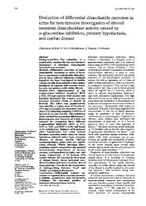
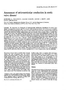
Figure I Case 1: (A) Initial biopsy specimen demonstrating subtotal villous atrophy, crypt hyperplasia, and dilated lymphatics in the lamina propria. (B) Specimen taken after restoration of the villous architecture to normal after gluten free diet. Dilated lymphatics are also present. (C) Specimen taken after gluten challenge showing shortened and blunted villi and hyperplastic crypts. Lymphangiectasia persists. (All haematoxylin and eosin stainx 100.)
blood T lymphocytes from case 2 (E rosette test) showed decreased values of 30 5 (normal range 60-80%). A gluten free, low milk, low fat, high protein diet with vitamin supplementation led to a gradual improvement during the next eight months in both patients. Diarrhoea ceased and they both reached an appropriate weight and height. Serum albumin and immunoglobulin concentrations returned to normal. After two years on a gluten free diet both children underwent gluten challenges after confirmation of histological remission. Histologically relapse occurred in each child after three months on gluten (fig 1B, IC and 2B, 2C). Small bowel biopsy specimens taken at follow up demonstrated the persistence of lymphangiectasia.
During a six year follow up, case 1 remained strict gluten free diet, but on several occasions ate large amounts of fatty foods. Her stools became voluminous and pale and her eyelids and legs swollen. At the time of her first relapse, at the age of 3 5 years, her serum albumin concentration decreased to 20 g/l, IgG to 2-6 g/l, and IgM to 0-17 g/l. During 2-5 years of follow up, case 3 has not relapsed clinically, has remained strictly on the recommended diet, and thrived. on a
t-
-
W
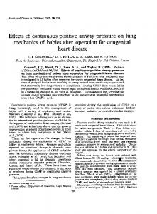
Figure 2 Case 2: (A) Initial biopsy specimen demonstrating subtotal villous atrophy and cystically dilated lymphatics. (B) Specimen taken after gluten free diet showing that villous architecture has returned almostto normal. Lymphangiectasia still present. (C) Specimen taken after gluten challenge showing evident total villous atrophy and lymphangiectasia. (Haematoxylin and eosin stain in (A) and (C) x 100, in
(B) x50.)
Discussion Severe intestinal protein loss and steatorrhoea are distinct features of a variety of gastrointestinal disorders including coeliac disease and intestinal lymphangiectasia.S Clinically, both diseases may present with chronic diarrhoea, failure to thrive, and oedema and thereby mimic one another.6 Moreover, appreciable hypoproteinaemia, steatorrhoea, hypocalcaemia, and abnormal laboratory indices of intestinal protein loss, for example raised faecal a1-antitrypsin clearance, may occur in both coeliac disease and intestinal lymphangiectasia. The distinctive small intestinal mucosal changes and the presence of lymphopenia, hypoimmunoglobulinaemia, and T cell depletion are helpful in differentiating intestinal lymphangiectasia from coeliac disease. Sometimes the normal villous architecture of the jejunal villi may be distorted appreciably by large dilated lacteals in intestinal lymphangiectasia, causing appearance of villous atrophy. Our patients suggest an association between intestinal lymphangiectasia and coeliac disease. The severe intestinal protein and fat losses, with associated mineral and vitamin deficiencies, which characterise intestinal lymphangiectasia may considerably exaggerate both the presentation and the course of coeliac disease. If not recognised promptly, intestinal lymphangiectasia may thereby cause treatment failure in
Perisic, Kokai
136
children with coeliac disease. Multiple small intestinal biopsy specimens, perhaps taken endoscopically, may be helpful in establishing the presence of intestinal lymphangiectasia in children with coeliac disease.3 6 As intestinal lymphangiectasia may be an incidental finding, additional data, for example the presence of hypoimmunoglobulinaemia and T cell depletion, may help in determining the clinical significance of lymphangiectatic mucosal changes.' 6 These patients with coeliac disease need lifelong diet management.
The authors thank Dr Ian W Booth for his contribution to this manuscript. 1 Cooke WT, Holmes GKT. Coeliac disease and associated disorders. In: Cooke WT, Holmes GKT, eds. Coeliac disease. Edinburgh: Churchill Livingstone, 1984:225-46. 2 Goodchild MC, Nelson R, Anderson CM. Cystic fibrosis and coeliac disease: coexistence in two children. Arch Dis Child
1973;48:684-91.
3 Meeuwisse GW. Diagnostic criteria in coeliac disease. Acta Paediatr Scand 1970;59:461-3. 4 King EJ. Microanalysis in medical biochemistry. London: Churchill, 1953;92-6. 5 Vardy PA, Lebenthal E, Shwachman H. Intestinal lymphangiectasia: a reappraisal. Pediatrics 1975;55:842-51. 6 Walker-Smith JA. Protein losing enteropathies. In: WalkerSmith JA, ed. Diseases of the small intestine in childhood. London: Butterworth, 1988:420-5.
Positive end expiratory pressure via a portable system in thoracic dystrophy S Edees, A Moulden, R J Winter Abstract The provision of positive end expiratory pressure, via a unique portable system, in the long term management of a child with thoracic dystrophy is reported. The system uses low gas flow enabling a reduction in equipment and simplification of the circuit as compared with a standard continuous positive airways pressure system. For a child requiring long term continuous positive airways pressure (CPAP), we devised a system for providing positive end expiratory pressure (PEEP) using low flows of oxygen. The system, initially employed to provide CPAP, was a standard high flow Bennett system necessitating that the patient be restricted to his cot on the infants' ward where there are piped supplies of oxygen and air. In order for him to become mobile around the hospital and ultimately at home, we devised a portable PEEP system.
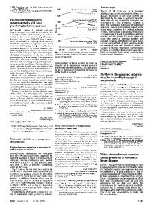
Description of the system Our system (figure) employed the same CPAP valve and T piece as the high flow pipeline
B University Hospital, Nottingham NG7 2UH, Department of Child Health S Edees A Moulden Department of Anaesthesia R J Winter Correspondence to: Dr Winter. Accepted 3 September 1991 (Arch Dis Child 1992;67:136-7)
system but required a mixer with a non-return valve to allow the use of low flow oxygen without loss of the PEEP from the valve and to prevent rebreathing. The system consisted of a T piece with an oxygen connector (A) in series with a 1950 non-return valve (B) on one limb, and the CPAP valve (C) on the other limb, the assembly being connected to the infant by a standard 15 mm Portex connector. The inspired oxygen delivered by the system depends upon the flow of oxygen into the circuit, the respiratory rate, and the inspiratory flow rate of the patient. An increase in the respiratory rate will reduce the time available for oxygen to accumulate in the wide bore tubing during expiration and therefore reduce the fractional inspired oxygen (FIo2). An increase in inspiratory flow will increase the volume of room air entrained via the 1950 one way valve reducing the FiO2. Therefore, the oxygen tension in the circuit cannot be accurately predicted from the present oxygen flow; however, we have found that a flow of 500 ml/min delivers approximately 28% oxygen to our patient. Increasing the flow of oxygen to the circuit up to 1 I/min increases the FIO2 to 36%, and 2 I/min to 48%. Rebreathing is prevented by the presence of the 1950 one way valve and also by the one way valve in the CPAP valve. As flow in the circuit is generated by the child's inspiratory effort the system provides PEEP, not CPAP.
AC
Diagram of system described.
Case report Soon after birth our patient, now aged 18 months, was noted to have a number of dysmorphic features including the Pierre Robin syndrome (cleft of the soft palate, micrognathia, and glossoptossis), macrocephaly, severe hyphoscoliosis, a small thorax, a penile web attached to 'the scrotum, and generalised hypotonia. Investigations were carried out to evaluate his