by 2-mercaptoethanesulfonic acid (coenzyme M) and some derivatives (acetyl ... converted to coenzyme M by enzyme systems present in the extracts. Methyl-.
Vol. 145, No. 1
JOURNAL OF BACTERIOLOGY, Jan: 1981, p. 27-34 0021-9193/81/010027-08$02.00/0
Coenzyme M Derivatives and Their Effects on Methane Formation from Carbon Dioxide and Methanol by Cell Extracts of Methanosarcina barkeri THEO J. HUTTEN, M. HENK DE JONG, BEN P. H. PEETERS, CHRIS VAN DER DRIFT, AND GODFRIED D. VOGELS Department of Microbiology, Faculty of Science, University of Nijmegen, Toernooiveld, NL-6525 Nijmegen, The Netherlands
Extracts of Methanosarcina barkeri reduced methanol and C02 to CH4 in the presence of H2 and converted methanol stoichiometrically into CH4 and C02 in the absence of H2. In dialyzed cell-free extracts these reactions were stimulated by 2-mercaptoethanesulfonic acid (coenzyme M) and some derivatives (acetyl and formylcoenzyme M and the oxidized form of coenzyme M), which could be converted to coenzyme M by enzyme systems present in the extracts. Methylcoenzyme M could not be used in these systems. P. Gunsalus and R. S. Wolfe, Fed. Proc. 35:1547, 1976). The reducing complex requires Mg2e ions and a catalytic amount of ATP for full activity (13). CH3-S-CoM was shown to be the first product that accumulated in substrate amounts in cell extracts or whole cells of M. bryantii (17). CHs-S-CoM strongly stimulates the reduction of C02 by H2 in cell-free extracts of Methanobacterium thermoautotrophicum (12). The rate of methane formation is enhanced 30-fold and, while CHs-S-CoM itself is converted to methane, 11 times more molecules of C02 are reduced simultaneously. This effect of CHa-S-CoM, called the RPG effect (27), is not exerted by HSCoM or (S-CoM)2. This paper deals with the different effects of HS-CoM, CH3-S-CoM, and other CoM derivatives on methanogenesis from C02 and methanol in dialyzed cell-free extracts of M. barkeri.
Unlike other methane-producing bacteria, which use only H2 plus C02 or, in some species, formate, Methanosarcina barkeri forms methane from H2 plus C02, methanol, acetate, methylamines (mono-, di-, and trimethylamine and ethyldimethylamine), and carbon monoxide (14, 16, 19, 20, 26; Ch. G. T. P. Schnellen, Ph.D. thesis, Technological University of Delft, Delft, The Netherlands, 1947). The phenotypic properties of M. barkeri differ much from those of other methanogens that this species has been placed in a different family or order (1); however, M. barkeri shares with the other methanogens some unique coenzymes, including coenzyme M (CoM). This compound was discovered by McBride and Wolfe (17; B. C. McBride and R. S. Wolfe, Fed. Proc. 29:344, 1970) as a new cofactor of methyl transfer reactions in methanogens, and Taylor and Wolfe (23) identified it as 2-mercaptoethanesulfonic acid (HS-CoM). This substance and its derivatives are found exclusively in methanogenic bacteria (3), and they are growth factors of Methanobrevibacter rumrantium (2, 17, 22). HS-CoM can be methylated by the methylcobalamin-CoM methyltransferase of Methanobacterium bryantii (24) and Methanospirillum hungatei (9), but a role for this enzyme in the reduction of C02 to CH4 is questionable (10, 27). HS-CoM can be formed by reduction of 2,2'dithiodiethanesulfonic acid [(S-CoM)2] in extracts of M. bryantii by means of an NADPHlinked oxidoreductase (17, 24). Methyl-CoM [2-(methylthio)ethanesulfonic acid (CH3-S-CoM)] is reduced to methane and HS-CoM by a reductase, which can be coupled to H2 in the presence of a hydrogenase and an as-yet-unknown factor, component B (10, 27; R. so
MATERIALS AND MErHODS Organism and growth conditions. M. barkeri
MS was kindly provided by R. S. Wolfe, Department of Microbiology, University of Illinois, Urbana. It was grown in a basal medium containing the following (in grams per liter): K2HPO4, 0.23; KH2PO4, 0.23; (NH4)2504, 0.23; NaCl, 0.46; MgSO4. 7H20, 0.09; CaCl2.
2H20,0.06;Na2S.9H20,0.24;L-cysteinehydrochloride.
H20, 0.18; and sodium resazurin, 0.001. The basal medium also contained 10 ml of vitamin solution (28) and 10 ml of trace mineral solution (28). One of the following carbon sources was used together with 2.5 g of NaHCO3 per liter: sodium acetate (1.5 g/liter) plus a gas mixture containing 80% H2 and 20% C02 at a pressure of 1.5 atmospheres (atm) (147 kPa); methanol (10 mi/liter, or sodium acetate (10 g/liter) plus a gas mixture containing 80% N2 and 20% C02. The H2-C02 mixtr was sparged through cultures (200 ml/14 liters per min), and the N2-CO2 mixture was applied as an 27
28
HUTTEN ET AL.
anaerobic atmosphere (at 1.5 atm) buffering the medium. The fermentors were inoculated with a 4% inoculum of cells grown previously with the same carbon source. Cells were grown at 37°C in 14- or 50-liter fermentors for 7 days (carbon source, C02 or methanol; cell yield, 2 g [wet weight] of cells per liter) or for 21 days (carbon source, acetate; cell yield, 1 g [wet weight] of cells per liter). Cells were harvested with a Sharples continuous centrifuge operating in an N2 atmosphere. After centrifugation, cells were kept frozen (-70°C) under an H2 atmosphere. Stock cultures were maintained essentially by the method of Balch and Wolfe (2) in 140-ml or 1-liter bottles closed with black butyl rubber stoppers and crimped aluminum seal caps or with screw caps with a central hole to accommodate syringe injections. Air was removed from the bottles by evacuation before sterilization of the media. After sterilization, the evacuated bottles were filled with the desired sterile gas mixture at 2 atm of pressure. The purity of each culture was checked by microscopy (7) and by inoculation into a medium which contained 5 g of yeast extract (Difco Laboratories) per liter and 5 g of trypticase (BBL Microbiology Systems) per liter; incubation was under an atmosphere containing 80% N2 and 20% C02. Preparation of cell-free extracts. All solutions used in the preparation of cell-free extracts were freed of oxygen by three cycles of evacuation and gassing with H2. The transfers of cells and cell-free extracts and dialysis were performed in a stainless steel anaerobic glove box (1.95 by 1.15 by 1.5 m) which was equipped with glass windows and contained a 97.5% N2-2.5% H2 atmosphere. The concentration of oxygen was kept below 1 id/liter (measured with a CSD type 20-120A couloximeter placed inside the anaerobic box), and the amount of moisture was kept constant at the dew point of 100C by circulating the gas over an external catalyst (BASF RO-20) and a heat exchanger. The external circuit was also equipped with a Domnick Hunter ultra-high-efficiency filter to reduce the number of particles in the atmosphere. The gas pressure was regulated at 1.2 to 2.5 mm of Hg above the outside pressure. Thawed cells were washed and suspended (50% wet cells) in 120 mM N-tris(hydroxymethyl) methyl-2aminoethanesulfonic acid (TES) buffer (pH 7.2) containing 15 mM MgCl2 and 2 mM dithiothreitol (DTT). The cell suspension was kept under an N2 atmosphere in a closed bottle and transferred twice anaerobicaUy through a gas-tight tube (Argyle MAR 2303) fitted with a syringe-tube connector to a French pressure cell. The broken cells obtained after application of a pressure of 138 MPa were returned similarly to a closed anaerobic bottle. This suspension was transferred to stainless steel centrifuge tubes, which were closed with stainless steel caps and centrifuged for 30 min at 30,000 x g and 40C. The supernatant fraction was used either in this crude cell-free form or after dialysis of a 25-ml sample for 16 h at room temperature against 2.5 liters of 50 mM TES buffer (pH 7.2) containing 15 mM MgCl2 and 2 mM DTT. The dialysis removed cofactors and the traces of methanol and acetate which were present in the extracts of cells grown with these substrates. Assay of methane formation. Incubations were
J. BACTERIOL.
performed in 10-ml bottles closed with black butyl rubber stoppers and crimped aluminum seal caps. The assay mixture (0.7 ml) contained 120 mM TES buffer (pH 7.2), 30 mM MgCl2, 7.5 mM ATP, 3 mM CoM derivatives, 2 mM DTT, and about 10 mg of protein, unless otherwise indicated. Incubation was at 370C. The gas phase was either a mixture of 80% H2 and 20% C02 (at 2 atm) or, when methanol was used, 100% N2 or 100% H2; the gas phase was applied by three cycles of evacuation and gassing before the extract was injected with a plastic syringe. The evacuation-gassing cycle was repeated once after the addition of the extract. Ethane (100 ,ul) was used as internal standard and did not influence CH4 production. Axalysis. Proton nuclear magnetic resonance spectra were recorded with Bruker WH90 and Varian EM 390 instruments. The nuclear magnetic resonance spectra in D2Q were consistent with the assigned structures of al components. Gas chromatographic analyses were performed with Pye Unicam model GCV and model GCD gas chromatographic systems equipped with flame ionization detectors and thermal conductivity detectors. Methane, hydrogen, and carbon dioxide concentrations were determined with a coupled flame ionization-thermal conductivity detector under the following conditions: Poropack Q (80/100) column at 1100C; injector and detector oven temperatures, 1500C; carrier gas, N2 (40 ml/min). Gas samples of 100 pl or less were analyzed. By feeding the data obtained to an LDC-computing model 304 integrator, which was programmed for comparing the amount of each gas with the amount of the internal standard ethane, the concentration of each gas was obtained directly as nanomoles per bottle. The response curve for methane was linear for the whole range of concentrations that could be produced under our conditions. Methanol concentrations were determined with a Pye Unicam model GCD gas chromatographic system equipped with a flame ionization detector at 1500C. The following conditions were used: a stainless steel column (6 feet [1.83 m] by 'A inch [3.2 mm]) containing 0.2% Carbowax 1500 on Carbopack C (80/100) at 1250C; injector oven temperature, 1500C; carrier gas, N2 (20 ml/min). The sample size was 1 pl, and propanol was used as an internal standard. Acetate concentrations were determined in samples acidified with 25% phosphoric acid with a Pye Unicam model GCD gas chromatographic system, using a glass column (30 inches [76.2 cm] by 0.25 inch [6.4 mm] by 4 mm) which contained 3% Carbowax 20 M and 0.5% phosphoric acid on Carbopack C (60/80). The temperatures of the column, detector, and injector ovens were 175, 200, and 2000C, respectively. N2 was the carrier gas (40 ml/ min). The sample size was 4 tl, and propionic acid was used as an internal standard. Benzylmercaptan, benzyl alcohol, and toluene concentrations were determined with a Pye Unicam model GCD gas chromatographic system equipped with a flame ionization detector. The stainless steel column (2 feet [61 cm] by 3/16 inch [4.8 mm]) contained SE 30 on WHP (60/80). Other conditions were as described above for the acetate analysis. Formic acid concentrations were determined with formate dehydrogenase by using the test system of Boehringer Mannheim. The concentrations of mercaptans were determined by the method of Ellman (8)
CoM DERIVATIVES
VOL. 145, 1981 in extracts prepared and tested in the absence of DTT. Protein concentrations were determined by the method of Lowry et al. (15), using bovine serum albumin as a standard. Chemicals. Table 1 shows elemental analytical data for the CoM derivatives synthesized as described below. Sodium HS-CoM was obtained from MerckSchuchardt AG, Darmstadt, Germany; the contaminating oxidized form [(S-CoM)2] was removed by filtration of a hot concentrated solution of HS-CoM in methanol, and HS-CoM was purified further by a twofold crystallization from methanol. Sodium CH3-SCoM was prepared essentially as described by Taylor and Wolfe (23). The crude product (ammonium salt) was evaporated to dryness and applied (as a concentrated solution in water) to an SP Sephadex C-25 (sodium form) column equilibrated with water. The eluted CH3-S-CoM (sodium salt) was recrystallized twice from methanol. The overall yield was 60%. Sodium 2-(benzylthio)ethanesulfonate (C6H5CH2-SCoM) was prepared in a way similar to the way in which CH3-S-CoM was prepared (yield, 80%). Potassium 2-(acetylthio)ethanesulfonate (CH3C0-S-CoM) was prepared by slowly adding a solution of 4 g of sodium 2-bromoethanesulfonate in 25 ml of water to a solution of 5 g of potassium thioacetate in 25 ml of water. After the reaction mixture was stirred at room temperature for 3 h, it was evaporated to dryness, and the residue was recrystallized twice from methanol (yield, 70%). Sodium 2-(formylthio)ethanesulfonate (HCO-S-CoM) was prepared in a way similar to the procedure described by Bax and Stevens (5) for the formylation of arenethiols. By mixing 4.1 g of acetic anhydride and 3.8 g of formic acid, the acetic-formic mixed anhydride was obtained. HS-CoM (2.0 g) and 2 drops of pyridine were added, and the reaction mixture was stirred for 24 h at room temperature. A large volume of ether was then added to precipitate the product, which was filtered, washed with ether, dried, and stored at -80°C (yield, 90%). This product decomposes partially when stored at -20°C for more than 1 month. 2-(Dimethylsulfonium)ethanesulfonate [(CH3)2-S+-CoM] was prepared essentially as described by Taylor and Wolfe (23). This compound was crystallized from methanol-water (yield, 60%). Sodium (S-CoM)2 was prepared by dissolving HS-CoM in methanol and oxidizing this solution with a solution of I2 in methanol. The reaction mixture was neutralized with solid NaHCO3 and evaporated to dryness after
TABLE 1. Elemental analytical data for coenzyme M derivatives Cation Cal-
tive
Found
culated
I(S-CoM)2 CH,-S-CoM IHCO-S-CoM
CH3CO-S-CoM I(CHa)2-S+-CoM C6H5CH2-S-
2Na+ Na+ Na+ K+ Na+
14.7 20.2 18.7 21.6 28.2 42.5
Cal-
Found
culated
14.7 19.9 18.3 21.7 28.4 42.3
2.5 4.0 2.6 3.2 5.9 4.4
removal of excess NaHCO3 by filtration; Nal was removed from the product by gel filtration over Sephadex G-10. Then the product was purified further by crystallization from methanol-water (yield, 30%). Methyl 2-(methylthio)ethanesulfonate (CH3SCH2CH2SO3CH3) was prepared by adding 0.02 mol of sulfuric acid to a solution of 3.56 g (0.02 mol) of CH3S-CoM (sodium salt) in water. The solution was freezedried for 48 h, and the residue was then stirred for 2 h with dry ethyl acetate to dissolve free sulfonic acid (CH5SCH2CH2SO3H). The undissolved material was removed by filtration, and an ethereal solution of diazomethane was added slowly at 00C until a yellow color persisted. After 2 h at room temperature, the solution was flushed with nitrogen (to remove the excess of diazomethane), diluted with ether, washed with water, and dried over MgSO4 at 00C. The product itself was unstable, but it could be stored in dilute solutions in ether at -80°C for 1 to 2 days. This product is a strong methylating agent and, when heated in solution or without solvent, undergoes a methyl transfer, leading to (CH3)2-S+-CoM. Similarly, methyl (methylthio)arenesulfonates were found to give a methyl transfer to a sulfonium salt (25). The structure of the product was confirmed by 'H nuclear magnetic resonance analysis. Because of its instability, no elemental analysis could be made. For biological experiments a solution of the product was evaporated quickly to dryness at 00C and used immediately. Methyl 2-(acetylthio)ethanesulfonate (CH3COSCH2CH2-SO3CH3) was prepared from CH3CO-S-CoM in a way similar to the way described above for CH3-SCH2-CH2-SO3CH3. A dried ethereal solution of the crude product was evaporated to dryness and applied to a Fluorasil column equilibrated with benzene. Elutions were performed with benzene, chloroform, and ether. The product obtained-was stable at room temperature and pure according to its 'H nuclear magnetic resonance spectrum. An elemental analysis was not made. For biological experiments, traces of chloroform were removed by dissolving the product in ether and subsequent evaporation to dryness. This operation was repeated several times. All other chemicals were obtained from E. Merck AG, Darmstadt, Germany, and were of analytical grade. Gases were obtained from Hoek Loos, Schiedam, The Netherlands. To remove traces of oxygen, the gases which contained hydrogen were passed over a catalyst (BASF RO-20) at room temperature, and those devoid of hydrogen were passed over a prereduced catalyst (BASF R3-11) at 1500C.
RESULTS
%H
%C CoM deriva-
29
2.3 4.1 2.5 3.2 6.0 4.3
CoM
, (CH3)2-S+-CoM forms an internal salt between the sulfonium and sulfonate moieties (21).
Methanogenesis by extracts of M. barkeri. Cell-free extracts prepared from cells grown on H2 plus C02, methanol, or acetate produced methane when incubated with 80% hydrogen-20% carbon dioxide or with methanol in the presence of either H2 or N2. Conversion of acetate to methane could not be obtained in these extracts. Dialysis of the extracts reduced the levels of activity to about one-half of the levels found with crude extracts, but this proce-
30 HUTTEN ET AL. dure was used routinely to reduce interference by residual and endogenous substrates. The dialyzed extracts were most active when the substrate was methanol in the presence of hydrogen (200 to 500 nmol of CH4 produced per mg of protein per h). The rate of methane formation was approximately 50% lower when the substrate was either methanol in the presence of nitrogen or H2 plus C02, and the rate decreased after 2 h. Methane production was approximately linearly proportional to protein concentration over a broad range tested (up to 15 mg/ ml), but at low protein concentrations (less than 5 mg/ml), a relatively small specific activity was observed. This activity was stimulated by increasing the pressure of the gases (Fig. 1). This stimulation was probably due to higher concentrations of dissolved gaseous substrates (especially hydrogen) for the enzymic system. The optimal concentration for methanol was 50 to 70 mM. Methane production from either C02 or methanol was optimal when the pH of the buffer was adjusted to 7.25; the pH changed to 6.6 when the mixture was incubated in the presence of 80% hydrogen-20% carbon dioxide at a pressure of 2 atm. Methane was not formed in the absence of ATP; its production was optimal at a concentration of 8 mM ATP, with half-maximal activity at 1 mM ATP. Mg2" ions stimulated methane fornation optimally at a concentration two- to four-fold higher than the concentration of ATP. DTT (2 mM) did not affect the initial reaction rate and was used as a reducing agent. HS-CoM stimulated methane production from methanol and 80% hydrogen-20% carbon dioxide when it was present at a concentration between 0.5 and 6 mM, and half-maximal activity was observed at 0.2 mM HS-CoM. The reactions were completely inhibited by 100 mM HS-CoM. Stoichiometry of methanol conversion. Methanol was converted stoichiometrically to methane and carbon dioxide, which were produced in a ratio of 3:1 (Table 2). This ratio remained constant in tests with different initial concentrations of methanol and also during the period of decreasing activity which occurred after 2 h. In the presence of hydrogen, methanol was converted completely to methane, and carbon dioxide could not be detected at any time during the incubation. Effects of CoM derivatives on methano-
J. BACTERIOL.
0
0.5
2.0 1.0 1.5 Partial pressure of the gasses(atm)
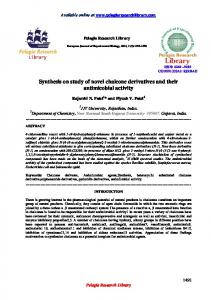
FIG. 1. Effect ofpartialpressure of gases on methane formation by dialyzed extracts of M. barkeri. The reaction components and conditions used were as described in the text, except for the gas phase (the gas phase was at a pressure of 2 atm its composition was changed); 5 mM HS-CoM was present in the incubation mixture. The activities were compared with the amount of methane (360 nmol of CH4 per h per mg of protein) produced in the presence of H2 plus CO2 (80: 20, vol/vol) at a pressure of 2 atm, which was taken as 100% activity. Symbols: [1, H2 pressure was varied between 0 and 1.6 atm in the presence of a constant pressure of CO2 (0.4 atm); *, CO2 pressure was varied between 0 and 0.4 atm in the presence of a constant pressure of H2 (1.6 atm); U, pressure of H2 plus CO2 (80:20, vol/vol) was varied; 0, methanol (50 mM) was used as the substrate and hydrogen pressure was varied between 0 and 1.6 atm. In all instances N2 was supplied to pressurize to 2 atm.
stimulated methane production from both C02 and methanol in dialyzed extracts of M. barkeri (Table 3). Some CoM derivatives [(S-CoM)2, genesis by dialyzed extracts. CH3-S-CoM, HCO-S-CoM, and CH3CO-S-CoM] replaced HSbut not HS-CoM, stimulates the reduction of CoM both in crude cell-free extracts (data not carbon dioxide to methane in undialyzed and shown) and in dialyzed extracts, but CH3-S-CoM dialyzed extracts of M. thermoautotrophicum was inactive and annulled the stimulatory effect (12; J. A. Romesser, Ph.D. thesis, University of exerted by HS-CoM (Table 3 and Fig. 2). With Illinois, Urbana, 1978). In contrast, HS-CoM methanol and CO2 as substrates, the stimulation
CoM DERIVATIVES
VOL. 145,1981
by HS-CoM was reduced to a half-optimal value by 0.06- and 0.7-fold molar concentrations of CH3-S-CoM, respectively. CH3-S-CoM was prepared from 2-bromoethanesulfonate, a potent inhibitor of methanogenesis when it is present at a 0.01-fold molar level compared with CH3-S-CoM (10); thus, the inhibitory effect of CH3-S-CoM might be due to residual traces of the halogen derivative in the CH3S-CoM sample. However, such traces could not be detected in the twice-crystallized preparation by various chromatographic, electrophoretic, and isotachophoretic techniques (data not shown). Moreover, our sample of CH3-S-CoM was active in the M. thermoautotrophicum system, and a sample obtained from another source (gift from R. S. Wolfe) gave the same results in the M. barkeri system. These results indicate that CH3-S-CoM had
31
to be activated in order to be reactive in methane
formation by extracts of M. barkeri, and it is possible that another methylated form of HSCoM or CH3-S-CoM was involved. Therefore, we tested the sulfonium derivative (CH3)2-S+CoM and the methyl sulfonate esters of CH3-SCoM and CH3CO-S-CoM. Both methyl esters were unstable in water, and even when tested at low concentrations (0.36 mM), they strongly inhibited methanogenesis, probably due to methylatton and denaturation of essential proteins. In view of these results, no tests were performed with the methyl esters of HS-CoM and HCO-SCoM. The sulfonium derivative inhibited methanogenesis (Table 3), in accordance with the results of Taylor and Wolfe (23). The inhibition was less (80% residual activity) when HS-CoM was present in an equimolar concentration (5 mM). C6H5CH2-S-CoM inhibited methanogenesis from all substrates tested and reduced the TABLE 2. Stoichiometry of methanol conversion in stimulatory effect of HS-CoM on methane forthe presence of nitrogen' mation from carbon dioxide and methanol to half-optimal values at concentrations 0.20 and Amt (umol) of the following Amt of 0.05 times, respectively, the concentration of comnpounds presnt after 5 h: Ratio of methanol added HS-CoM (Fig. 2). None of the CoM derivatives CH4 to CO2 CR, (moI)b MethaC02 listed in Table 3 could be reduced to methane by the dialyzed extracts of M. barkeri under the 2 1.35 0.43 _C 3.1 conditions used. Moreover, C6H5CH2-S-CoM -C 4 2.38 0.78 3.0 was not reduced or hydrolyzed to HS-CoM, 8 4.2 2.62 0.90 2.9 methylmercaptan, benzylmercaptan, or toluene, 10 6.5 2.42 0.86 2.8 a Incubations were performed with a dialyzed cell- and CH3-S-CoM was not hydrolyzed to methafree extract of M. barkeri grown with methanol as nol and HS-CoM. Conversion of CoM derivatives by exdescribed in the text; 3 mM HS-CoM was present in tracts. The stimulating effects exerted by the the incubation mixture. b No methane or carbon dioxide was formed in the acyl derivatives of HS-CoM and its oxidized form on methanogenesis (Table 3) might have absence of methanol. - , Methanol concentration was below the limit of been due to conversion of these compounds to detection (0.4 ,mol). HS-CoM by components of the incubation mixTABLE 3. Effects of CoM derivatives on methanogenesis by extracts of M. barkeri Methane production (nmol/mg of protein per h) with the following CoM derivative
substrates and gas phases:.
Concn (MM)
H
vol/vol)
HS-CoM (S-CoM)2
H2
Methanol, H2
Methanol, N2
400 12 480 100 320 12 480 13 CH6-S-CoM 50 8 20 15 HCO-S-CoM 400 12 NTb NT CH3CO-S-CoM 350 8 NT NT (CH3)2-S+-CoM 10 12 10 10 CIsHCH2-S-CoM NDc NT 5 1 3.6d ND CH3S-CH2-CH2-SO3CH6 NT ND ND 3.6d CH6CO-S-CH2-CH2-SO3CH3 ND NT ND ND None 50 12 25 17 a Dialyzed extracts were tested as described in the text. Similar results were obtained with extracts obtained from cells grown with either C02, acetate, or methanol as the carbon source. bNT, Not tested. 'ND, Not detectable (specific activity less than 0.5 nmol of CH4 per mg of protein per h). d Similar results were obtained with a concentration of 0.36 mM. 5 2.5 5 5 5 5 5
32
HUTTEN ET AL.
J. BACTERIOL.
of either H2 or N2. In the presence of N2, methanol is converted according to the following reaction: 4CH30H 3CH4 + C02 + 2H20 Since this reaction proceeds even in dialyzed extracts, the coenzymes involved in the reduction and oxidation steps apparently are firmly bound to compounds of high molecular weight or are present in the crude extracts in abundant amounts. This does not hold true for ATP, Mg2+, and CoM derivatives, which must be added for optimal activity. Since we observed only minor differences among the reaction systems as to activities, cofactor requirements, and sensitivities to inhibitors, the reactions for the reduction of C02 and methanol appear to be determined by some common and probably rate-limiting presence
0
2
4
6
a
10
Concentration (mM)
FIG. 2. Effect of CH3-S-CoM and C6H5CH2-S-CoM on methane formation by dialyzed extracts of M. barkeri in the presence of 5 mM HS-CoM. The tests were performed as described in the text. Methane was produced from H2 plus CO2 (80:20, vol/vol) (solid symbols) or methanol in the presence of hydrogen (open symbols). The concentrations of CH3-S-CoM (0, U) and C6H5CH2-S-CoM (0, @) were varied; the amounts of methane formed in the absence of these compounds were 320 (C02 as substrate) and 500 (methanol as substrate) nmol/h per mg ofprotein.
ture. HCO-S-CoM was rapidly hydrolyzed to formic acid and HS-CoM under acidic and alkaline conditions (Table 4). Under neutral conditions hydrolysis slowed down. In the presence of extracts, HCO-S-CoM was rapidly and completely converted to formic acid and HS-CoM, as shown by the Ellman test (8) for mercaptans and the formate dehydrogenase test for formic acid. CH3CO-S-CoM was rather stable to chemical hydrolysis, but in the presence of extract and under the conditions described above about 30% was hydrolyzed to acetate and HS-CoM within 30 min, as shown by the Ellman test (8) and a gas chromatographic analysis of the acetate formed. These results demonstrated that the stimulating effects of CH3CO-S-CoM (and probably also HCO-S-CoM) were due to enzymatic hydrolysis of these compounds to HSCoM. (S-CoM)2 was completely converted to HS-CoM within 30 min under the conditions used for methane formation in the presence of hydrogen. The stimulating effect of (S-CoM)2 on methane formation in the presence of hydrogen (Table 3) was probably due to its enzymatic reduction to HS-CoM. DISCUSSION Cell-free extracts of M. barkeri produce methane from H2 plus C02 or from methanol in the
steps. The effects exerted by CoM derivatives deTABLE 4. Chemical stability of CoM derivatives CoM derivative tested'
HCO-S-CoM
Conditionsb Solvent Temp (°C)
Rate of hydrolYawc
NaOH 37 and 65 Very high 25%in 15 TES 37 mind
HCl CH3CO-S-CoM
37 65
T,/2 = 12 min T1/2 = 2 min
NaOH 37 and 65 Very high
No hydrolysis (2 h) 37 HCI T,/2 = 10 h 65 T1/2 =40 min a CH3-S-CoM was stable toward hydrolysis for 24 h under all conditions tested. bCoM derivatives were dissolved in D20 and investigated at 37 and 65°C under neutral (0.125 M TES, pH 7.2), acidic (1 N HCI), and alkaline (1 N NaOH) conditions by using 'H nuclear magnetic resonance spectroscopy. 'The indications used were as follows: very high (instantaneous hydrolysis); half-lives (T,/2) for reactions with pseudo-first order kinetics; percentage of compound hydrolyzed within a certain period for reactions with aberrant kinetics; and the period of time (in parentheses) over which no hydrolysis was observed. Unless otherwise indicated, the products formed were HS-CoM and either formic acid or acetic acid. d Besides formic acid and HS-CoM, a third product was present according to the 'H nuclear magnetic resonance spectra; this product had one singlet absorption at 0.12 ppm downfield from the singlet from formic acid, and it could not be identified, as the ABCD pattern of CH2 units was too complex to be interpreted. The overall reaction of hydrolysis slowed down after about 15 min. TES
37 and 65
VOL. 145, 1981
further attention here. CH3-S-CoM can be reduced to HS-CoM and methane by cell-free extracts of various methanogenic bacteria (1, 10, 11, 12, 17, 23). CH3-S-CoM replaces the growth requirement for HS-CoM in M. ruminantium strain Ml, is transported as easily as HS-CoM, and is one of the major forms of CoM found in cells after transport of HS-CoM or CH3-S-CoM (4). Recently, Shapiro and Wolfe (18) demonstrated the formation of CH3-S-CoM from methanol and the reduction of CH3-S-CoM with hydrogen in crude cell-free extracts of M. barkeri. Therefore, the role of CH3-S-CoM as a precursor of methane formation and as a physiologically active derivative of HS-CoM seems well proven. Moreover, CH3-S-CoM exerts an effect (the RPG effect), which is not shown by HS-CoM; this effect involves stimulating CO2 reduction to methane, which indicates that the methylreductase reaction is coupled to the activation of C02 (12, 27). This effect was observed with M. thernoautotrophicum, M. bryantii, and M. hungatei and, to a lesser extent, with M. bryantii strain M.o.H.G. and Methanobacterium fornicicum, but not with M. barkeri (J. A. Romesser, R. P. Gunsalus, and R. P. Wolfe, Fed. Proc. 36: 713, 1977; Romesser, Ph.D. thesis). Whereas CH3-S-CoM is most active in extracts of other methanogens, dialyzed cell-free extracts of M. barkeri use HS-CoM in the conversion of C02 and methanol to methane, and CH3-S-CoM abolishes the stimulating effect of HS-CoM. The inability of CH3-S-CoM to act as a precursor of methane in dialyzed extracts of M. barkeri and its inability to stimulate the conversion of C02 and methanol to methane indicate that CH3-S-CoM is not readily converted to HS-CoM. This could be due to lack of a coenzyme or a cofactor in the dialyzed extract; such factors were probably present in the undialyzed extracts of M. barkeri which converted CH3-S-CoM to methane (18). We considered the possible role of other methylated intermediates, with negative results. Activation of CH3-S-CoM in methanogenesis by M. barkeri may involve as yet-unknown CoM derivatives which were reported in previous studies. In transport studies with H 3S-labeled CoM, cells of M. ruminantium accumulated the label mainly as a heterodisulfide of unknown composition (4). Moreover, in studies on the short-tern fixation products of '"C02 and '4CH30H in whole cells of M. barkeri, Daniels and Zeikus (6) observed '4CH3S-CoM and an unknown compound which was designated C1-X-T. This compound was also found in studies with Methanobrevibacter smithii and M. thermoautotrophicum; in the latter case it was the most strongly labeled compound serve
CoM DERIVATIVES
33
found. Its relationship to CoM derivatives was evident, since it was also labeled in 'S incorporation studies and it was active as a growth factor of M. ruminantium. CH3CO-S-CoM and HCO-S-CoM stimulated methane formation in M. barkeri. These compounds are not substrates for methanogenesis, as reported by Romesser (Ph.D. thesis). The timulating effects are probably due to enzymatic conversions of these acyl derivatives to HS-CoM. Similar conversions may account for the fact that these compounds are as effective as HS-CoM for growth of M. ruminantium (3). CH3CO-S-CoM is transported as easily as HSCoM by cells of this organism (4). The stimulating effect of (S-CoM)2 can be explained by the presence of (S-CoM)2 reductase, which has been found in various methanogens (10, 11). Although the reduction of (S-CoM)2 can also occur with chemical reductants, like DTT, which was present in our test system, its reduction is most probably enzymatic in nature, since the amount of DTT applied was much lower than the amount required for substantial chemical reduction (4) and since (S-CoM)2 can replace HSCoM as a cofactor only when hydrogen is present in the system. ACKNOWVLEDGMENTS This study was supported by fellowship 14-35-09 from the Netherlands Organization for the Advancement of Pure Research (Z.W.O.). We thank P. Maas for synthesizing some of the CoM derivatives and B. Zwanenburg for advice concerning the syntheses and for helpful discussions.
LITERATURE CITED 1. Balch, W. E., G. E. Fox, L. J. Magrum, C. R. Woese, and R. S. Wolfe. 1979. Methanogens: reevaluation of a unique biological group. Microbiol. Rev. 43:260-296. 2. Balch, W. E., and R. S. Wolfe. 1976. New approach to the cultivation of methanogenic bacteria: 2-mercaptoethanesulfonic acid (HS-CoM)-dependent growth of Methanobacterium runinantium in a pressurized atmosphere. Appl. Environ. Microbiol. 32:781-791. 3. Balch, W. E., and R. S. Wolfe. 1979. Specificity and biological distribution of coenzyme M (2-mercaptoethanesulfonic acid). J. Bacteriol. 137:256-263. 4. Balch, W. E., and R. S. Wolfe. 1979. Transport of coenzyme M (2-mercaptoethanesulfonic acid) in Methanobacterium ruminantium. J. Bacteriol. 137:264-273. 5. Bax, P. C., and W. Stevens. 1970. Mized carboxylic anhydrides. VIII. Synthesis of aryl thiolformates. Recl. Trav. Chim. Pays-Bas 89:265-269. 6. Daniels, L., and J. G. Zeikus. 1978. One-carbon metabolism in methanogenic bacteria: analysis of short-term fixation products of "4CO2 and '4CH30H incorporated into whole cells. J. Bacteriol. 136:75-84. 7. Doddema, IL J., and G. D. Vogels. 1978. Improved identification of methanogenic bacteria by fluorescence microscopy. Appl. Environ. Microbiol. 36:752-754. 8. ElIman, G. L 1958. A colorimetric method for determining low concentrations of mercaptans. Arch. Biochem. Biophys. 74:443-450. 9. Ferry, J. G., and R. S. Wolfe. 1977. Nutritional and biochemical characterization of Methanospirillum hun-
34 10.
J. BACruOL.
HUTTEN ET AL. gatii. AppL Environ. MicrobioL 34:371-376. D. Eirich, J. Romesser, W. Balch, S. Shapiro, and R. S. Wolfe. 1976. Methyl-traer and methane formation, p. 191-198. In H. G. Schlegel, G. Gottschalk, and N. Pfennig (ed.), Prceedin of the Symposium on Microbial Production and Utilization of Gases (H2, C02, CO). Akademie der Wiseenchaften Goittingen, Goltze KG, Gottingen. Gunalus, R. P., J. A. Romessar, anid R. S. Wolfe. 1978. Preparation of coenzyme M analogues and their activity in the methyl coenzyme M reductase system of Methanobacterium thenmoaatotrophicun. Biochemistry 17:2374-2377. G Rlus, P., and R. S. Wolfe. 1977. Stimulation of CO. reduction to methane by methyl-coenzyme M in extracts of Methanobacterium. Biochem. Biophys. Res. Commun. 76:790-796. Gunsalus, R. P., and R. S. Wolfe. 1978. ATP activation and properties of the methyl coenzyme M reductase system in Methanobacterium thermoautotrophicum. J. Bacteriol. 135:861-867. Hippe, H, D. Caspai, Fiebig, and G. Gottschalk 1979. Utilization of trimethylamine and other N-methyl compounds for growth and methane formation byMethanosarcina barkeri. Proc. Natl. Acad. Sci. U.S.A. 76: Gunaalus, IL,
zu
11.
12.
13.
14.
494498. 15. Lowry, 0. H, N. J. Rosebrough, A. L Farr, and R. J. Ran4lalL 1951. Protein measurement with the Folin phenol reagent. J. Biol. Chem. 193:266-275. 16. Mah, R. A., M. R. Smith, and L. Baresi. 1978. Studies on an acetate-fermenting strain of Methanosarcina. Appl. Environ. Microbiol. 35:1174-1184. 17. McBride, B. C., and R. S. Wolfe. 1971. A new coenzyme of methyl transfer, coenzyme M. Biochemistry 10:23172324. 18. Shapiro, S., and R. S. Wolfe. 1980. Methyl-coenzyme M, an intermediate in methanogenic disimiltion of C, compounds by Methanosarcina barkeri. J. Bacteriol. 141:728-734.
19. Smith, IL R., and I. A. Mal. 1978. Growth and methanogenes by Methano ecina strain 227 on acetate and methanol. Appl. Environ. MicrobioL 36:870-879. 20. Stadtman, T. C., and He A. Barker. 1961. Studies on the methane fermentation. IX. The origin of methane in the acetate and methanol fermentation by Methanosarcina. J. Bacteriol. 61:81-86. 21. Taylor, C. D. 1976. Structure and methylation of a new cofactor (coenzyme M) in methyl-transfer reactions, p. 181-190. In H. G. Schlegel, G. Gottechalk, and N. Pfennig (ed.), Proceeding of the Symposium on Microbial Production and Utilization of Gasme (H2, CO2, CO). Akademie der Wisenschaften zu Gottingen, Goltse KG,
Gttingen.
22. Taylor, C. D., B. C. McBride, R. S. Wolfe, and ML P. Bryant. 1974. Coenzyme M, esential for growth of a rumen strain of Methanobacteruwn ruimantiwm. J. Bacteriol. 120:974-975. 23. Taylor, C. D., and R. S. Wolfe. 1974. Structure and methylation of coenzyme M (HSCH2CH.9032). J. Biol. Chem. 249:48794885. 24. Taylor, C. D., and R. S. Wolfe. 1974. A simplified assay for coenzayme M (HSCH2CH2SO3). Resolution of methylcobalamin-coenzyme M methyltransferam and use of sodium borohydride. J. Biol. Chem. 249:48864890. 25. Tenud, L, S. Farooq, J. Seible, and A. Eachamosr. 1970. Endocycliwche Sw-Reaktionen am gesattigten Kohlenstoff? Helv. Chim. Acta 63:2059-2069. 26. Weimer, P. J., and J. G. Zeikus. 1978. One carbon metabolism in methanogenic bacteria: cellular characterization and growth of Methanosarcia barkeri. Arch. Microbiol. 119:49-57. 27. Wolfe, R. S., and L.J. HEiggin. 1979. Microbial biochemistry of methane-a study in contrasts, p. 267-300. In J. R. Quayle (ed.), Microbial biochemistry. University Park Press, Baltimore. 28. Wolin, E. A., M. J. Wolin, and R. S. Wolfe. 1963. Formation of methane by bacterial extracts. J. Biol. Chem. 238:2882-2886.