Comparative study of morphological and histological changes between differently structured expanded polytetrafluoroethylene implants in an animal model Ji Heui Kim, M.D., Ph.D.,1,2 Jung Min Lee, B.S.,2 Hyung Woo Ju, B.S.,2 Jun Ho Lee, M.D., Ph.D.,1 Ah Young Jun, M.D.,3 and Chan Hum Park, M.D., Ph.D.1,2
Y P
ABSTRACT
Background: Expanded polytetrafluoroethylene (e-PTFE) is a popular graft material used in augmentation rhinoplasty. New e-PTFE has thicker fibrils and is more compact than first developed e-PTFE. This study aimed to compare morphological and histological changes between differently structured e-PTFE implants in a rat model. Methods: Two types of e-PTFE were implanted in the cranial region of 30 adult male rats. En bloc specimens containing the implants and surrounding soft tissues were sampled 1, 3, and 6 months after implantation. We measured the three-dimensional size of the implants over time and evaluated histological changes using light and electron microscopy. Results: Grossly, no implants were extruded, and there was no evidence of wound infection. All first developed e-PTFE samples were fixed to surrounding tissues after 1 month, whereas new e-PTFE samples tended to migrate and were easily separated from surrounding tissues until 3 months after implantation. The first developed e-PTFE height diminution rate was 14.7% of the initial value after 6 months; however, new e-PTFE size was not changed. Diameter and height diminution rates for first developed e-PTFE were significantly greater than those for new e-PTFE after 6 months. Histologically, connective tissue in growth was observed in first developed e-PTFE after 1 month, and the internodal space decreased over time; however, connective tissue did not infiltrate into new e-PTFE until 6 months and the internodal space was not significantly changed. Conclusion: First developed e-PTFE should be carefully trimmed in augmentation because of its potential to decrease in size over time, whereas new e-PTFE is more likely to show migration and instability. (Am J Rhinol Allergy 27, 162–167, 2013; doi: 10.2500/ajra.2013.27.3902)
D
orsal augmentation is the main procedure in most rhinoplasty procedures in Asian patients. To achieve this, several types of implants are used for augmentation rhinoplasty. Autologous cartilage and bone have been considered to be the safest and most preferred implants in augmentation rhinoplasty or nasal reconstruction1 because they have low infection rates, low resorption rates, high viability, and are easy to handle.2–5 However, autologous tissue can cause donor site morbidity over time. Limited amounts of septal and conchal cartilage can also be problematic.5 Therefore, several alloplastic implants have been developed. Gore-Tex (W. L. Gore Associates, Inc., Phoenix, AZ), which is an expanded polytetrafluoroethylene (e-PTFE), initially developed by Gore in 1969, has recently been used for rhinoplasty augmentation.6 Clinically, many authors have indicated that Gore-Tex has a low complication rate, high biocompatibility, solidity similar to that of adjacent tissue, structural rigidity, is easy to handle and remove, and has high patient satisfaction when used for augmentation rhinoplasty.4,7–9 There have been reports of morphological and histological
O D
O N
From the 1Department of Otorhinolaryngology–Head and Neck Surgery, Chuncheon Sacred Heart Hospital, Hallym University College of Medicine, Chuncheon, Republic of Korea, 2Nano-Bio Regenerative Medical Institute, Hallym University, Chuncheon, Republic of Korea, and 3Department of Rehabilitation Medicine, Chuncheon Sacred Heart Hospital, Hallym University College of Medicine, Chuncheon, Republic of Korea Funded by a grant from the Hallym University Medical Center Research Fund (01-2011-16) and Bio-industry Technology Development (111100-03-2-CG000), Ministry for Food, Agriculture, Forestry and Fisheries, Republic of Korea The authors have no conflicts of interest to declare pertaining to this article Address correspondence and reprint requests to Chan Hum Park, M.D., Ph.D., Department of Otorhinolaryngology–Head and Neck Surgery, Chuncheon Sacred Heart Hospital, Hallym University College of Medicine, 153, Kyo-Dong, Chuncheon, 200704, Republic of Korea, or to Ah Young Jun, M.D., Department of Rehabilitation Medicine, Chuncheon Sacred Heart Hospital, Hallym University College of Medicine, 153, Kyo-Dong, Chuncheon, 200-704, Republic of Korea E-mail address:
[email protected];
[email protected] Published online April 4, 2013 Copyright © 2013, OceanSide Publications, Inc., U.S.A.
162
T
O C
changes in surrounding tissues associated with Gore-Tex implantation in animal models. Mass et al. indicated that an e-PTFE patch implanted in a rabbit showed high biocompatibility, low tissue reactivity, and increasing stability over time, even in 12-month implants.10 We previously reported that the thickness, width, and height of reinforced and nonreinforced Gore-Tex implants diminished in a rabbit model.11,12 Reinforced Gore-Tex placed into the subperiosteal pocket of rabbits showed a thinner capsule and more neovascularization when compared with silicone.13 In patients who had undergone augmentation rhinoplasty with nonreinforced e-PTFE, the postoperative thickness of the implants was decreased by 29%.14 The Surgiform implant (Surgiform Technology, Ltd., Lugoff, SC) is another e-PTFE product that is currently used in augmentation rhinoplasty. Compared with Gore-Tex, it has thicker fibrils and a much more compact microstructure. Although Surgiform is widely used in rhinoplasty, there are no studies on morphological and histological changes associated with Surgiform use. In this study, we evaluated the morphological and histological changes between implanted GoreTex and Surgiform in a rat model over the course of 6 months.
MATERIALS AND METHODS Experimental Materials and Methods Thirty adult male Sprague-Dawley rats weighing 350 g were purchased from the animal center of Hallym University in South Korea. The rats were individually housed, and their environment was maintained at room temperature. Standard laboratory rat food and water were freely provided to the rats during the experiments. The study was approved by the Institutional Review Board of Hallym University, Chuncheon, Korea. After anesthesia with i.p. xylazine (5–10 mg/kg) and intramuscular ketamine (40–80 mg/kg), the cranium hair was shaved and sterilized using povidone-iodine. A reinforced Gore-Tex block and Surgiform facial augmentation material (SFAM) were used as implants for augmentation. Implant site was determined as the cranium connected with the nasal bones, which is too small for e-PTFE block implanta-
May–June 2013, Vol. 27, No. 3
Delivered by Publishing Technology to: Kosin University IP: 218.38.200.169 On: Mon, 13 May 2013 11:28:42 Copyright (c) Oceanside Publications, Inc. All rights reserved. For permission to copy go to https://www.oceansidepubl.com/permission.htm
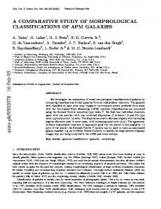
Figure 1. Scanning electron micrograph of normal reinforced (A) Gore-Tex and (B) Surgiform (scale bar: 10 m; ⫻3000). Gore-Tex micropores range from 10 to 40 m, whereas Surgiform micropores range from 1 to 15 m. Surgiform has thicker fibrils and a much more compact microstructure when compared with Gore-Tex. Internodal lengths (white lines) were measured to evaluate morphological changes of Gore-Tex and Surgiform.
Y P
Figure 2. Gross appearance of inserted Gore-Tex and Surgiform after 6 months. There was no evidence of wound infection, hematoma, or seroma formation in any specimen. Both (A) Gore-Tex and (B) Surgiform were adhered to the soft tissue.
tion. Implant blocks (4 mm in diameter and 3 mm in height) were measured, cut, and gas sterilized by ethylene oxide. Epinephrine (1:100,000) containing 1% lidocaine was injected into the cranial region and a vertical incision was made on the cranium. Minimal blunt dissection was used to create a subperiosteal pocket, and the implant was inserted into the pocket. Fifteen rats were implanted with GoreTex and 15 rats with SFAM. The skin incision was closed with 4-0 nonabsorbable sutures. After implantation, antibiotic ointment was applied to the cranial wound, and an i.v. antibiotic (cefazolin sodium, 15 mg/kg) was injected at regular intervals to prevent infection. Three groups of five rats each were killed using pentobarbital sodium at 1, 3, and 6 months after implantation. All rats survived the experiments. After specimen sampling, we examined the soft tissue surrounding the implant for inflammation and evaluated the relationship and extent of adhesion between the implant and surrounding soft tissue. Implant size changes (diameter and height) over time were measured blindly by two authors (J.H.K. and J.M.L.) using digital calipers (Digimatic caliper; Mitutoyo, Nakatsugawa, Japan).
O D
O N
T
Histological Evaluation
The sampled implant and surrounding soft tissues were fixed in 10% buffered formalin, embedded in paraffin, and sectioned to a 5-m width. Samples were stained with hematoxylin and eosin (H&E) and Masson’s trichrome and evaluated under a light microscope. Two authors (J.H.K. and C.H.P.) performed blind evaluations for reliable measurements of histological changes as described in previous studies.11,12 In terms of foreign body reactions, weak reactions were classified as moderate (⫹), strong reactions as severe (⫹⫹), and no reaction as negative (⫺). The extent of connective tissue ingrowth and the deposition of collagen between the specimens and surrounding tissues were scored as moderate (⫹) if found only in the upper portion of the specimens, severe (⫹⫹) if found in the center portion as well, and negative (⫺) if not observed at all. Slight neovascular proliferation was classified as moderate (⫹), prominent proliferation as severe (⫹⫹), and no proliferation as negative (⫺). The degree of calcification was classified as moderate (⫹) if there were
American Journal of Rhinology & Allergy
O C
fewer than two responses, severe (⫹⫹) if there were more than three responses, and negative if there were none (⫺). Samples were also observed using a scanning electron microscope (SEM, Zeiss SUPRA-55 VP field-emission scanning electron microscope; Carl Zeiss AG, Oberkochen, Germany) as described previously.12 The internodal length was measured blindly by two authors (J.H.K. and J.M.L.) to evaluate morphological changes (Fig. 1).
Statistical Analysis Comparisons of size changes between Gore-Tex and SFAM over time were statistically analyzed using a Wilcoxon’s signed-rank test. All data were analyzed using SPSS for Windows, Version 12.0 (SPSS, Inc., Chicago, IL), and values of p ⬍ 0.05 were considered statistically significant.
RESULTS Gross Findings None of the 30 rats had specific complications, such as extrusion, acute inflammation, abscess formation, seroma accumulation, or surrounding soft tissue necrosis associated with the implants. All Gore-Tex samples were fixed to surrounding tissues after 1 month, and the degree of adhesion to the surrounding soft tissue increased with time after insertion of Gore-Tex; however, separation of the block from the surrounding soft tissue was not difficult. In contrast, Surgiform samples did not adhere to the surrounding tissue and tended to migrate from the original implantation site at the end of 3 months. Surgiform was not fixed to the surrounding tissue until 6 months after insertion. Grossly, there was no inflammation or infection at the time of sampling of both Gore-Tex and Surgiform. The shapes of almost all Gore-Tex and Surgiform were preserved (Fig. 2).
Morphological Changes Almost all Gore-Tex implants decreased in size from their initial size (p ⬍ 0.05; Fig. 3). The mean diameters of the Gore-Tex were
163
Delivered by Publishing Technology to: Kosin University IP: 218.38.200.169 On: Mon, 13 May 2013 11:28:42 Copyright (c) Oceanside Publications, Inc. All rights reserved. For permission to copy go to https://www.oceansidepubl.com/permission.htm
tation. Microvessel proliferation was also observed mainly 3 months after implantation and regressed after 6 months. Partial calcification was observed on the contact surface 3 and 6 months after implantation. SEM revealed that the surrounding tissue was fully integrated with the Gore-Tex at 6 months. Internodal spaces were filled with connective tissue and deposited collagen fiber with time (Fig. 6). The internodal length progressively decreased with time and showed significant reductions 6 months after implantation (Table 3). Surgiform. Unlike Gore-Tex, surrounding tissues were separated from the Surgiform implant 3 months after implantation (Figs. 4 and 5; Table 4). Slight ingrowth of connective tissue was found 6 months after implantation. In addition, neovascularization was not observed, even at 6 months. Calcification was identified on the contact surface of bone similar to Gore-Tex. There was no integration between surrounding tissue and the Surgiform based on SEM (Fig. 6), but rather fibrous capsules surrounding the Surgiform were found at 1 and 3 months. At 6 months, there was minimal infiltration of connective tissue into the surface of the Surgiform. The internodal space was not significantly changed (Table 3).
Y P
Figure 3. Changes in Gore-Tex and Surgiform sizes with time. Table 1 Changes in Gore-Tex and Surgiform diameter (mm) according to the duration of insertion (mean ⴞ SD, diminution rate) Duration 1 mo 3 mo 6 mo
Gore-Tex
Surgiform
p Value
3.78 ⫾ 0.12 (5.60%) 3.77 ⫾ 0.07 (5.65%) 3.76 ⫾ 0.10 (5.90%)
3.93 ⫾ 0.11 (1.85%) 3.93 ⫾ 0.07 (1.85%) 3.95 ⫾ 0.07 (1.25%)
0.095 0.032 0.016
O C
DISCUSSION Table 2 Changes in Gore-Tex and Surgiform height (mm) according to the duration of insertion (mean ⴞ SD, diminution rate) Duration 1 mo 3 mo 6 mon
Gore-Tex
Surgiform
p Value
2.79 ⫾ 0.11 (7.00%) 2.73 ⫾ 0.09 (9.00%) 2.56 ⫾ 0.13 (14.67%)
2.90 ⫾ 0.13 (3.27%) 2.92 ⫾ 0.11 (2.60%) 2.91 ⫾ 0.12 (2.80%)
0.151 0.049 0.008
O D
Histological Evaluation Gore-Tex. In H&E (Fig. 4) and Masson’s trichrome (Fig. 5)–stained sections, there were mild inflammatory reactions around the surface of the Gore-Tex 1 month after implantation. Foreign body reactions were not found in Gore-Tex–implanted specimens 1, 3, and 6 months after implantation. Ingrowth of connective tissue into the Gore-Tex increased with time after implantation. Collagen was infiltrated into the central part of the Gore-Tex in all samples 6 months after implan-
164
T
O N
3.78 ⫾ 0.12 mm (5.6% diminution rate), 3.77 ⫾ 0.07 mm (5.7%), and 3.76 ⫾ 0.10 mm (5.9%) at 1, 3, and 6 months after implantation, respectively. The diameters of the Gore-Tex were significantly decreased at 1 month (p ⫽ 0.008), but did not significantly decrease with time (p ⬎ 0.05). The mean heights of the Gore-Tex were 2.79 ⫾ 0.11 mm (7.0% diminution rate), 2.73 ⫾ 0.09 mm (9.0%), and 2.56 ⫾ 0.13 mm (14.7%) at 1, 3, and 6 months after implantation, respectively. There was no statistical change in the height of Gore-Tex between 1 and 3 months (p ⫽ 0.690); however, there was a statistically significant decrease in height between 3 and 6 months (p ⫽ 0.032). In contrast with Gore-Tex, the sizes of the Surgiform implants were not changed when compared with their preimplantation size (p ⬎ 0.05). The mean diameters of the Surgiform were 3.93 ⫾ 0.11 mm (1.85% diminution rate), 3.93 ⫾ 0.07 mm (1.85%), and 3.95 ⫾ 0.07 mm (1.25%) at 1, 3, and 6 months after implantation, respectively. The mean heights of the Surgiform were 2.90 ⫾ 0.13 mm (3.3% diminution rate), 2.92 ⫾ 0.11 mm (2.6%), and 2.91 ⫾ 0.12 m (2.8%) at 1, 3, and 6 months after implantation, respectively. When comparing diameter and height changes between Gore-Tex and Surgiform implants 6 months after implantation, the values of Gore-Tex implants were significantly reduced as compared with those of Surgiform (p ⬍ 0.05; Tables 1 and 2).
Gore-Tex has been a popular augmentation material since the United States Food and Drug Administration approved it as an implantation material for cosmetic surgery in 1993.8 It is chemically safe, highly biocompatible, and nondegradable.4,7–9,15 Gore-Tex has a microstructure typified by nodes interconnected by fine fibrils and multiple pores. The pore diameter ranges from 10 to 40 m, and moderate ingrowth of fibroblasts, capillaries, and collagen into the pores allow Gore-Tex implants to be fixed in position and stable. In addition, contracture is minimized. However, the implant size can be decreased because of reductions in pore size caused by pressure.12,14 On the other hand, the Surgiform micropores range from 1 to 15 m and do not easily allow infiltration of surrounding tissue. In addition, it is suspected that the size of Surgiform does not change considerably, similar to silicone. During augmentation rhinoplasty using ePTFE it is important that the height of augmentation is determined based on the morphological and histological changes of e-PTFE. Although there have been a few studies on e-PTFE, there have been no reports specifically on Surgiform. e-PTFE elicited a significantly thinner capsule and more neovascularization in a rabbit model when compared with silicone.13 In addition, in e-PTFE removed from the human body, the degree of foreign body reaction and collagen deposition varied with each specimen and had no apparent relationship with the duration. Neovascularization was observed in specimens with durations of ⬎12 months, and partially degenerative calcification was observed in a specimen with a 36-month duration. Capsule formation was not observed in removed e-PTFE.16 In this study, adhesion of the Gore-Tex to the surrounding tissue increased with time, and strong capsular formation was not observed at the time that the inserted Gore-Tex was harvested, and removal of blocks was relatively easy. Furthermore, the structure of the Gore-Tex was preserved without inflammation or infection based on gross findings. These gross findings were similar to those of previous studies.10,15 The size of the Gore-Tex changed with time, which differs from the results of other studies,16,17 indicating that overcorrection or undercorrection is not required in facial augmentation surgery. The height decreased to 14.7% of the initial value after 6 months, whereas the diameter decreased to 5.9%, suggesting that horizontal pressure on the Gore-Tex block is relatively weaker than vertical pressure. The decrease in height may have resulted from the initial reduction of the Gore-Tex pores by surrounding pres-
May–June 2013, Vol. 27, No. 3
Delivered by Publishing Technology to: Kosin University IP: 218.38.200.169 On: Mon, 13 May 2013 11:28:42 Copyright (c) Oceanside Publications, Inc. All rights reserved. For permission to copy go to https://www.oceansidepubl.com/permission.htm
Figure 4. Hematoxylin and eosin (H&E) staining of (A, C, and E) Gore-Tex and (B, D, and F) Surgiform (scale bar: 50 m; ⫻800). (A) Connective tissue was incorporated into the Gore-Tex implant and moderate ingrowth was observed 1 month after Gore-Tex implantation. (B) Surrounding connective tissue was separated from Surgiform 1 month after implantation. (C) Incorporation of connective tissue into the Gore-Tex implant was stronger and connective tissue ingrowth was severe 3 months after Gore-Tex implantation. Microcalcification (arrow) and neovascular structures (arrowheads) were found within the Gore-Tex block. (D) Integration of connective tissue with the Surgiform implant was not found 3 months after implantation. (E) Collagen fibers were deposited on the inside of Gore-Tex and microcalcification had progressed 6 months after implantation. Neovascular structures were not remarkable. (F) Connective tissue was slightly integrated into the surface of the Surgiform implant, and degenerative changes of Surgiform were also found.
Figure 5. Masson’s trichrome (MT) staining of (A) Gore-Tex and (B) Surgiform (scale bar: 50 m; ⫻800) 6 months after implantation. (A) Connective tissue was incorporated with the Gore-Tex implant and collagen fibers were diffusely deposited within the Gore-Tex. (B) Ingrowth of connective tissue was insignificant and collagen deposition was still not found within Surgiform.
O D
Y P
T
O C
O N
sure.12,14 Jung et al showed that the positive correlation between initial Gore-Tex thickness and the degree of thickness change may support that.14 Therefore, the height of Gore-Tex for augmentation rhinoplasty should be determined with consideration of its height reduction with time. In our histological evaluation, foreign body reactions were not apparent, and ingrowth of connective tissue into the Gore-Tex increased with time. Collagen infiltration, neovascularization, and microcalcification were found in the 6-month specimens. These results differed from our human study in terms of time16; however, ingrowth of connective tissue into the inner portion of the Gore-Tex supported the postoperative fixation and stability of Gore-Tex and minimized contracture by reducing the surrounding capsule.18 There was no macroscopic inflammation or infection in the Surgiform-implanted specimens, as with Gore-Tex. In contrast to Gore-Tex, Surgiform was not fixed to the surrounding tissue, but
American Journal of Rhinology & Allergy
easily separated from the tissue until 6 months after implantation at the time of harvest. Furthermore, the height and diameter of Surgiform were not significantly decreased from their initial sizes, even after 6 months. These findings were confirmed by H&E and SEM evaluation, which showed that surrounding tissue never merged into the inner portion of Surgiform, but rather the surface of the Surgiform was covered with a strong capsule. Surrounding tissue was finally integrated with the surface of the Surgiform after 6 months of implantation. Because Surgiform has dense pores, it may be resistant to pressure, as is silicone, and tissue ingrowth into dense pores is difficult, resulting in a high risk of capsule formation, instability, and migration. In addition, collagen infiltration and neovascularization were not observed in the Surgiform-implanted specimens. These findings suggest that overcorrection does not need to be considered in augmentation using Surgiform, but implant migration should be kept in mind.
165
Delivered by Publishing Technology to: Kosin University IP: 218.38.200.169 On: Mon, 13 May 2013 11:28:42 Copyright (c) Oceanside Publications, Inc. All rights reserved. For permission to copy go to https://www.oceansidepubl.com/permission.htm
Y P
O C
T
Figure 6. Scanning electron micrograph of inserted (G) Gore-Tex and (S) Surgiform (scale bars: 10 m; ⫻3000). Integration of connective tissue with Gore-Tex progressed with time after implantation. However, a capsule had formed between connective tissue and Surgiform 1 and 3 months after implantation. Slight integration between the surrounding tissue and surface of the Surgiform implant was found 6 months after implantation.
O N
Table 3 Changes in Gore-Tex and Surgiform intermodal length (m) according to the duration of insertion (mean ⴞ SD, diminution rate)
O D
Duration
Gore-Tex
28.37 ⫾ 5.66 26.39 ⫾ 7.24 (7.07%) 24.49 ⫾ 5.53 (13.77%) 14.60 ⫾ 2.66 (48.58%)
Control 1 mo 3 mo 6 mo
p Value
Surgiform
p Value
0.961 0.961 0.015
4.72 ⫾ 0.39 4.65 ⫾ 0.87 (1.39%) 4.35 ⫾ 0.87 (7.79%) 4.25 ⫾ 0.67 (9.86%)
0.961 0.462 0.078
The p value compared with the control.
Table 4 Light microscopic findings according to the duration of Gore-Tex and Surgiform implantation Duration
1 mo 3 mo 6 mo
Foreign Body Reaction
Connective Tissue Ingrowth
Collagen Deposit
Neovascularization
Calcification
G
S
G
S
G
S
G
S
G
S
⫺ ⫺ ⫺
⫺ ⫺ ⴚ
⫹ ⫹⫹ ⫹⫹
⫺ ⫺ ⫹
⫺ ⫹ ⫹⫹
⫺ ⫺ ⫺
⫺ ⫹ ⫺
⫺ ⫺ ⫺
⫺ ⫹ ⫹
⫺ ⫹ ⫹⫹
G ⫽ Gore-Tex; S ⫽ Surgiform; ⫺ ⫽ negative; ⫹ ⫽ moderate; ⫹⫹ ⫽ severe.
166
May–June 2013, Vol. 27, No. 3
Delivered by Publishing Technology to: Kosin University IP: 218.38.200.169 On: Mon, 13 May 2013 11:28:42 Copyright (c) Oceanside Publications, Inc. All rights reserved. For permission to copy go to https://www.oceansidepubl.com/permission.htm
6.
Microcalcification was found at the implant–bone interface of both Gore-Tex– and Surgiform-implanted specimens after 6 months. Thus, partial degenerative changes were observed over time in both Gore-Tex and Surgiform implants, suggesting that the stability of e-PTFE may not be perfect. Further specific evaluations regarding the stability of e-PFTE are needed. Additionally, we must beware of the risk of postoperative infection and extrusion of e-PTFE in rhinoplasty. Winkler et al. recently reported that in patients in whom e-PTFE was used during rhinoplasty, the postoperative infection rate was 5.3% and the extrusion rate was 2.7%.19 The rate of these complications is higher than that in rhinoplasty using autologous cartilage.20
7.
8.
9.
10.
11.
CONCLUSIONS In this study, we observed that the height of Gore-Tex was reduced to 14.7% of the initial value after 6 months. Connective tissue ingrowth and collagen deposition increased with time after Gore-Tex implantation. Therefore, Gore-Tex should be carefully trimmed during augmentation because of its potential to decrease in size over time. On the other hand, the size of the Surgiform was not significantly changed when compared with its initial value. In the Surgiform-implanted samples, surrounding tissues were not integrated with the surface of the Surgiform until 6 months after insertion, and neovascularization was not clearly found. Accordingly, migration of Surgiform should be carefully considered.
12.
13.
14.
15.
T
REFERENCES 1.
2. 3.
4. 5.
O D
16.
17.
Owsley TG, and Taylor CO. The use of Gore-Tex for nasal augmentation: A retrospective analysis of 106 patients. Plast Reconstr Surg 94:241–248, 1994. Silver WE, and Goldberg J. Nasal grafts and implants. Facial Plast Surg Clin North Am 2:477–499, 1994. Jung DH, Jung YK, Lee WW, and Paik SI. A study on grafts and implants materials in augmentation rhinoplasty. Korean J Otolaryngol 39:250–257, 1996. Maas CS, Monhian N, and Shah SB. Implants in rhinoplasty. Facial Plast Surg 13:279–290, 1997. Lovice DB, Mingrone MD, and Toriumi DM. Grafts and implants in rhinoplasty and nasal reconstruction. Otolaryngol Clin North Am 32:113–141, 1999.
O N
American Journal of Rhinology & Allergy
Rothstein SG, and Jacobs JB. The use of Gore-Tex implants in nasal augmentation operations. Entechnology Sep:40–45, 1989. Queen TA, and Palmer FR III. Gore-Tex for nasal augmentation: A recent series and a review of the literature. Ann Otol Rhinol Laryngol 104:850–852, 1995. Godin MS, Waldman SR, and Johnson CM. The use of expanded polytetrafluoroethylene (Gore-Tex) in rhinoplasty. A 6-year experience. Arch Otolaryngol Head Neck Surg 121:1131–1136, 1995. Sclafani AP. Clinical and histological response of subcutaneous expanded polytetrafluoroethylene (Gore-Tex) and porous high-density polyethylene (Medpore) implants to acute and early infection. Arch Otolaryngol Head Neck Surg 121:328–336, 1997. Maas CS, Gnepp DR, and Bumpous J. Expanded polytetrafluoroethylene (Gore-Tex soft-tissue patch) in facial augmentation. Arch Otolaryngol Head Neck Surg 119:1008–1014, 1993. Park CH, and Chun JH. Histological and morphological change of implanted reinforcement Gore-Tex in nasal dorsum of rabbit. Korean J Otorhinolaryngol Head Neck Surg 51:705–711, 2008. Kim JH, Park CH, Lee OJ, et al. Histologic changes in transplanted expanded polytetrafluoroethylene in an animal model. Laryngoscope 122:17–22, 2012. Batniji RK, Hutchison JL, Dahiya R, et al. Tissue response to expanded polytetrafluoroethylene and silicone implants in a rabbit model. Arch Facial Plast Surg 4:111–113, 2002. Jung YG, Kim HY, Dhong HJ, et al. Ultrasonographic monitoring of implant thickness after augmentation rhinoplasty with expanded polytetrafluoroethylene. Am J Rhinol Allergy 23:105–110, 2009. Cohen MS, Constantino PD, and Friedman CD. Biology of implants used in head and neck surgery. Facial Plast Surg Clin North Am 7:17–33, 1999. Park CH. Histological study of expanded polytetrafluoroethylene (Gore-Tex) implanted in the human nose. Rhinology 46:317–323, 2008. Singh S, and Baker JL. Use of expanded polytetrafluoroethylene in aesthetic surgery of the face. Clin Plast Surg 27:579–593, 2000. Lewis RP, Schweitzer J, Odum BC, et al. Sheets, 3-D strands, trimensional (3-D) shapes, and sutures of either reinforced or nonreinforced expanded polytetrafluoroethylene for facial soft-tissue suspension, augmentation, and reconstruction. J Long Term Eff Med Implants 8:19–42, 1998. Winkler AA, Soler ZM, Leong PL, et al. Complications associated with alloplastic implants in rhinoplasty. Arch Facial Plast Surg. 27: 1–5, 2012. Lee MR, Unger JG, and Rohrich RJ. Management of the nasal dorsum in rhinoplasty: A systematic review of the literature regarding technique, outcomes, and complications. Plast Reconstr Surg 128:538e– 50e, 2011. e
18.
19.
20.
Y P
O C
167
Delivered by Publishing Technology to: Kosin University IP: 218.38.200.169 On: Mon, 13 May 2013 11:28:42 Copyright (c) Oceanside Publications, Inc. All rights reserved. For permission to copy go to https://www.oceansidepubl.com/permission.htm