Comparative study of the electrochemical behavior ...
Recommend Documents
Dec 12, 2000 - Duros de Carbonitreto de Titânio. D.Sc. Thesis, Uni- versidade Federal do Rio de Janeiro, 1998. 13.Achete, C.A.; Senna, L.F.; Freire Jr, F.L.; ...
Jun 12, 2016 - 1 School of Applied Physics and Materials, Wuyi University, Jiangmen, Guangdong 529020, China. 2Department of Physics, Hi-Tech.
Jaeshin Kima, Gregory V. Korshina,Ð. , Alexander B. Velichenkob ... sediments or sludge (Montgomery-Brown and Reinhard. 2003); however, the water column ...
Gray FM, Connor JA (eds) (1997) Polymer electrolytes (RSC ma- ... Croce F, Appetecchi GB, Persi L, Scrosati B (1998) Nanocomposite polymer electrolytes for ...
emission bands has been very important in order to elucidate their chemistry in ... Pyridoxal-5'-phosphate (PLP) and pyridoxal (PL) are able to catalyse a great ...
Aug 2, 2012 - The zinc metal treated with ATSC at 5% has showed good corrosion resistance ..... [16] M. Ihara, H. Nishihara, and K. Aramaki, âMechanism of.
Mar 24, 2011 - International Journal of Analytical Chemistry. Volume 2011 ... Analytical Detection of the p-Nitrophenol Using Silver. Solid Amalgam .... from the city, is relatively free from urban or industrial pollution. .... the electron transfer
Jan 7, 2011 - On Au(111), a (31/2 Р31/2)R30° arrangement of the FSN SAMs is ... potential range where the redox reaction of FSN molecules does not occur ...
of accumulated H2O2 concentration were pH = 3, j = 1.28 mA cm-2 and 0.1 M ... hydrogen peroxide electrochemically produced since nearly twice H2O2 was ...
Key Words: BDD cathode, graphite cathode, reduction oxygen, hydrogen peroxide. ABSTRACT .... Samples were withdrawn from the reactor at regular time intervals and the ... -2.7 x 10⁻⁵ mA and a cathodic potential (Epc) -1.0 V vs. Ag/AgCl.
Nov 2, 2014 - Statistical analysis such as noise resistance, lo- calization index, skewness and kurtosis has been evaluated. Noise resistance showed a good.
Mar 30, 2017 - high energy and power density at low cost [4]. For instance, with .... 10% fluoroethylene carbonate (FEC) as the electrolyte. Pure Li foil.
Jul 13, 2012 - A novel comparative force degradation ultra-performance liquid chromatographic assay method was developed and validated for Telmisartan ...
nica intima, the tunica media and the tunica adventitia). This dilatation may multiply the risk of developing some pathology such as: the aneurysm and the ...
Background: Electrochemical oxidation considered to be among the best methods in ... Commons Public Domain Dedication waiver (http://creativecommons.org/.
ELICO (CL-25D) polarograph with chart recorder (LR-101P) were used. pH were maintained using ELICO pH meter (LI-120) and temperature of the solution ...
Sep 17, 2018 - Cite this article as: Fida Hussain, Ghazanfar Abbas, M. Ashfaq Ahmad, Rizwan. Raza ... This is a PDF file of an unedited manuscript that has been accepted for ... aDepartment of Electrical Engineering, COMSATS University, ..... fundame
... A RECTANGULAR. DUCT IN THE PRESENCE OF THREE PLANAR BAFFLES ... Department of Mechanical Engineering, Institute of Technology. University ...
Turbulent kinetic energy. L. Channel length, m l1,l2,l3. The three inter fin spacing, m. Nu. Nux. Averaged Nusselt number. Local Nusselt number. P. Pressure, Pa.
seguida o deslocamento da onda para potenciais mais negativos. O manganês(II), de comportamento diferente dos demais metais estudados, mostra apenas ...
Mar 31, 2017 - composites: Effect of type of fiber and matrix ... of two laminated composites at different fiber and matrix system. ..... Creep Behavior of Basalt.
of flavin adenine dinucleotide (FAD) and flavin mononucleotide (FMN) ..... voltamograms were not well defined, showing that the interaction is not exactly the ...
Comparative study of the electrochemical behavior ...
Oct 29, 2013 - applications of (bio)sensing platforms based on the use of multi-walled ... E-mail addresses: [email protected] (E.N. Primo), [email protected] (G.A. Rivas). 0003-2670/$ .... mers for solar cells and drug-delivery.
Comparative study of the electrochemical behavior and analytical applications of (bio)sensing platforms based on the use of multi-walled carbon nanotubes dispersed in different polymers E.N. Primo a,∗ , F.A. Gutierrez a , G.L. Luque a , P.R. Dalmasso a , A. Gasnier a , Y. Jalit a , M. Moreno b , M.V. Bracamonte a , M. Eguílaz Rubio a , M.L. Pedano a , M.C. Rodríguez a , N.F. Ferreyra a , M.D. Rubianes a , S. Bollo c , G.A. Rivas a,∗ a
INFIQC, Departamento de Fisicoquímica, Facultad de Ciencias Químicas Universidad Nacional de Córdoba, Ciudad Universitaria, 5000 Córdoba, Argentina Departamento de Química Analítica y Análisis Instrumental, Facultad de Ciencias, Universidad Autónoma de Madrid, Espa˜ na c Laboratorio de Bioelectroquímica, Facultad de Ciencias Químicas y Farmacéuticas, Universidad de Chile, Santiago, Chile b
h i g h l i g h t s
g r a p h i c a l
a b s t r a c t
• CNT-polymers fully cover the GCE delimiting areas with higher density of CNT. • Sonication of CNT and ethanol are necessary to obtain efficient CNTs dispersions. • CNTs dispersion depends on the polymer, sonication time, solvent, CNT/ratio. • CNT-polymer is successfully used as platform to build biosensors.
a r t i c l e
i n f o
Article history: Received 22 July 2013 Received in revised form 29 September 2013 Accepted 21 October 2013 Available online 29 October 2013 Keywords: Carbon nanotubes dispersion Glassy carbon electrodes Electrochemical (bio)sensors Biopolymers Bioanalytes Non-covalent functionalization
Emiliano N. Primo obtained his degree in chemistry (2010) from Córdoba National University (Córdoba, Argentina), graduating with an Honor Award. At present, he is a Ph.D. student at the Physical Chemistry Department, Faculty of Chemical Sciences (Córdoba National University) where he holds an auxiliary professor position. His research interests focus on the functionalization of carbon nanotubes, graphene and graphene oxide with nucleic acids for the design of electrochemical and plasmonic bio-interfaces aimed to the development of genosensors.
Pablo R. Dalmasso received his Ph.D. in chemistry from the National University of Córdoba (Córdoba, Argentina) in 2009. He did the postdoctoral training in the Biosensors Group at the Physical Chemistry Department, Faculty of Chemical Sciences (National University of Córdoba) under the supervision of Prof. Gustavo A. Rivas between 2010 and 2012. He is currently assistant researcher at Argentine Research Council (CONICET).
Fabiana A. Gutierrez obtained her Ph.D. in chemistry (2009) from the Córdoba National University (Argentina). During her Ph.D. in the group of electrochemistry and surfaces, she focused on the functionalization of metal surfaces. She did the postdoctoral training in the group of Biosensors at the Physical Chemistry Department, Faculty of Chemical Sciences (Cordoba National University), she focused on electrochemical biosensors based on the functionalization of carbon nanotubes and magnetic nanoparticles. At present, she is assistant researcher at CONICET and assistant professor at Córdoba National University. She is an active member of the Biosensors Group at the Physical Chemistry Department, and her research interests focus on electrochemical and plasmonics genosensors and immunosensors: design, characterization and analytical applications for the quantification of clinical and environmental biomarkers.
Aurelien Gasnier joined the group of Pr. Audebert in 2005 and received his M.S. in organic synthesis from Université Paris-Sud (France). During his Ph.D. of organic chemistry at Université Joseph Fourier (France) he developed soluble coordination polymers based on electroactive terpyridine-cyclam and characterized their gel state under the co-direction of Profs. Moutet, Royal and Térech. In 2009 he performed one year of postdoct at University of Chicago (U.S.) in the group of Pr. Luping Yu where he synthesized polymers for solar cells and drug-delivery. Since 2010, he is dedicated to the development of nanostructured and polymer functionalized graphene and carbon nanotube electrodes in the group of Prof. Gustavo Rivas at Universidad Nacional de Córdoba (Argentina). He is assistant researcher at CONICET and assistant professor at Córdoba National University.
Guillermina L. Luque graduated in chemistry en 2003. She received her PhD in 2009 from National University of Cordoba. Her scientific activity is carried out at the National Institute of Physical-Chemistry of Córdoba of the National University of Cordoba (Argentine). Her current research interest includes theoretical and experimental studies of graphene and its application in electrochemical sensors and biosensors, lithium batteries, and hydrogen storage.
Yamile Jalit obtained her Ph.D. in chemistry (2013) from the Córdoba National University (Argentina). She did the doctoral training in the group of Biosensors at the Physical Chemistry Department, Faculty of Chemical Sciences (Cordoba National University), and she focused on Electrochemical and Optical Biosensors based on the surface biofunctionalization with nanomaterials to design new platforms for the detection of DNA, oligonucleotides and aptamers.
Dr. Mónica Moreno obtained her B.A. of science, section chemistry (June 1999) and her Ph.D. in chemistry (December 2006) from Autonomous University of Madrid (Spain). Since 2003 and to date she has been employed at this university. She is currently hired doctor teacher LOU. Her research is focused on capillary electrophoresis, development of new sensors based on carbon and their coupling to FIA systems, HPLC or capillary electrophoresis and new electroanalytical applications in environmental and food (herbicides, pollutants and natural products, mainly polyphenols).
E.N. Primo et al. / Analytica Chimica Acta 805 (2013) 19–35
Victoria Bracamonte obtained her PhD degree in Chemistry (2012) from National University of Córdoba (Córdoba, Argentina). At present, Dra. Victoria Bracamonte is a postdoc student at the Institute of Physics “Enrique Gaviola”, Faculty of Mathematic, Astronomy and Physicand she holdsan auxiliary professor position at the Physical Chemistry Department, Faculty of Chemical Sciences (National University of Córdoba). Her research interests focus on the synthesis and characterization of graphene based nanomaterials and liquid phase exfoliation of graphene in different media.
Marcos Eguílaz Rubio received his degree in chemical sciences with a specialization in Environment from the Complutense University of Madrid (Madrid, ˜ in 2006 and his Ph.D. in analytical chemistry Espana) in 2012 from the same university. He performed postdoctoral research in the group of Electroanalysis and Electrochemical (bio)sensors of the Complutense University of Madrid between 2012 and 2013. At present, he is a postdoctoral fellow of CONICET in the Biosensor’ Group at the Physical Chemistry Department, Faculty of Chemical Sciences, National University of Cordoba, Argentina. His postdoctoral research is focused on the design and characterization of biofunctional analytical platforms based on carbon nanotubes and graphene functionalized with polymers.
María Laura Pedano obtained her Ph.D. in chemical sciences (2006) from the National University of Córdoba (Córdoba, Argentina). She was awarded the Prize Enrique Herrero Ducloux, from the Argentinean Chemical Association to the best doctoral thesis in Physical Chemistry in the period 2004–2006. She performed postdoctoral research at Joseph Fourier University (Grenoble, France) during 2007 and at Northwestern University (Chicago, U.S.A) between 2008 and 2010. At present, she is assistant professor at the Department of Physical Chemistry, Faculty of Chemical Sciences, National University of Córdoba; and associate researcher at the Argentine Research Council (CONICET). She is an active member of the Biosensor’s Group at the Physical Chemistry Department, and her research now is focusing in the development of plasmonic nanostructures and its application to optical and electrochemical biosensors.
Marcela C. Rodríguez studied biochemistry at the National University of Córdoba, Argentina. After completing her Ph.D. thesis under the supervision of Professor Gustavo Rivas in 2003, she was a postdoctoral fellow in the group of Professor Joseph Wang from 2004 to 2005 at the Bioelectronics and Biosensors Center of Arizona State University, USA. Her postdoctoral studies were focused on the design of aptasensing devices for protein detection and the coupling of nanomaterials to sensing platforms. She is the recipient of the José A. Catoggio Award from Argentine Association of Analytical Chemists (2005). She is Independent Researcher of Argentine Research Council (CONICET) and since 2009 she is professor of chemistry and electroanalytical chemistry at the National University of Córdoba Professor Rodríguez’ research interests include the development of novel optical and electrochemical nanomedical-theranostics-biosensing systems involving specific nucleic acids, aptamers and nanostructured materials.
21
Nancy F. Ferreyra obtained her Ph.D. in chemistry in 2002 from the National University of Córdoba (Argentina). She performed her postdoctoral training at Université Joseph Fourier (Grenoble, France). In 2004 she joined the team headed by Prof. Rivas at Physical Chemistry Department, Faculty of Chemical Sciences, National University of Córdoba, where she is currently Professor and Independent Researcher at National Research Council (CONICET, Argentina). Her research interests are focused on the development of electrochemical biosensors based on carbon nanotubes and self-assembled structures of functionalized gold nanoparticles and biomolecules.
María D. Rubianes obtained her Ph.D. in chemistry (2005) from the Córdoba National University (Argentina). She did the postdoctoral training in the group of Polymers at the Organic Chemistry Department, Faculty of Chemical Sciences (Cordoba National University). At present, she is professor at the Department of Physical Chemistry, Faculty of Chemical Sciences of Cordoba National University and Adjunt Researcher at Argentine Research Council (CONICET). She is an active member of the Biosensors Group at the Physical Chemistry Department, and her research interests focus on the synthesis and characterization of modified polymers for the functionalization of nanostructured materials.
Soledad Bollo obtained her Ph. D. in chemistry (1998) from University of Chile (Santiago, Chile). She did the postdoctoral training at New Mexico State University, Las Cruces (USA) in 1998. At present, she is full professor at University of Chile. Since 2008, she heads the Department of Pharmacological and Toxicological Sciences at the Faculty of Chemical and Pharmaceutical Sciences. Her research interests focus on the design and characterization of electrochemical (bio)sensors based on carbon nanotubes and the study of DNA damage. She has over 60 peer-reviewed papers and one book chapter.
Gustavo A. Rivas obtained his Ph.D. in chemistry (1991) from Cordoba National University (Cordoba, Argentina). He did the postdoctoral training at University of Valence, Valence (Spain) in 1994 and 1995 and at New Mexico State University, Las Cruces (USA) between 1995 and 1996. At present, he is full professor at Cordoba National University and Superior Researcher at Argentine Research Council (CONICET). He was the head of the Department of Physical Chemistry at the Faculty of Chemical Sciences between 2008 and 2010. He was President of the Argentinean Society of Analytical Chemists between 2005 and 2007. He is the recipient of the Ranwell Caputto Award from National Academy of Sciences of Argentina (2001), Rafael Labriola Award from Argentinean Society of Chemistry (2004), and Konex Award in Nanotechnology (2013). His research interests focus on the design and characterization of electrochemical (bio)sensors based on nanostructured materials, the development of new bioanalytical platforms for biosensing in batch and flow systems, the study of DNA damage, and the design of new strategies for nanomaterials and polymers modification. He has over 120 peer-reviewed papers and two book chapters. He is Editor of Sensors and Actuators B: Chemical and belong to the Editorial Board of Analytical Letters and Electroanalysis.
22
E.N. Primo et al. / Analytica Chimica Acta 805 (2013) 19–35
1. General concepts about CNT and CNT-based electrochemical (bio)sensors
2. Distribution of CNT at glassy carbon surfaces modified with CNT-polymers and electrochemical reactivity
Carbon nanotubes (CNTs) have been largely used for electrochemical (bio)sensing due to their outstanding mechanical, electronic and structural properties [1–13]. CNTs are one of the allotropes of carbon and consist of carbon atoms with sp2 hybridization arranged in graphene sheets rolled up in a tube [14]. According to the number of walls, they can be divided in singlewall (SWCNTs) and multi-wall (MWCNTs) CNTs [14]. Depending on the presence of side-walls defects, MWCNTs can be separated in hollow type MWCNTs (hCNTs) which present ideally defect-free side walls, and bamboo-like MWCNTs (bCNTs), that are characterized by having transverse walls regularly located along the tubes resulting in edge planes of graphene material at regular intervals along the walls [14]. Due to the strong – interactions between their aromatic rings, CNTs tend to aggregate in aqueous solutions, making difficult their dispersion and further use for the development of electrochemical (bio)sensors [6,7,9,14,15]. Therefore, several alternatives have been proposed to circumvent this problem. Among them, the non-covalent functionalization has been largely used since it does not disturb the unique electronic structure of CNTs [15,16]. Polymers [17–30] have allowed the successful dispersion of CNTs mainly by wrapping the nanostructures through stacking with their surface. Table 1 summarizes the most representative electrochemical (bio)sensors obtained by immobilization of CNT dispersed in polymers reported in the last two years. Different conductive substrates have been used for the preparation of CNT-based electrochemical sensors; glassy carbon, gold, pyrolytic graphite, graphite paste, indium tin oxide (ITO) and Pt being the most representative [31–65]. The dispersions were obtained by using polymers like chitosan, poly(diallyldimethylammonium) chloride (PDDA), poly(3,4-ethylenedioxythiophene), osmium derived polymers, poly-(4-vinylpiridine), poly(aniline), poly(1,2diaminobenzene), cyclodextrin, single stranded DNA, poly(allylamine) hydrochloride, poly(acrylic acid), Nafion, poly(ethylenimine) (PEI), poly(styrene) sulfonate, among others [31–65]. They were used for the detection of pharmaceutical products, biomarkers, cells, phenolic compounds either in standard solutions or in complex matrices, like, soil, tissue, wax, foods and medicines [31–65]. The first contribution of our group in the field of CNTs was the dispersion of MWCNT in mineral oil for the development of highly sensitive, selective and versatile (bio)sensors. Since 2007, our group reported the use of new polymers for dispersing MWCNT, PEI [66–70], PEI functionalized with dopamine (PEI-Do) [71], poly(lysine) (Polylys) [72,28,73], poly(histidine) (Polyhis) [74–76], glucose oxidase (GOx) [77,78] and double stranded calf-thymus DNA (dsDNA) [79], polymers that allow not only the dispersion of CNTs, but also the development of new electrochemical (bio)sensors. In this review, we present some new insights and a critical comparison of the electrochemical behavior of glassy carbon electrodes (GCE) modified with the CNT-polymers dispersions previously indicated. The comparison was focused on the analysis of the dispersions preparation conditions, the influence of the sonication time, solvent, polymer/CNT ratio and nature of the polymer on the efficiency of the dispersions, on the behavior of the resulting modified electrodes and (bio)sensing applications. The electrochemical response of the different CNT-polymers modified GCE was comparatively evaluated using ascorbic acid as redox marker. The analytical applications of the resulting (bio)sensors for the quantification of biomarkers of clinical relevance like glucose, NADH, dopamine and DNA are also discussed in the following sections.
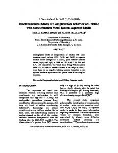
To know the distribution of the (bio)sensing elements/modifiers on the surface of the electrodes when developing (bio)analytical platforms is a very important aspect, since most of the properties of the modified surfaces will depend on the efficiency of this surface coverage. Therefore, to evaluate the distribution of CNT at the surface of GCE modified with CNT dispersed in polymers is a crucial aspect. Fig. 1 compares Scanning Electron Microscopy (SEM) micrographs of GCE modified with different CNTs-polymer dispersions: (A) CNT-GOx; (B) CNT-PEI; (C) bCNT-dsDNA; and (D) bCNT-Polyhis. The carbon nanostructures were hollow-type MWCNTs with exception of the dispersion with dsDNA, where the MWCNTs were bamboo-like MWCNTs. The comparative analysis of the images demonstrates that in all cases the surface of GCE is completely covered by the CNT-dispersions although this coverage is not homogeneous (Fig. 1A–D). In general, there is are pattern of a full covered surface with areas containing different density of CNTs, which depend on diverse factors such as the solvent used to prepare the dispersion, the nature of the polymer selected to disperse the CNTs and the CNTs/polymer ratio. Fig. 1C shows one example about the influence of the solvent on the efficiency of the dispersion and the distribution of the dispersed CNTs on the glassy carbon surface. This picture displays a GCE modified with bCNT-dsDNA prepared in 50:50 (v/v) ethanol/water (main picture) and water (inset). It is clear that the presence of ethanol largely improves the quality of the dispersion and its deposition on the glassy carbon surface. On the contrary, the use of water as solvent produces a deposit with an important number of aggregates. From the analysis of these images and those for other CNT-polymer dispersions previously reported [66–72,28,73–79], it is possible to conclude that, the distribution of the CNTs-polymer dispersion at the electrode surface highly depends on the solvent, and, in general, the best coverage is obtained using ethanol as co-solvent. Another interesting aspect to consider is the influence of the polymer concentration on the quality of the dispersion and further deposition at the glassy carbon surface. Fig. 1D shows images of GCE modified with 1.00 mg mL−1 CNT dispersed in 0.25 mg mL−1 Polyhis (main picture) and 2.00 mg mL−1 Polyhis (inset), both solutions prepared in 75/25 (v/v) ethanol/acetate buffer pH 5.00. The comparison of these images reveals that when the concentration of Polyhis increases, there is a segregation of the CNTs entrapped in the polymer net. After the critical analysis of these results, it is possible to conclude that, when preparing dispersions of CNTs in polymer solutions is necessary to take into account that: (a) ethanol has to be present in the polymer solution to obtain a more efficient dispersion and better distribution of the wrapped CNTs on the electrode surface due to the decrease in the viscosity of the medium and the improvement in the mobility of the polymeric chains, and (b) the nature and the amount of the polymer are critical factors, since the concentration has to be high enough to ensure the dispersion of the nanostructures, but not too high that blocks the electron transfer. To know the correlation between the information given by the distribution of CNTs on the electrode and the local electroactivity of the resulting surface is very useful when developing electrochemical (bio)sensors Fig. 2 displays Electrochemical Scanning Microscopy (SECM) images obtained at bare GCE and at GCE modified with bCNT-dsDNA, CNT-PEI, and CNT-Polylys using ferrocene methanol as redox marker. In all cases is possible to observe that the average currents are similar to the one for GCE, although these values depend on the region of the electrode under evaluation. In fact, there are areas with higher density of CNTs that produces local increments in the currents, in agreement with SEM images. This effect is more pronounced at GCE/CNT-PEI. On the
Table 1 Electrochemical (bio)sensors based on CNTs dispersed in different polymers: analytical parameters and applications in real samples. Modified electrode
Detection
Analyte
Real sample
Analytical performance
Ref.
GCE/MWCNT-CHIT/MIP
DPV (5 min accumulation at o.c.p.)
Pork samples
LR: 2.0 × 10⁻⁶ –1.0 × 10⁻³ M DL: 4.4 × 10⁻⁷ M
[31]
PGE/MWCNT-CHIT/dsDNA
DPV (5 min accumulation at o.c.p.)
quinoxaline-2-carboxylic acid (residue marker for quinoxaline veterinary medicines of illegal use) Amitrol
LR1 : 0.1–10 M LR2 : 10–500 M DL: 0.03 M Sensitivity: 174.0 mA mM−1 cm−2 LR: 5–85 mM DL: 0.72 mM Sensitivity: 174.0 mA mM−1 cm−2 LR: 8 × 10⁻⁸ –2 × 10⁻⁵ M DL: 3 × 10⁻⁸ M Sensitivity: 0.46 mA mM−1 LR: 0.1–1.0 mM DL: 0.031 mM
[41]
Sensitivity: (817.3 ± 185.8) nA mM−1 LR: up to 7 mM DL: 1 M tR ∼ 5 s Interferences: AA:7.9%; UA:13.7%; acetaminophen: 7.2% Sensitivity: (45.6 ± 10.8) nA mM−1 LR: up to 510 M DL: 2 M tR ∼ 8 s Sandwich type biosensor: after interaction with the sample, the platform was left incubate with anti-IgG secondary antibody labeled with HRP LR: 5.0 × 10⁻¹⁰ –10.0 × 10⁻⁸ M DL: 5.0 × 10⁻¹⁰ M 93% of regeneration of the sensing surface after five regenerationhybridization-intercalation cycles RSD: 4.4% (n = 5) Sensitivity:0.056 A mM−1 cm−2 LR: up to 5.3 mM DL: 0.05 mM
Sensitivity: 2.3 A mM−1 LR: 0.02–1.2 mM DL: 4.4 M tR ∼ 30–35 s RSD: 2.8% (n = 5) Storage or operational stability: 7 days (88%) Sensitivity: 18.6 A mM−1 LR: 0.5–22 M DL: 0.4 M tR ∼ 15–20 s Storage or operational stability: 4 weeks (89%) RSD intraday: 1.4% (n = 10)
E.N. Primo et al. / Analytica Chimica Acta 805 (2013) 19–35
25
contrary, at GCE/bCNT-dsDNA the surface looks more homogeneous, suggesting an important ordering effect of the biomolecule in the multistructure. Therefore, the deposition of CNT-polymer dispersions at the electrodes surfaces generate areas with different density of CNTs, that are associated with heterogeneous local increments in the redox probes currents transduced in SECM electroactive spots.
3. Influence of the preparation conditions on the electrochemical behavior of GCE modified with CNT-polymers 3.1. Effect of the sonication time Chen et al. [80] reported that the cavitation produced as a consequence of the ultrasonication is the responsible for debundling and dispersing the nanotubes. Thus, the time of ultrasonication is a critical parameter to obtain an efficient dispersion of CNT in the polymer matrix and a highly stable functionalization of the carbon nanostructures. The effect of this parameter on the final dispersion of the nanostructures and further activity of the resulting CNT-modified GCE is comparatively evaluated here using CNT dispersions involving polyelectrolytes with clearly different properties: (i) a positively charged homopolymer, Polyhis; (ii) a negatively charged enzyme, GOx; and (iii) a negatively charged polyelectrolyte with bioaffinity properties, dsDNA. The analysis was performed from the oxidation of a redox marker (GCE/CNTPolyhis); the oxidation of the final product of an enzymatic reaction (GCE/CNT-GOx); and the intrinsic electrochemical signal of the dispersing agent that support the CNTs (GCE/CNT-dsDNA). Fig. 3A shows the influence of the sonication time used for dispersing CNT in Polyhis on the sensitivity for the oxidation of the redox marker ascorbic acid (AA) obtained from amperometric experiments performed at 0.000 V. The sensitivity increases with the sonication time and reaches a maximum value after 30 min. Fig. 3B depicts the sensitivities obtained with GCE/CNT-GOx from amperometric experiments performed at 0.700 V for successive additions of glucose as a function of the sonication time used for preparing the dispersion. It is important to remark that, in this case, GOx works not only as dispersing agent but also as biorecognition element; therefore, the information was obtained from the oxidation of hydrogen peroxide enzymatically generated in the presence of glucose and oxygen. The sensitivity increases for electrodes prepared with dispersions sonicated between 5.0 and 30.0 min, decreasing after that. Fig. 3C shows the influence of the sonication time on the electrochemical behavior of GCE modified with bCNT dispersed in dsDNA without adding any redox marker. In this case, the electrochemical signal was obtained from the guanine electrooxidation current, which increases with the sonication time up to 45 min. It is important to remark that the use of electroactive polymers as dispersing agents offers the advantage of monitoring the immobilization of the dispersion from the evaluation of the intrinsic redox signal of the polymer. Spectrophotometric experiments demonstrated that the ethanolic medium (50/50 (v/v) ethanol/water) produced a decrease in the wavelengths for the maximum absorption of the enzymatic cofactor of GOx, FAD, (compared to the spectrum in water), indicating that GOx is partially denatured by the action of ethanol [77], being this effect more pronounced when the sonication time increases. This partial denaturation explains the decrease in sensitivity observed when increasing the sonication time (Fig. 3B). In the case of dsDNA [79], there is an increase in the absorbance at 260 nm in the ethanolic solution, compatible with some grade of denaturation, which is even more pronounced under sonication. Therefore, is clear that the presence of ethanol produces
26
E.N. Primo et al. / Analytica Chimica Acta 805 (2013) 19–35
Fig. 1. SEM images of glassy carbon disks modified with different dispersions of CNT: (A) 1.00 mg mL−1 CNT in 1.00 mg mL−1 GOx solution prepared in 50:50 (v/v) ethanol/water; (B) 1.00 mg mL−1 CNT in 1.00 mg mL−1 PEI solution prepared in 50:50 (v/v) ethanol/water; (C) 1.00 mg mL−1 bCNT in 100 ppm dsDNA solution prepared in 50:50 (v/v) ethanol/water (inset: 1.00 mg mL−1 bCNT in 1.00 mg mL−1 dsDNA solution prepared in water); and (D) 1.00 mg mL−1 CNT in 0.25 mg mL−1 Polyhis solution prepared in 75:25 (v/v) ethanol/acetate buffer solution pH 5.00 (inset: CNT in 2.00 mg mL−1 Polyhis).
a partial denaturation of the double helix which increases with the sonication time. The enhancement of the guanine oxidation signal with the sonication time discussed in Fig. 3C, indicates that the association of the ethanolic medium and the ultrasound facilitates the interaction between the dsDNA bases and the sidewalls of the bCNTs due to a partial denaturation and a decrease in the length of dsDNA, making possible a more efficient dispersion even when the viscosity of the solvent decreases [80]. The effect of the sonication time on the efficiency of the different CNTs dispersions and on the electrochemical behavior of GCE modified with these dispersions can be summarized as follows: (a) the sonication of the mixture CNTs-polymer is absolutely necessary to obtain an efficient dispersion of CNTs and (b) the optimum sonication time depends on the polymer used for dispersing the nanotubes and this time is even more critical when using biomolecules.
3.2. Effect of the CNT/polymer ratio CNTs deposited at the modified GCE produce an increase in the electroactive area and, consequently, in the electrochemical response of the given compound; however, from the analytical point of view, the amount of CNTs should not be too high since it would produce important increments in the capacitive currents that would increase the detection limits. On the other hand, the presence of the polymer is necessary not only to functionalize the CNTs and to allow their dispersion, but also to provide them of new properties. Nonetheless, the amount of polymer should not be too high since it could produce diffusional problems or changes in the charge transfer rate. Some examples are presented in Tables 2a and 2b. Table 2a shows the variation of the peak current
for guanine electro-oxidation at GCE modified with bCNT-dsDNA dispersions obtained by mixing different amounts of bCNT with a constant amount of dsDNA. The results show that the incorporation of bCNTs above 1.00 mg mL−1 does not produce additional increment in the oxidation current, due to a limited capability of the dsDNA present in the medium to disperse the bCNTs. The shifting in the overvoltages for the oxidation of guanine towards more positive values, suggests a less efficient dispersion of the nanostructures and, consequently, a less effective oxidation of guanine residues.
Table 2a Peak current (ipa ) and peak potential (Epa ) for guanine electrooxidation obtained at GCE modified with bCNT-dsDNA by mixing different amounts of bCNT in a 100 ppm solution of dsDNA. Amount of bCNT (mg mL−1 )
ipa (A)
Epa (V)
0.50 1.00 1.50 2.00 2.50
8.35 82.43 35.76 32.81 28.25
1.060 1.100 1.140 1.140 1.150
Table 2b Sensitivities obtained from amperometric measurements performed at 0.000 V for successive additions of AA using GCE modified with CNT-Polyhis prepared with 1.00 mg mL−1 CNTs and different concentrations of Polyhis. Polyhis concentration (mg mL−1 )
E.N. Primo et al. / Analytica Chimica Acta 805 (2013) 19–35
27
Fig. 2. SECM surface-plot images of: GCE; GCE/bCNT-dsDNA (1.00 mg mL−1 bCNT in 100 ppm dsDNA solution prepared in 50:50 (v/v) ethanol/water); GCE/CNT-PEI (1.00 mgmL−1 CNT in 1.00 mg mL−1 PEI solution prepared in 50:50 (v/v) ethanol/water); and GCE/CNT-Polylys (1.00 mg mL−1 CNT in 1.00 mg mL−1 Polylys solution prepared in 50:50 (v/v) ethanol/water). The experiments were carried out in 0.100 M phosphate buffer solution pH 7.40 using 5.0 × 10⁻⁴ M ferrocene methanol (FcOH) as redox mediator. The tip potential was held at 0.500 V to produce the oxidation of FcOH, while the substrate potential was kept at 0.000 V to allow the feedback between the electrodes.
Table 2b shows the sensitivities obtained from amperometric determinations of AA at GCE modified with CNT dispersed in different amounts of Polyhis from 0.12 to 3.00 mg mL−1 . Even when Polyhis efficiently disperses CNT, as the concentration of Polyhis increases, the blocking effect of the polymer [74] becomes more important than the catalytic activity of CNT and the sensitivity largely decreases. The effect of the GOx concentration used to disperse CNTs was also evaluated from the amperometric response of GCE/CNT-GOx to successive additions of glucose [77]. The sensitivity to glucose increased almost linearly with the increment in the GOx concentration, indicating that the presence of higher amounts of polymer produces a more efficient dispersion of CNTs. From the comparison of these results, is evident that the ratio CNTs/polymer and the nature of the polymer itself have to be critically evaluated to obtain efficient CNTs dispersions and sensitive electrochemical responses of the electrodes modified with these dispersions. The polymers have to contain hydrophobic regions to easily interact with CNT, and hydrophilic ones to ensure an efficient dispersion in aqueous media. As it was previously discussed, the polymer concentration has to be selected to allow an efficient dispersion of CNTs and an effective electron transfer once the dispersion modified the electrodes.
4. Electrochemical behavior of GCE/CNT-polymer: ascorbic acid as redox marker Different markers have been used for evaluating the electrochemical behavior of GCE/CNT-polymer such as potassium ferricyanide, ferrocene methanol, dopamine, ascorbic acid, among others. In this case, the redox marker selected for the comparison was AA since this electrochemical behavior is sensitive to changes in the electroactive surface. Fig. 4 displays the voltammetric behavior of the selected redox marker AA (pKa = 4.1) at different platforms: GCE/CNT-Plys (A), GCE/CNT-PEI-Do (B), GCE/CNT-PEI (C), GCE/CNT-dsDNA (D), GCE/CNT-Polyhis (E) and GCE (F), while Table 3 compiles the corresponding voltammetric parameters for the different platforms. Compared to GCE, the overvoltage for AA oxidation decreases at all the GCE/CNT-polymer evaluated here, although this decrease depends on the nature of the dispersing agent. In fact, the oxidation overvoltages decrease 433, 428, 406 and 356 mV at GCE modified with CNTs dispersed in the polycations Polylys, PEI, PEI-Do, and Polyhis compared to bare GCE. However, the decrease in the overvoltage is smaller when the polycation used for dispersing CNTs is Polyhis, due to its blocking activity, as it was previously discussed [74]. Therefore, the improved electrochemical activity for
28
E.N. Primo et al. / Analytica Chimica Acta 805 (2013) 19–35
AA at GCE/CNT-Polyhis even despite the blocking effect of Polyhis, is attributed to the catalytic activity of CNTs and the most favorable interaction between the negatively charged redox marker and the electrode surface that minimizes the blocking effect of the polycation. In the case of the dispersion prepared with dsDNA, the overvoltage for AA decreases compared to bare GCE (218 mV), although this decrease is not as pronounced as in the case of GCE modified with CNT-polycations due to the repulsive effect of the polyanion. Once more, the significant decrease in the overvoltage for the oxidation of AA, demonstrate that the catalytic activity of CNTs plays a fundamental role even considering the repulsive interaction between the polymer that supports the bCNTs and the redox marker. Thus, (a) the incorporation of CNTs at the glassy carbon surfaces always produces a decrease in the overvoltage for AA oxidation due to their catalytic activity; (b) the magnitude of the decrease in the overvoltage depends on both, the catalytic activity of CNTs and the nature of the polymer (net charge, secondary and tertiary structure, degree of cross-linking and length of the polymeric chains). 5. Analytical applications of GCE/CNT-polymer The strategy of using polymers to disperse CNTs is connected not only with the possibility to find new alternatives to disperse CNTs, but also, and even more important, to give to the resulting supramolecular architectures new properties from the synergism between CNTs and the polymers used to disperse them. In this sense, the (bio)sensing applications of GCE/CNT-polymers were explored for the detection of different biomarkers like glucose, dopamine, NADH, among others. 5.1. Quantification of glucose
Fig. 3. Effect of the sonication time on the electrochemical response of GCE modified with different dispersions. (A) Sensitivities obtained from amperometric recordings at GCE/CNT-Polyhis (1.00 mg mL−1 :0.25 mg mL−1 ) for successive additions of 2.0 × 10⁻⁶ M AA at 0.000 V as a function of the sonication time during the preparation of the dispersion. (B) Sensitivities obtained from amperometric recordings at GCE/CNT-GOx (1.00 mg mL−1 :1.00 mg mL−1 ) for successive additions of 1.0 × 10⁻³ M glucose as a function of the sonication time during the preparation of the dispersion, working potential: 0.700 V. (C) Peak currents obtained from the voltammograms shown in the inset for GCE modified with bCNT-dsDNA dispersion (1.00 mg mL−1 :100 ppm) prepared with 5 min (a); 30 min (b); 45 min (c) and 60 min (d) sonication. v = 0.100 V s−1 . The voltammograms are presented previous baseline substraction. Supporting electrolyte: A, 0.050 M phosphate buffer solution pH 7.40; B, 0.100 M phosphate buffer solution pH 7.40; C, 0.200 M acetate buffer solution pH 5.00.
GCE/CNT-PEI [66], GCE/CNT-Polylys [72] and GCE/CNT-Polyhis [75] were used as platforms for developing glucose biosensors by self-assembling GOx at CNTs-polymers modified GCE mainly through electrostatic interaction between the negative charge of the enzyme and the positive charge of the polymers that supports the CNTs Table 4 summarizes the analytical parameters obtained from amperometric determinations of the hydrogen peroxide enzymatically generated at 0.700 V after successive additions of glucose. The analysis of these parameters demonstrates that GOx can be successfully adsorbed at GCE/CNT-polycation and that the response to glucose at GCE/CNT-PEI/GOx is slightly more sensitive. Another interesting approach was based on the use of GCE/CNTpolycations as platform to build supramolecular architectures for glucose biosensing by self-assembling of multilayers in order to allow a rational design of the bioanalytical layers and a more efficient transduction of the biorecognition event. Fig. 5A and B shows the dependence of the sensitivity towards glucose obtained from amperometric experiments as a function of the number of GOx layers using GCE/CNT-Polylys (A) and GCE/CNT-Polyhis (B). Higher sensitivities were obtained when using CNT-polycations instead of Polyhis or Polylys as connectors, demonstrating that CNT-polymer dispersions are useful not only Table 3 Voltammetric parameters obtained from the cyclic voltammograms for 1.0 × 10⁻³ M AA shown in Fig. 4. Electrode
E.N. Primo et al. / Analytica Chimica Acta 805 (2013) 19–35
29
Fig. 4. Cyclic voltammograms for 1.0 × 10⁻³ M AA obtained at different electrodes: (A) GCE/CNT-Polylys, (B) GCE/CNT-PEI-Do, (C) GCE/CNT-PEI, (D) GCE/bCNT-dsDNA, (E) GCE/CNT-Polyhis and (F) GCE. Supporting electrolyte: 0.050 M phosphate buffer solution pH 7.40. Scan rate: 0.100 V s−1 .
Fig. 5. (A) Sensitivities obtained from amperometric recordings at 0.700 V after successive additions of glucose as a function of the number of GOx layers immobilized at GCE/CNT-Polylys. The connection between GOx layers is performed using (a) 1.00 mg mL−1 Polylys or (b) 1.00 mg mL−1 CNT/Polylys. Conditions: GOx adsorption time: 20.0 min; GOX concentration: 2.50 mg mL−1 ; (a) Polylys adsorption time: 20.0 min; (b) CNT-Polylys adsorption conditions: 20 L until dryness. (B) Sensitivities obtained from amperometric recordings at 0.700 V after successive additions of glucose as a function of the number of GOx layers immobilized at GCE/CNT-Polyhis. The connection between GOx layers is performed using (a) 0.25 mg mL−1 Polyhis or (b) 1.00 mg mL−1 :0.25 mg mL−1 CNT-Polyhis. Conditions: GOx adsorption time: 5.0 min; GOx concentration: 2.00 mg mL−1 ; (a) Polyhis adsorption time: 15 min; (b) CNT-Polyhis adsorption conditions: 10 L until dryness. (C) Amperometric recording at 0.700 V for successive additions of glucose at GCE/(CNT-Polyhis/GOx)5 /Nafion. Nafion concentration: 2.50% (w w−1 ) prepared in ethanol.
30
E.N. Primo et al. / Analytica Chimica Acta 805 (2013) 19–35
Table 4 Comparison of the analytical parameters obtained from amperometric experiments for successive additions of glucose using different GCE/CNT-polymers modified with glucose oxidase (GOx). Working potential: 0.700 V. Electrode
due to the catalytic activity of the nanostructures and the charge of the polymer, but also because they facilitate the building of the multilayers system due to the obvious increment of the electroactive area, and/or higher robustness, and/or more efficient adsorption of GOx, and/or a better communication between the enzyme and the electroactive areas. Thus, CNT-Polyhis makes possible the construction of supramolecular architectures more sensitive than the corresponding for GCE/CNT-Polylys, suggesting a better connection of the different layers and/or a better adsorption of the enzyme at the multistructure. At variance with these examples, in the case of GCE/CNT-PEI [66], the sensitivity did not show the expected linear increment after assembling two bilayers of (CNTPEI/GOx), indicating that the second layer of CNT-polycation was not efficiently immobilized, and/or the second layer was less permeable to the diffusion of the substrate. As in many other designs, a barrier is necessary to ensure the selectivity of glucose quantification. In the case of GCE/(CNTPolyhis)-GOx)5 , Nafion was used as barrier platform for further analytical applications. Fig. 5C shows the amperometric recording obtained at 0.700 V for the oxidation of the enzymatically generated hydrogen peroxide at GCE/(CNT-Polyhis/GOx)5 /Nafion after successive additions of glucose. The amperometric profile shows a well-defined response, with a linear relationship between oxidation current and glucose concentration in the range from 2.50 × 10⁻⁴ to 5.00 × 10⁻³ M, an average sensitivity of (1.94 ± 0.03) mA M−1 , and a detection limit of 2.2 M. The electrode was used to quantify glucose in baby milk samples with excellent agreement.
Table 5 summarizes the analytical parameters of the most representative glucose biosensors based on the immobilization of GOx at electrodes modified with CNT reported since 2011 [81–83,57,84–91]. Compared to these biosensors, GCE/CNTPolyhis/GOx)5 /Naf demonstrated to be highly competitive, with detection limits better than most of the detection limits reported for other CNTs-based electrochemical biosensors [82,85–91]. Another interesting and innovative alternative to develop glucose biosensors was the use of GCE modified with CNT dispersed in GOx [77]. The enzyme retains its biocatalytic activity even after dissolution in 50:50 (v/v) ethanol/water solution and sonication for 15 min using either ferrocene methanol or oxygen as redox mediators [77]. The intimate contact of CNT and GOx makes possible the efficient electron transfer as well as an excellent biocatalytic activity, allowing the highly sensitive detection of glucose. The sensitivity is 27.3, 17.9, and 17.9 folds smaller than those obtained at GCE/CNT-PEI and GCE/CNT-Plys and GCE/CNT-Polyhis, respectively. However, despite this smaller sensitivity, the use of GCE/CNT-GOx for quantifying glucose is an interesting strategy since the dispersing agent itself works as biorecognition element without additional reagents or immobilization steps. 5.2. Quantification of dopamine The electrochemical quantification of Do in the presence of AA is an important challenge in the electroanalytical field since both compounds are oxidized at very close potentials at most of the electrodes. Do can be easily adsorbed at GCE/CNT-PEI [67], even
Table 5 Analytical parameters of electrochemical glucose (bio)sensors based on CNTs. Platform
Interferences analyzed: UA, AA Interferences analyzed: UA, AA Real sample: fetal bovine and blood serum Interferences analyzed: AA Real sample: mouse serum Interferences analyzed: AA, Do, UA
CV (scan rate: 0.05 V s−1 ) Amperometry (E = −0.100 V)
0.127 A M−1 cm−2 77.9 A mM−1 cm−2
17.5 3.1
Up to 0.8 mM 10 M–1.1 mM
Amperometry (E = 0.650 V)
2.44 A mM−1 cm−2
5
0–6 mM
Amperometry (E = 0.600 V)
31.13 A mM−1 cm−2
25
0.16–5.6 mM
cm
[82]
[57] [84]
[87]
[90] [91]
Symbols: ICPTES: isocyanatopropyltriethoxysilane; Nf: Nafion; hPtCo: hollow PtCo nanochain; hPtNPs: hollow Pt nanoparticles; AuNPs: Au nanoparticles; AgNPs: Ag nanoparticles; L-cys: L-cysteine; Ppy: poly(pyrrole); MGCE: magnetic glass carbon electrode; HCNTs: Helical carbon nanotubes; PDo: poly(dopamine); CHIT: Chitosan; AuNRs: Au nanorods; PVA: poly(vinylalcohol); PVIOs: poly(vinylimidazole) functionalized with Os redox centers; AuPCB: Au printed circuit board; MPA: mercaptopropionic acid; ACS: alumina-coated silica; PB: Prussian blue; DAR-CHIT: diazoresin–chitosan; PDDA: poly(diallyldimethylammonium chloride).
E.N. Primo et al. / Analytica Chimica Acta 805 (2013) 19–35
31
Table 6 Analytical parameters for Do electrochemical sensors based on CNTs: analytical parameters, operative conditions and samples investigated. Platform
Detection
Sensitivity (A M−1 )
DL (M)
Linear range (M)
Analytical performance
Ref.
GCE/MWCNT-CD/PLuAuNPs
DPV
–
0.38
1.0–50.0
Eox ∼ 0.100 V Interferences analyzed: AA and UA Real sample: pharmaceutical formulation
[92]
Amperometry (E = 0.161 V) LSV (scan rate: 0.100 V s−1 )
– –
0.19 1.05
1.0–56.0 7.35–833
[93]
Au/MWCNTCHIT/PAMAM/DNA
DPV (5 s accumulation at o.c.p.)
–
0.03
0.2–10, 10–100
GCE/SWCNT-PtNPsDNA/Nf GCE/PMT/SWCNT-Nf
Amperometry (E = 0.230 V)
–
0.8
Up to ∼315
DPV
0.42
–
1.5–20
Eox ∼ 0.500 V Simultaneous determination of Do, AA, UA and Trp Real sample: spiked human serum and urine. Eox ∼ 0.150 V Determination of Do and UA in the presence of AA Selective determination of Do in the presence of AA and UA Eox ∼ 0.370 V Determination of Do in the presence of AA and UA Real sample: pharmaceutical formulations
GCE/MWCNT-CHIT
DPV (50 s accumulation at o.c.p.)
0.14 0.543
0.19
20–50 2.0–100
Eox ∼ 0.150 V Simultaneous determination of Do and morphine
[97]
GCE/HCNT-PAH
Amperometry (E = 0.550 V) DPV
0.3281 0.69
0.35 0.8
0.5–280 2.5–105
[98]
GCE/MWCNT-PolyDoPtNPs
DPV
1.03
0.08
0.25–20
GCE/GS-MWCNT-PTCA-IL* *IL: [BMIM] [BF6 ]
DPV (1 min accumulation at 0.200 V)
0.0150
0.0012
0.03–3820
GCE/MWCNT-PEI-AuNPs
DPV
5.2
0.00656
0.5–4.0
Eox ∼ 0.100 V Determination of Do in the presence of AA and UA Eox ∼ 0.250 V Detection of Do in the presence of 4 M UA Eox ∼ 0.400 V Detection of Do in the presence of 0.5 mM AA and 0.33 mM UA Real sample: pharmaceutical preparation Eox ∼ 0.150 V Interference essays: with 0.2 mM glycine, glucose, urea, AA and cysteine Real sample: human blood serum spiked with Do
when the surface is positively charged, indicating that, as in previous cases, the main responsible for this facilitated electrooxidation is the catalytic activity and adsorptive properties of CNTs. The electrocatalytic activity of CNT deposited at glassy carbon surfaces produces a preferential decrease in the overvoltage for the oxidation of AA and makes possible a clear definition of Do and AA oxidation processes [67]. Based on this important decrease in the overvoltage for the oxidation of AA, it was possible to detect Do in the presence of AA. The calibration plot for Do in the presence of 1.0 × 10⁻³ M AA gave a sensitivity of 1.0 × 10⁷ A M−1 cm2 with a detection limit for Do of 9.2 × 10⁻⁷ M, which was similar to the one obtained when AA is present, clearly indicating the usefulness of the methodology [67]. GCE/CNT-Polyhis was also used for the determination of Do in the presence of 1.0 × 10⁻³ M AA [74]. As in the case of GCE/CNT-PEI, the important decrease in the AA oxidation overvoltage obtained in the presence of CNTs, made possible the separation of AA and Do oxidation processes, with peaks currents at −0.060 and 0.140 V for the oxidation of AA and Do, respectively. The corresponding calibration plot for Do gave a sensitivity of 6.91 × 10⁷ A M−1 cm2 , with a detection limit of 15 nM. Compared to the performance of other electrochemical sensors for Do reported in the last two years (see Table 6), the sensors
evaluated here represent an interesting alternative, with detection limits lower than in most of the cases [92–95,97–99], and selective Do quantification even in high excess of AA or even serotonine [67]. 5.3. Quantification of NADH Table 7 compiles the sensitivities for the amperometric quantification of NADH obtained at −0.025 V at GCE, GCE/CNT-PEI and GCE/CNT-PEI-Do [71]. The sensitivity obtained at GCE/CNT-PEI-Do is 810 and 13 times higher than those obtained at GCE and at GCE/CNT-PEI, respectively, clearly demonstrating the advantages of using PEI functionalized with Do to disperse CNTs, since the quinone molecules generated after the activation of the electrode are the responsible for the improved oxidation of NADH at such low Table 7 Sensitivities for NADH obtained from amperometric experiments at -0.025 V at GCE and GCE modified with CNT dispersed in PEI or PEI-Do. Electrode
The platform was used for the biosensing of ethanol by adding to the dispersion ADH and monitoring the EQ response of its reduced cofactor, NADH No interference from UA and AA No interference from UA, AA and Do – No interferences from UA, Do and L-cysteine. Interference of AA The platform was also used to the H2 O2 sensing No interference from SO4 2− , Na+ , K+ and I− No interference from AA
[109]
GCE/SiNps-AZU-MWCNTCHIT
0.400
2.38 × 10⁵ A M−1 cm−2
2
2.5–100 × 10−5
GCE/MWCNT-CHIT
0.327
7.9 × 10³ A M−1
3
5–7500 × 10−7
GCE/oxMWCNT-CHIT (boiled)
−0.050
3.2 × 10³ A M−1 cm−2
4
1–10 × 10−5
The platform was used to immobilize glutathione reductase for the biosensing of GSSG by monitoring the consumption of NADH, the cofactor of the enzyme A similar platform was used for the biosensing of Glu, by adding GDH and NAD+ to the modifying film The platform was used for the biosensing of L-glutamate by adding to the assay solution GltDH and monitoring the EQ response of its reduced cofactor, NADH No interference from UA, acetaminophen. Some interference from AA
1.2 × 10⁴ A M−1 cm−2
2
5
−1
−2
cm
[103] [104] [105] [106]
[107] [108]
[110]
[111]
[112]
GCE/oxMWCNT-CHIT (microwaved) GCE/SWCNT-HA/TB-Nf
0.000
2.87 × 10⁴ A M−1 cm−2
119
–
The platform was used for the biosensing of D-sorbitol in a FIA configuration by immobilizing over the CHIT platform SDH and monitoring the EQ response of its reduced cofactor, NADH
No interference of UA and AA The platform was used for the biosensing of ethanol by adding to the dispersion ADH and monitoring the EQ response of its reduced cofactor, NAD‘ Real samples: white and red wine, liqueur and vodka The platform was also used for the H2 O2 sensing
potential [71]. The difference in sensitivity between the first and the tenth calibration obtained from amperometric experiments with the same electrode surface was just 12%, proving the robustness, stability and reproducibility of the platform.
Table 8 summarizes the analytical parameters for the electrochemical detection of NADH reported in the literature in the last two years for CNT-based electrodes [102–116]. It is important to remark that the detection limit reached with this sensor was
E.N. Primo et al. / Analytica Chimica Acta 805 (2013) 19–35
33
better than those reported in [102,108,112–114,116] and comparable to those obtained in [109–111]. 5.4. Quantification of other bioanalytes
Fig. 6. (A) Base line substracted linear scan voltammograms in 0.020 M acetate buffer solution pH 5.00 after accumulation of 50 ppm dsDNA in acetate buffer solution for 5.0 min at open circuit potential, followed by 5 s rinsing in acetate buffer and medium exchange in clean buffer solution using different platforms: (a) solid line: GCE/CNT-Polylys; (b) dashed line: GCE/CNT-GOx/PDDA. Inset: magnification of voltammogram (b). (B) Guanine oxidation signals (represented as a percentage of the signal of un-hybridized sequence) obtained from voltammetric experiments performed at GCE/CNT-Polylys/oligo(dG)11 after 5.0 min hybridization using different concentrations of oligo(dC)11 : 20.0, 50.0, 75.0 and 100.0 mg L−1 . (C) Oxidation peak currents obtained from voltammetric profiles in 0.020 M acetate buffer solution pH 5.00 over (a) GCE/CNT-Polylys/oligo(dG)11 and after interaction with (b) a non-complementary sequence (50 mg L−1 of oligo(dA)20 ) and (c) its complementary sequence (50 mg L−1 of oligo(dC)11 ).
GCE/CNT-polymers were also used for the sensitive and selective (bio)sensing of other bioanalytes like uric acid in serum samples [67], paracetamol in pharmaceutical formulations [76], methylene blue [78], serotonin [67], phenolic compounds [70], and pesticides like amitrol [70]. The bioanalytical platforms were used as detectors not only in batch experiments but also as detectors in flow systems like Flow Injection Analysis and Capillary Electrophoresis [70] with excellent sensitivity and reproducibility. DNA was another bioanalyte successfully detected with GCE/CNT-dispersion, either from the direct adsorption of dsDNA, or from the hybridization of short oligonucleotides. Fig. 6A shows the voltammetric adsorptive stripping with medium exchange in a 0.200 M acetate buffer solution pH 5.00 after the adsorption of 50 ppm dsDNA at open circuit potential at GCE modified with CNT-Polylys (solid line) and CNT-GOx/PDDA (dashed line). At both surfaces there is an important decrease in the overvoltages for the oxidation of guanine residues (peak potentials (Ep ) of 0.726 V and 0.700 V at GCE/CNT-Polylys and GCE/CNT-GOx/PDDA, respectively) compared to bare GCE (Ep = 1.000 V), evidencing an important improvement in the electrooxidation due to the catalytic activity of CNTs and the facilitated adsorption of the polycation. By comparing the adsorption of dsDNA at GCE/CNT-Polylys and GCE/CNT-GOx/PDDA, there is a dramatic enhancement in the oxidation current in the case of GCE/CNT-Polylys, even when the charge of PDDA is also positive, attributed to the limitation in the amount of polycation adsorbed at CNT-GOx layer. The biorecognition properties of the DNA layer immobilized at GCE/CNT-Polylys were also evaluated in connection with the biosensing of the hybridization event taking oligo(dG)11 (probe) and oligo(dC)11 (target) as model. Fig. 6B shows the percentage of change in the guanine oxidation signal obtained at GCE/CNT-Polylys/oligo(dG)11 after hybridization with different concentrations of target. There is a decrease in the guanine oxidation current with the increment in the concentration of oligo(dC)11 due to the hybrid formation that turns more difficult the accessibility of guanine residues for electrooxidation [117]. Electrochemical Impedance Spectroscopy experiments using potassium ferrocyanide/potassium ferricyanide as redox probes demonstrated that there is an increase in the Rct , attributable to the electrostatic repulsion between the redox marker and the immobilized oligonucleotide and the additional blockage of the charge transfer when the double helix is formed [72]. Fig. 6C displays the guanine oxidation signal obtained in the absence of target (a), and in the presence of 50.0 mg L−1 oligo(dA)20 (b) or 50.0 mg L−1 oligo(dC)11 (c). The expected decrease in the guanine oxidation signal is observed after the double helix formation between oligo(dG)11 and oligo(dC)11 , while in the presence of the non-complementary sequence, the voltammetric signal is almost the same as in the absence of the target, indicating that GCE/MWCNT-Plys/oligo(dG)11 represents a selective platform for detecting the hybridization event. Therefore, the platforms of GCE/CNT-polymer have demonstrated their versatility for building electrochemical (bio)sensors that allow the quantification of different analytes of clinical relevance. 6. Conclusions and perspectives This review present a critical discussion of different strategies for preparing electrochemical (bio)sensors based on the
34
E.N. Primo et al. / Analytica Chimica Acta 805 (2013) 19–35
dispersion of multi-walled carbon nanotubes in polymeric matrices. In all cases the ratio CNT/polymer, the sonication time and the nature of the polymer have demonstrated to be critical for the efficiency of the dispersion and the behavior of the resulting modified electrodes. The rational selection of the dispersing agent and dispersing conditions have made possible to obtain nanostructures with particular properties, that can be successfully used for the construction of efficient electrochemical (bio)sensors, either due to the charge or to the bioaffinity properties, opening the doors to the development of sensitive and selective electrochemical biosensors using different biorecognition elements. Even when the field of electrochemical (bio)sensors based on CNTs has received enormous attention in the last two decades, there are still important aspects on this area that requires further work, like the incidence of the 3D structure of the polymer/dispersing agent on the interaction with CNT surfaces and on the efficiency of the resulting dispersions/modified electrodes; the development of more reproducible strategies for the synthesis of CNTs and more controllable deposition strategies of CNT-dispersions at the electrodes surfaces, among others. Acknowledgements The authors thank CONICET, SECyT-UNC, ANPCyT, MINCyTCórdoba, CONICYT (Chile research grant 1080526), Ministerio de Ciencia e Innovación (Spain, Grant CTQ2009-09791), and the agreement between Universidad Nacional de Córdoba (Córdoba, Argentina) and Universidad Autónoma de Madrid (Madrid, Spain) for the financial support. E.N.P., M.V.B., Y.J., F.A.G., and M.E. acknowledge CONICET for the fellowships. References [1] M. Pumera, S. Sánchez, I. Ichino, J. Tang, Sens. Actuators B: Chem. 123 (2007) 1195. [2] I. Willner, B. Willner, Nano Lett. 10 (2010) 3805. [3] M. Tunckol, J. Durand, P. Serp, Carbon 50 (2012) 4303. [4] B. Jacobs, M.J. Peairs, B.J. Venton, Anal. Chim. Acta 662 (2010) 105. [5] P. Cheng Ma, N. Siddiqui, G. Marom, J.K. Kima, Composite: Part A 41 (2010) 1345. [6] G.A. Rivas, M.D. Rubianes, M.C. Rodríguez, N.F. Ferreyra, G.L. Luque, M.L. Pedano, S.A. Miscoria, C. Parrado, Talanta 74 (2007) 291. ˜ ˜ J.M. Pingarrón, Anal. Chim. Acta 622 (2008) 11. [7] L. Agüí, P. Yánez-Sede no, [8] C.B. Jacobs, M.J. Peairs, B.J. Venton, Anal. Chim. Acta 662 (2010) 105. ˜ ˜ J.M. Pingarrón, J. Riu, F.X. Rius, Trends Anal. Chem. 29 (2010) [9] P. Yánez-Sede no, 939. [10] B. Pérez-López, A. Merkoci, Microchim. Acta 179 (2012) 1. [11] R.S. Dey, R.K. Bera, C.R. Raj, Anal. Bioanal. Chem. 405 (2013) 3431. [12] T.H. Park, Biotechnol. J. 6 (2011) 1310. [13] G. Dreselhaus, M.A. Pimenta, R. Saito, J.-C. Charlier, S.D.M. Brown, P. Corio, A. Marucci, M.S. Dresselhaus, Tomanek, Enbody (Eds.), Science and Applications of Nanotubes, Kluwer Academic, New York, 2000, p. 275. [14] K.J. Cash, H.A. Clark, Trends Mol. Med. 16 (2010) 584. [15] C. Gao, Z. Guo, J.-H. Liu, X.-J. Huang, Nanoscale 4 (2012) 1948. [16] Y.Y. Huang, E.M. Terenjev, Polymer 4 (2012) 275. ˜ ˜ [17] K. González-Segura, P. Canete-Rosales, R. del Rio, Claudia Yánez, N.F. Ferreyra, G.A. Rivas, S. Bollo, Electroanalysis 24 (2012) 2317. [18] L. Jiu, X. Gao, L. Wang, Q. Wu, Z. Chen, X. Liu, J. Electroanal. Chem. 692 (2013) 1. [19] M.L. Polo-Luque, B.M. Simonet, M. Valcarcel, TrACS 47 (2013) 99. [20] G.A. Rivas, S.A. Miscoria, J. Desbrieres, G.D. Barrera, Talanta 71 (2007) 270. [21] E.D. Belashova, N.A. Melnik, N.D. Pismenskaya, K.A. Shevtsova, A.V. Nebavsky, K.A. Lebedev, V.V. Nikonenko, Electrochim. Acta 59 (2012) 412. [22] S. Bollo, N. Ferreyra, G.A. Rivas, Electroanalysis 19 (2007) 833. [23] M.A. Pantoja-Castro, J.F. Pérez-Robles, H. González-Rodríguez, Y. VorobievVasilievitch, H.V. Martínez-Tejada, C. Velasco-Santos, Mater. Chem. Phys. 140 (2013) 458. [24] S. Mallakpour, A. Zadehnazari, Carbon 56 (2013) 27. [25] M. Tunckol, S. Fantini, F. Malbosc, J. Durand, P. Serp, Carbon 57 (2013) 209. [26] R. Olivé-Monllav, M.J. Esplandiú, M.J. Esplandiú, M.J. Bartroli, M. Baeza, F. Céspedes, Sens. Actuators B: Chem. 146 (2010) 353. [27] J.E. Oliveira, V. Zucolotto, L.H.C. Mattoso, E.S. Medeiros, J. Nanosci. Nanotech. 12 (2012) 2733. [28] M.C. Rodríguez, J. Sandoval, L. Galicia, S. Gutiérrez, G.A. Rivas, Sens. Actuators B: Chem. 134 (2008) 559.
[29] A. Combessi, C. Mazel, M. Maugin, L. Frandin, J. Appl. Polym. Sci. 130 (2013) 1778. [30] F. Jakubka, S.P. SchieBl, S. Martin, J.M. Englert, F. Hauke, A. Hirsch, J. Zaum Seil, ACS Macro Lett. 1 (2012) 815. [31] Y. Yang, G. Fang, G. Liu, M. Pan, X. Wang, L. Kong, X. He, S. Wang, Biosens. Bioelectron. 47 (2013) 475. [32] A.A. Ensafi, M. Amini, B. Rezaei, Colloids Surf., B 109 (2013) 45. [33] P. Wang, H. Chen, J. Tian, Z. Dai, X. Zou, Biosens. Bioelectron. 45 (2013) 34. [34] A.A. Ensafi, E. Heydari-Bafrooei, B. Rezaei, Biosens. Bioelectron. 41 (2013) 627. [35] B. Zhang, D. Huang, X. Xu, G. Alemu, Y. Zhang, F. Zhan, Y. Shen, M. Wang, Electrochim. Acta 91 (2013) 261. [36] G. Xu, B. Li, X. Luo, Sens. Actuators B: Chem. 176 (2013) 69. [37] V. Andoralov, S. Shleev, T. Arnebrant, T. Ruzgas, Anal. Bioanal. Chem. 405 (2013) 3871. [38] H. Ghadimi, R.M.A. Tehrani, A.S.M. Ali, N. Mohamed, S. Ab Ghani, Anal. Chim. Acta 765 (2013) 70. [39] I. Cesarino, F.C. Moraes, M.R.V. Lanza, S.A.S. MacHado, Food Chem. 135 (2012) 873. [40] N. Chauhan, A. Singh, J. Narang, S. Dahiya, C.S. Pundir, Analyst 137 (2012) 5113. [41] R. Rawal, S. Chawla, Devender, C.S. Pundir, Enzyme Microb. Technol. 51 (2012) 179. [42] H. Ahmar, A. Reza Fakhari, M. Reza Nabid, S.J. Tabatabaei Rezaei, Y. Bide, Sens. Actuators B: Chem. 171–172 (2012) 611. [43] L. Wenjing, H. Jiadong, Y. Jinghua, Z. Xiuming, L. Qing, H. Xiaorui, X. Xianrong, L. Su, Food Control. 26 (2012) 620. [44] G. Zhu, X. Zhang, P. Gai, X. Zhang, J. Chen, Nanoscale 4 (2012) 5703. [45] T.T. Nguyen, D.T. Phuong, A.T. Mai, L. Anh-Tuan, T.T. Le, V.T. Vu, V.H. Nguyen, D.C. Nguyen, Curr. Appl. Phys. 12 (2012) 1553. [46] H.H. Takeda, B.C. Janegitz, R.A. Medeiros, L.H.C. Mattoso, O. Fatibello-Filho, Sens. Actuators B: Chem. 161 (2012) 755. [47] S. Yeong-Tarng, C.M. Wen, R.H. Lin, T.L. Wang, C.H. Yang, Y.K. Twu, Int. J. Electrochem. Sci. 7 (2012) 8761. [48] G. Ran, W.J. Yi, Y. Li, H.Q. Luo, N.B. Li, Anal. Methods 4 (2012) 2929. [49] Y. Li, P. Cheng, J. Gong, L. Fang, J. Deng, W. Liang, J. Zheng, Anal. Biochem. 421 (2012) 227. [50] A. Erdem, M. Muti, H. Karadeniz, G. Congur, E. Canavar, Colloids Surf. B 95 (2012) 222. [51] L. Li, B. Liang, J. Shi, F. Li, M. Mascini, A. Liu, Biosens. Bioelectron. 33 (2012) 100. [52] Y. Xu, F. Wang, L. Wang, F. Zhao, B. Yang, B. Ye, J. Solid State Electrochem. 16 (2012) 1473. [53] B. Unnikrishnan, P.C. Hsu, S.M. Chen, Int. J. Electrohem. Sci. 7 (2012) 11414. [54] Y. Zhang, H. Chen, X. Gao, Z. Chen, X. Lin, Biosens. Bioelectron. 35 (2012) 277. [55] S. Hou, Z. Ou, Q. Chen, B. Wu, Biosens. Bioelectron. 33 (2012) 44. [56] A. Yu, Q. Wang, J. Yong, P.J. Mahon, F. Malherbe, F. Wang, H. Zhang, J. Wang, Electrochim. Acta 74 (2012) 111. [57] R. Cui, Z. Han, J. Pan, E.S. Abdel-Halim, J.J. Zhu, Electrochim. Acta 58 (2011) 179. [58] L. Li, C. Bu, Y. Zhang, J. Du, X. Lu, X. Liu, Electrochim. Acta 58 (2011) 105. [59] A.M. Parra-Alfambra, E. Casero, M.A. Ruiz, L. Vázquez, F. Pariente, E. Lorenzo, Anal. Bioanal. Chem. 401 (2011) 883. [60] J. Shi, T.G. Cha, J.C. Claussen, A.R. Diggs, J.H. Cho, D. Marshall Porterfield, Analyst 136 (2011) 4916. [61] M.G. Villa, C. Jiménez-Jorquera, I. Haro, M.J. Gomara, R. Sanmartí, C. Fernández-Sánchez, E. Mendoza, Biosens. Bioelectron. 27 (2011) 113. [62] Q. Wang, J. Shi, J. Ni, F. Gao, F. Gao, W. Weng, K. Jiao, Electrochim. Acta 56 (2011) 3829. [63] R. Pauliukaite, A.P. Doherty, K.D. Murnaghan, C.M.A. Brett, J. Electroanal. Chem. 656 (2011) 96. ˜ ˜ J.M. Pingarrón, J. Elec[64] M. Eguílaz, R. Villalonga, L. Agüí, P. Yánez-Sede no, troanal. Chem. 661 (2011) 171. [65] E.R. Sartori, F. Campanhã Vicentini, O. Fatibello-Filho, Talanta 87 (2011) 235. [66] M.D. Rubianes, G.A. Rivas, Electrochem. Commun. 9 (2007) 480. [67] M.C. Rodríguez, M.D. Rubianes, G.A. Rivas, J. Nanosci. Nanotechnol. 8 (2008) 6003. [68] F. Gutierrez, G. Ortega, J.L. Cabrera, M.D. Rubianes, G.A. Rivas, Electroanalysis 22 (2010) 2650. [69] G.L. Luque, A. Granero, S. Bollo, N.F. Ferreyra, G.A. Rivas, Electrochim. Acta 56 (2011) 9121. [70] A. Sánchez Arribas, E. Bermejo, M. Chicharro, A. Zapardiel, G.L. Luque, N.F. Ferreyra, G.A. Rivas, Anal. Chim. Acta 596 (2007) 183. [71] A. Gasnier, M.L. Pedano, F. Gutierrez, P. Labbe, G.A. Rivas, M.D. Rubianes, Electrochim. Acta 71 (2012) 73. [72] Y. Jalit, M.C. Rodríguez, M.D. Rubianes, S. Bollo, G.A. Rivas, Electroanalysis 20 (2008) 1623. [73] Y. Jalit, M. Moreno, F.A. Gutierrez, A. Sanchez Arribas, M. Chicharro, E. Bermejo, A. Zapardiel, C. Parrado, G.A. Rivas, M.C. Rodríguez, Electroanalysis 25 (2013) 1116. [74] P. Dalmasso, M.L.P. Gustavo, A. Rivas, Anal. Chim. Acta 710 (2012) 58. [75] P. Dalmasso, M.L. Pedano, G.A. Rivas, Biosens. Bioelectron. 39 (2013) 76. [76] P. Dalmasso, M.L. Pedano, G.A. Rivas, Sens. Actuators B: Chem. 173 (2012) 732. [77] F.A. Gutiérrez, M.D. Rubianes, G.A. Rivas, Sens. Actuators B: Chem. 161 (2012) 191. [78] F. Gutierrez, M.D. Rubianes, G.A. Rivas, Electroanalysis 25 (2013) 1135.
E.N. Primo et al. / Analytica Chimica Acta 805 (2013) 19–35 ˜ [79] E.N. Primo, P. Canete-Rosales, S. Bollo, M.D. Rubianes, G.A. Rivas, Colloid. Surf. B 108 (2013) 329. [80] Q. Chen, S. Debnath, E. Gregan, H.J. Byrne, J. Phys. Chem. C 114 (2010) 8821. [81] M. Zhang, R. Yuan, Y. Chai, W. Li, H. Zhong, C. Wang, Bioprocess. Biosyst. Eng. 34 (2011) 1143. [82] G. Fu, X. Yue, Z. Dai, Biosens. Bioelectron. 26 (2011) 3973. [83] X. Che, R. Yuan, Y. Chai, J. Li, Z. Song, W. Li, X. Zhong, Colloid Surf. B 84 (2011) 454. [84] Y. Wang, R. Yuan, Y. Chaia, W. Li, Y. Zhuo, Y. Yuan, J. Li, J. Mol. Catal. B: Enzym. 71 (2011) 146. [85] Y.L. Wang, L. Liu, M.G. Li, S.D. Xu, F. Gao, Biosens. Bioelectron. 30 (2011) 107. [86] H. Zhang, Z. Meng, Q. Wang, J. Zheng, Sens. Actuators B: Chem. 158 (2011) 23. [87] Y. Li, F. Wang, F. Huang, Y. Li, S. Feng, J. Electroanal. Chem. 685 (2012) 86. [88] W.C. Wu, J.L. Huang, Y.C. Tsai, Mater. Sci. Eng., C 32 (2012) 983. [89] G. Fu, Z. Dai, Talanta 97 (2012) 438. [90] X. Li, J. Zang, Y. Liu, Z. Lu, Q. Li, C. Ming, Anal. Chim. Acta 771 (2013) 102. [91] F. Huang, F. Wang, S. Feng, Y. Li, S. Li, Y. Li, J. Solid State Electrochem. 17 (2013) 1295. [92] J. Dong, D. Jianyuan, Y. Hongyan, L. Ling, X. Dan, Talanta 85 (2011) 2344. [93] M. Noroozifar, M. Khorasani-Motlagh, R. Akbari, M.B. Parizi, Biosens. Bioelectron. 28 (2011) 56. [94] X. Liu, Y. Peng, X. Qua, S. Ai, R. Han, X. Zhu, J. Electroanal. Chem. 654 (2011) 72. [95] S.S. Jyothirmayee Aravind, S. Ramaprabhu, Sens. Actuators B: Chem. 155 (2011) 679. [96] D.P. Quana, D.P. Tuyen, T.D. Lam, P.T.N. Tram, N.H. Binh, P.H. Viet, Colloid Surf. B 88 (2011) 764.
35
[97] A. Babaei, M. Babazadeh, H.R. Momeni, Int. J. Electrochem. Sci. 6 (2011) 1382. [98] R. Cui, X. Wang, G. Zhang, C. Wang, Sens. Actuators, B: Chem. 161 (2012) 1139. [99] M. Lin, H. Huang, Y. Liu, C. Liang, S. Fei, X. Chen, C. Ni, Nanotechnology 24 (2013) 065501. [100] X. Niu, W. Yang, H. Guo, J. Ren, J. Gao, Biosens. Bioelectron. 41 (2013) 225. [101] L. Jin, X. Gao, L. Wang, Q. Wu, Z. Chen, X. Lin, J. Electroanal. Chem. 692 (2013) 1. [102] J. Yuan, J. Chen, X. Wu, K. Fang, L. Niu, J. Electronal. Chem. 656 (2011) 120. [103] J.-M. You, S. Jeon, Electrochim. Acta 56 (2011) 10077. [104] F.S. Saleh, T. Okajima, F. Kitamura, L. Mao, T. Ohsaka, Electrochim. Acta 56 (2011) 4916. [105] K.-C. Lin, C.-Y. Yin, S.-M. Chen, Analyst 137 (2012) 1378. [106] K.-C. Lin, Y.-S. Li, S.-M. Chen, Sens. Actuators B: Chem. 184 (2013) 212. [107] T.-Y. Huang, J.-H. Huang, H.-Y. Wei, K.-C. Ho, C.-W. Chu, Biosens. Bioelectron. 43 (2013) 173. [108] T. Hoshino, H. Mugumura, Electrochemistry 80 (2012) 85. [109] C.C. Correa, M. Santhiago, A.L. Barboza Formiga, L.T. Kubota, Electrochim. Acta 90 (2013) 309. [110] S. Karra, M. Zhang, W. Gorski, Anal. Chem 85 (2013) 1208. [111] L. Cai, Y. Xie, L. Li, H. Li, X. Yang, S. Liu, Colloid Surf. B 81 (2010) 123. [112] M. Wooten, W. Gorski, Anal. Chem. 82 (2010) 1299. [113] J. Sefcovicova, J. Filip, P. Tomcik, P. Gemeiner, M. Bucko, P. Magdolen, J. Tkac, Microchim. Acta 175 (2011) 21. [114] Y. Sun, Q. Ren, X. Liu, S. Zhao, Y. Qin, Biosens. Bioelectron. 39 (2013) 289. [115] J. Kochana, J. Adamski, Cent. Eur. J. Chem. 10 (2012) 224. [116] K.-C. Lin, X.-C. Jian, S.-M. Chen, Int. J. Electrochem. Sci. 6 (2011) 3427 [105]. [117] J. Wang, X. Cai, C. Jonsson, M. Balakrishnan, Electroanalysis 8 (1996) 20.