an enzyme-linked immunosorbent assay (ELISA) ... stock solution was diluted with blocking buffer to the ... mosquito was triturated in 50 M1 of blocking buffer.
Bulletin of the World Health Organization, 65 (1): 39-45 (1987)
Comparative testing of monoclonal antibodies against Plasmodium falciparum sporozoites for ELISA development* R. A. WIRTZ,' F. ZAVALA,2 Y. CHAROENVIT,3 G. H. CAMPBELL,4 T. R. BURKOT, I. SCHNEIDER,' K. M. ESSER,' R. L. BEAUDOIN,3 & R. G. ANDRE' Ten monoclonal antibodies developed against Plasmodium falciparum sporozoites at four institutions were evaluated for use in an enzyme-linked immunosorbent assay (ELISA). Four of the antibodies were eliminated because of their low sensitivity or requirement for high concentrations of capture antibody, while an additional four were rejected because they exhibited cross-reactivity with P. berghei sporozoites. Of the two remaining monoclonal antibodies, that designated 2A10 had the highest sensitivity, a requirement for lower concentrations of capture antibody, and had been tested successfully against sporozoites from a wider range ofgeographical areas than the others. Use of this monoclonal antibody in a standardized ELISA method gave a test ten times more sensitive than previously reported for P. falciparum sporozoites and its detection limit was less than 100 sporozoites per mosquito.
A two-site immunoradiometric assay has been reported for Plasmodium spp. sporozoites in anopheline mosquitos (9). The method employs monoclonal antibodies that recognize the repetitive epitope of the circumsporozoite protein (4, 10), and, by analogy, * The views of the authors do not purport to reflect the position of the Department of the Army, Navy, or the Department of Defense (Para. 4-3, AR 360-5). ' Department of Immunology, Division of Communicable Diseases and Immunology, Walter Reed Army Institute of Research, Washington, DC, 20307-5100 USA. Requests for reprints should be sent to Dr R. A. Wirtz at this address. 2 Division of Parasitology, New York University Medical Center, New York, NY, USA. 3Malaria Branch, Naval Medical Research Institute, Bethesda, MD, USA. 4Division of Parasitic Diseases, Centers for Disease Control, Atlanta, GA, USA.
4748
an enzyme-linked immunosorbent assay (ELISA) to detect mosquitos infected with Plasmodium falciparum has also been developed (1). For field work, ELISA has distinct advantages over immunoradiometric (3) and immunofluorescent methods (7): stable, easily transportable reagents that avoid the disposal problems associated with radioisotopes; and the results can be obtained visually, thereby facilitating routine use of the method in laboratories that have no -y-counters of fluorescence microscopes. Two assays to detect P. falciparum sporozoites that are based on monoclonal antibodies (1, 3) have been described, and greater diversity can be expected as more laboratories develop their own antibodies. Furthermore, use of methods that employ monoclonal antibodies that may recognize different
-39-
R. A. WIRTZ ET AL.
40
epitopes on the circumsporozoite protein will make it difficult to compare results. Selection and use of a standard ELISA method and monoclonal antibody would facilitate comparison of data, and the availability of the method as a kit would make it suitable for workers who lack the resources to develop monoclonal antibodies. In November 1984, research workers from New York University (NYU), the United States Naval Medical Research Institute (NMRI), the US National Institutes of Health, and WHO met with investigators at the Walter Reed Army Institute of Research (WRAIR) to discuss the development of ELISA kits for P. falciparum sporozoites based on a standardized method and on a single monoclonal antibody for each malaria species. It was agreed to submit candidate monoclonal antibodies against P. falciparum sporozoites for comparative testing and these were screened using the following selection criteria: specificity for P. falciparum sporozoites; ability to recognize such sporozoites from all the geographical regions tested; ability to retain activity after conjugation to horseradish peroxidase; and better sensitivity than existing assays (1). The participants also agreed that the cell line producing the selected monoclonal antibody be placed in the American Type Culture Collection (Rockville, MD) for unrestricted distribution.
MATERIALS AND METHODS
Monoclonal antibodies Monoclonal antibodies against P. falciparum sporozoites were contributed by the Centers for Disease Control (CDC 58-159-2), the NMRI (NFS 1 and NFS 2), NYU (2Cll and 2A10), and WRAIR (1B2.2, 1G3.4, 5G5.3, 5A4.1, and 5C1.l) for comparative testing. Antibodies were purified by proteinA column chromatography (5) and conjugated to horseradish peroxidase (6) by a commercial laboratory.a Conjugated and unconjugated monoclonal antibodies were divided into 0.5-mg aliquots, lyophilized, and coded. The lyophilized aliquots were then dissolved in distilled water to yield working stock solutions containing 0.5 g/l monoclonal antibody and stored at 4 °C; the aliquots were ranked and selected before the code was broken. Antigens
Salivary gland sporozoites were used for all comparative tests. The following cultured strains of
P. falciparum sporozoites were produced in Anophelesfreeborni: NF54 (the Netherlands), T4 (Thailand), the 7G8-clone of IMTM 22 (Brazil). Sporozoites were also produced from Anopheles dirus mosquitos infected on gametocyte-carrying humans in Thailand (Th 15). After isolation and trituration of the glands, sporozoites were counted using a haemacytometer, and stored at -70 °C in culture medium 199. Working stock solutions, containing approximately 40 000 sporozoites per ml, were prepared in blocking buffer (1.0%o bovine serum albumin (BSA), 0.50/o casein, 0.01lo thiomersal, and 0.002% phenol red made up in 0.01 mol/l phosphatebuffered saline (PBS), pH 7.4) containing 0.5% Nonidet P-40 (NP-40). b Immediately before use the stock solution was diluted with blocking buffer to the desired concentration. Mosquito triturate from uninfected insects was also used to dilute sporozoites for ELISA tests: each mosquito was triturated in 50 M1 of blocking buffer containing 0.5% NP-40, and 150 1l of blocking buffer containing the required number of sporozoites added.
ELISA methods Direct ELISA was used to determine the antibody and peroxidase activity of the conjugated monoclonal antibodies. The capture antigen employed (R32tet32) was a purified P. falciparum-circumsporozoite recombinant construct that contained 30 Asn-Ala-Asn-Pro and two Asn-Val- Asp-Pro tetrapeptide repeats fused to 32 amino acids derived from the tetr region of the PAS1 plasmid (8). All ELISA incubations were carried out at 24-26 IC. Aliquots (50 1J) of the capture antigen (2 ,g/ml PBS) were pipetted into the wells of flexible poly(vinyl chloride) (PVC) U-shaped, microtitration plates,' which were covered and stored overnight at room temperature. The contents of the wells were aspirated, and the wells then filled with blocking buffer and left for 1 hour. After aspiration of blocking buffer, 50 yd of each peroxidase-conjugated monoclonal antibody (2 tg/ml blocking buffer) was added to each well and the plate covered and stored for 1 hour. The contents of the wells were subsequently aspirated, the wells washed three times with PBS-0.05% Tween 20 (PBS-Tw), and 100 Id of peroxidase substrated was added to each well. The absorbance of solutions at X = 414 nm was determined 30 minutes after the addition of substrate using an ELISA plate reader.e b '
d
0
Kirkegaard & Perry Laboratories, Inc., Gaithersberg, MD,
USA.
'
Sigma Chemical Co., St. Louis, MO, USA. Dynatech Laboratories, Inc., Alexandria, VA, USA. See footnote a. Titertek Multiskan, Flow Laboratories Inc., McLean, VA,
USA.
MONOCLONAL ANTIBODIES AGAINST PLASMODIUM FALCIPARUM SPOROZOITES
Prior to comparative testing of the monoclonal antibodies, a basic ELISA method was selected after evaluating available microtitration plates (Dynatech PVC flexible, Immulon 1, Immulon 2, Linbro, and Costar), well shapes (U-shaped and flat bottom), blocking buffers (BSA, casein, defatted powdered milk, and Tween 20), reaction volumes and times, enzyme systems (peroxidase and phosphatase), and substrates. The modified two-site "sandwich" ELISA procedure (1) described below was used for comparative testing. Each well of a flexible PVC microtitration platef was coated with 50 1l of a PBS solution containing the capture antibody, covered, and stored overnight. After approximately 16 hours, the solution containing the monoclonal antibody was aspirated, the wells filled with blocking buffer, and the plates stored for 1 hour. Subsequently, the well contents were aspirated and 50 11 of sporozoite solution was added to the appropriate well. After incubation for 2 hours, the plate was washed twice with PBS-Tw solution, 50 Il of the homologous peroxidase-conjugated antibody diluted in blocking buffer was added to each well, and the plate then covered and stored for 1 hour. The wells were then washed three times with PBS-Tw solution and 100 1I of peroxidase substrate was added to each well. Finally, the absorbance at X = 414 nm was read at the designated times.
Immunofluorescence antibody assays Sporozoites from salivary glands were isolated in medium 199, counted using a haemacytometer, and diluted to a concentration of 2000-4000 sporozoites per 5 1l of medium 199 containing 0.01% BSA. An aliquot (5 Al) thus prepared was spread on to each well of multi-well, printed immunofluorescence antibody (IFA) slides, which were then air-dried at room temperature and stored at - 70 °C until used. The IFA assays were initiated by spreading 20 Al of monoclonal antibody diluted in blocking buffer on to the well of an assay slide. After incubation for 20 minutes in a moist chamber at room temperature, solutions were aspirated, and the spots washed with two drops of PBS. An aliquot (20 AI) of goat antimouse antibody conjugated to fluorescein isothiocyanateg (diluted 1:40 with blocking buffer containing a solution of Evans blue (3 g/l)) was then added to each spot. After a second 20-minute incubation, the spots were washed with three drops of PBS, mounted in glycerol, and examined under ultraviolet light at 500 x magnification for fluorescence. f See footnote c. 9 See footnote a.
41
RESULTS
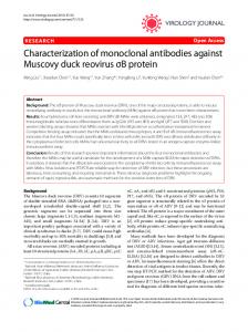
Absorbance values for all conjugated monoclonal antibodies were greater than 2.0 for direct ELISA tests with R32tet32 as the capture antigen, except for the CDC 58-159-2 monoclonal antibody (0.13+ 0.02). The mean absorbance of concurrently run negative controls with an anti-P. vivax monoclonal antibody was 0.02±0.01. The CDC 58-159-2 conjugated antibody was also negative in an IFA assay, but all other peroxidase monoclonal antibodies were positive (Table 1). Addition of peroxidase substrate to aliquots of all conjugated monoclonal antibodies produced strong, uniform colour changes. For initial comparative ELISA tests, a uniform concentration of capture (0.5 jig per well; 10 mg/l PBS) and peroxidase-conjugated monoclonal antibodies (0.25 tg per well; 5 mg/l blocking buffer) was used against the 7G8 and T4 strains of P. falciparum sporozoite (Fig. 1). Five of the 10 monoclonal antibodies tested gave absorbance values for 1000 sporozoites that were greater than 1.0 at 15 minutes. In order to determine the optimum concentration of capture monoclonal antibodies, four different dilutions were tested using 7G8 and NF54 sporozoites (500 per well) with a fixed concentration of peroxidase-conjugated antibody (0.25 ug per well). Results were similar for both sporozoites, with the antibodies divided into three distinct groups: those
765 T4
I
1.57
0i
159-2 CDC5S
2C1 2A10 FS I NFB2 antibody 132.2 102.4 Moroclonal
505. 5A4.1
SC1.1
Fig. 1. ELISA absorbance values (X=414 nm) for 10 peroxidase-labelled monoclonal antibodies tested against the 7G8 and T4 strains of Plasmodium falciparum sporozoites using the following conditions: concentration of capture monoclonal antibody 0.5 ji per well; peroxidase-monoclonal antibody level 0.25 jig per well; 1000 sporozoites per well; 15-minute reaction time. Values shown are the mean of 3 tests ± standard deviation < 5%.
42
R. A. WIRTZ ET AL.
Table 1. Results of the immunofluorescent antibody (IFA) assay for Plasmodium falciparum sporozoite peroxidaseconjugated and unconjugated monoclonal antibodies Sporozoite and antibody concentration (jcg/ml)
Monoclonal antibody 2C11
2A10
NFS 1
NFS 2
1B2.2
1G3.4
5G5.3
5A4.1
5C1.1
+4
+4
+4
+4
+4
+4
+4
+4
+4
+4 +2
+4 +2
+4 +4
+4 +4
+4 +4
+4 +2
+4
+4 +4
+4
+4
+3
+2
+3
P. berghei 5.00 0.05
-
+3 +1
-
-
+4 +2
-
+3 -
+4 -
-
P. cynomolgi 5.00
-
P. knowlesi 5.00
-
CDC 58-159-2
Peroxidase-conjugated monoclonal antibody P. falciparum 5.00
Unconjugated monoclonal antibody P. falciparum 5.00 0.05
P. vivax 5.00 P. yoe/ii 5.00
-
-
-
-
-
-
-
-
-
-
-
-
-
-
-
-
-
-
-
-
-
_
_
_
_
_
_
_
_
_
_
_
_
_
_
_
_
_
_
Values indicated with a dash were negative.
with low sensitivity for all capture concentrations tested (CDC 58-159-2, 1G3.4, and 5C 1. 1); those with maximum sensitivity at high capture concentrations (0.1 zg or 0.5 ug per well) (2Cl1, 2A10, NFS 1, NFS 2, and 5G5.3); and those (1B2.2 and 5A4. 1) with maximum sensitivity at low capture concentrations (0.004 or 0.02 Ag per well) (Fig. 2). With the optimum concentration of capture monoclonal antibody and a fixed concentration of homologous peroxidase monoclonal antibody (0.2 Ag per well), the ELISA test was run against four strains of sporozoites (7G8, NF54, T4, and Thi5). The absorbances at X = 414 nm for 500 sporozoites per well 15 minutes after the addition of substrate are shown in Fig. 3. Negative control values for each assay (in the absence of sporozoites) are shown in the histogram as solid areas. The cross-reactivity of the antibodies with other species of human and non-human sporozoites was studied using an IFA assay. All the antibodies displayed strong reactions with P. falciparum but were negative for sporozoites from P. cynomolgi, P. knowlesi, P. vivax, or P. yoelii. The following antibodies cross-reacted with P. berghei sporozoites: 2C11, 1B2.2, 5G5.3, and 5A4.1 (Table 1).
1.0
05
0
0.004
Q02
0.5 0.004 0.1 0D2 Concentration of capture antibody tpg per well)
0.1
0.5
Fig. 2. ELISA absorbance values (X = 414 nm) at various concentrations of capture antibody for 10 monoclonal antibodies tested against (a) the 7G8 or (b) the NF54 strain of Plasmodium falciparum sporozoites using the following conditions: concentration of peroxidasemonoclonal antibody 0.25 Mg per well; 500 sporozoites per well; 30-minute reaction time. Values shown are the mean ± standard deviation of 3 tests. 0 =2A10; O =CDC 58-159-2; * = 1G3.4; * =5A4.1; 0=2C11;
*1=11B2.2;
A=5G5.3;
0=5C1.1.
*=NFS 2; A=NFS 1;
43
MONOCLONAL ANTIBODIES AGAINST PLASMODJUM FALCIPARUM SPOROZOITES 2.0 I 234
1 234
1
1 234
Frw
c
1 234
2.0 r
1 234
rhi-
-
1 234
1 23 4__
0
1.5 t-
1-I
.0 < 0.5 0
C
3
h.dE.
COCSC 2C 11 2A10 1582
0.1
0.1
NFS 1 0.5
_!
*
0.1
0.004
1.01-
234
NFS 2 1B 2.2 1G 14 0.1
SG 5.3 0.1
M 4.1 0.02
5C 1.1 0.1
D
0.5
Optimum concentration of capture monoclonal antibody (pg per well)
Fig. 3. ELISA absorbance values (X-=414 nm) at the optimum concentration of capture monoclonal antibodies (pg per well) tested against four strains of Plasmodium falciparum sporozoites using the following conditions: concentration of peroxidase-monoclonal antibody 0.2 pg per well; 500 sporozoites per well; 15minute reaction time. Values shown are the mean of 3 tests ± standard deviations < 5%; solid area is background reading.
1=7G8; 2=NF54; 3=T4; 4=Thl5.
The optimum concentrations of capture monoclonal antibody for the 2A10 and NFS 2 monoclonal antibodies were determined more precisely. The absorbance values at X = 414 nm for 200 sporozoites per well are shown in Fig. 4 for both the 7G8 and Thl5 sporozoites, 15 minutes after the addition of substrate. The optimum concentrations of capture monoclonal antibody for 2A10 and NFS 2 were 0.1 and 0.2 pg per well, respectively. The optimum concentrations for peroxidase monoclonal antibodies for 2A10 and NMRI 2 were determined using 200 sporozoites per well either with or without mosquito triturate. For both 2A10 and NFS 2 the optimum concentration of conjugated antibody was 0.05 pg per well (1.0 ipg/ml blocking buffer). While absorbance values were consistently lower for 2A10 and NFS 2 when mosquito triturate was used to dilute the sporozoites, the difference was not statistically significant (PS0.05) at the optimum antibody concentration. The ELISA tests based on 2A10 and NFS 2 were then run concurrently, using the optimum reaction concentrations and a serial dilution of 7G8 sporozoites (Fig. 5). The concentration of capture monoclonal antibodies for 2A10 and NFS 2 was 0.1 and 0.2 pg per well, respectively, with a peroxidase-conjugate level of 0.05 ug per well for both antibodies. DISCUSSION
Antibody activity
was
exhibited in the direct
0.51F 0
la
a
I
a
0.4 0.2 0.1 0.05 Concentration of capture antibody (pg per well)
Fig. 4. ELISA absorbance values (X = 414 nm) at various concentrations of 2A10 or NFS 2 capture monoclonal antibody tested against the 7G8 or Thailand (Thi5) strain of Plasmodium falciparum sporozoites using the following conditions: concentration of peroxidasemonoclonal antibody 0.2 pig per well; 200 sporozoites per well; 15-minute reaction time. Values shown are the mean ± standard deviation of 3 tests.
*=2A10,Thl5; O=2A10,7G8; *=NFS2,Thl5; A = NFS 2, 7G8.
2.0 r 0 1.5
-
,
1< 1.0
I
0,
I
I I
0.5 0
I
4 O
J
6
12
',
a
25
No. of Pb,smodium fakiparum
50
100
sporozoite
per well
I
200
400
Fig. 5. Sensitivity of the ELISA for Plasmodium falciparum sporozoites. ELISA with monoclonal antibodies, 2A1 0 and NFS 2, respectively, using the following conditions: concentration of capture monoclonal antibody, 0.1 pg and 0.2 pg per well, respectively; peroxidasemonoclonal antibody 0.05 pg per well; 1-hour reaction time. Values shown are mean ± standard deviation of 3 tests.
A=2A10;
A=NFS2.
44
R. A. WIRTZ ET AL.
ELISA and IFA tests by all the conjugated monoclonal antibodies, except that from the Centers for Disease Control, and all displayed peroxidase activity. Loss of antibody activity upon conjugation of monoclonal antibodies to periodate-oxidized horseradish peroxidase has been discussed by Burkot et al. (2). The importance of optimum concentration of capture monoclonal antibody in a double-sided ELISA for the detection of a repeating epitope becomes apparent upon comparison of Fig. 1 and 3. In this respect, the most striking effect was observed with the 1B2.2 and 5A4.1 monoclonal antibodies, which essentially did not function at high antibody concentrations (Fig. 1), but at the lower, optimum levels were the most sensitive of those tested (Fig. 3). Because of their low sensitivity (Fig. 1-3), the 1G3.4 and 5ClI. monoclonal antibodies were eliminated at the initial stages of the selection procedure. Preliminary evidence indicates that the 1G3.4 antibody recognizes an Asn-Val-Asp-Pro tetrapeptide of the P.falciparum circumsporozoite protein. Since this particular tetrapeptide represents only four of the 41 tetrapeptide repeats on the protein (4), its lower sensitivity is not unexpected. The 2Cll, 1B2.2, 5G5.3, and 5A4.1 monoclonal antibodies were rejected because of their crossreactivity with P. berghei sporozoites (Table 1). An ELISA test using the 1B2.2 monoclonal antibody
functioned well with P. berghei sporozoites, indicating that an epitope similar to that in P. falciparum is repeated in the P. berghei circumsporozoite protein. Also, the NFS 1 antibody was eliminated because it was less sensitive than 2A10 or NFS 2 (Fig. 3) and required larger amounts of capture antibody
(Fig. 2).
Both 2A10 and NFS 2 were selected as excellent candidate monoclonal antibodies, 2A10 being more sensitive in the initial comparative testing (Fig. 3 and 4). Furthermore the optimum concentration of capture material for 2A10 was half that required by NFS 2 (Fig. 4), although the optimum concentrations of the peroxidase-conjugated antibodies were similar (0.05 gg per well) for both antibodies. By using either 2A10 or NFS 2 at the optimum concentration, the method described was ten times more sensitive than existing ELISA tests for P. falciparum sporozoites and was associated with a 50% reduction in background absorbance (1). This permitted the detection of less than 25 sporozoites per 50 ul of test solution (Fig. 5). Trituration of a mosquito in 200 ld of solution would therefore allow detection of fewer than 100 sporozoites per insect. On the basis of the results described, the 2A10 monoclonal antibody was selected as the best candidate for development of a standardized ELISA test, and in a more extensive study it recognized P. falciparum sporozoites from 15 isolates from different geographical regions (11).
RESUMt tTUDE COMPARATIVE D'ANTICORPS MONOCLONAUX ANTI-SPOROZOTTES DE PLASMODIUM FALCIPARUM EN VUE DE LA MISE AU POINT D'UNE tPREUVE IMMUNO-ENZYMATIQUE ELISA On a evalue 10 anticorps monoclonaux en vue de leur utilisation dans une epreuve de detection des sporozoites de Plasmodiumfalciparum. Ces anticorps ont e purifies, leur reactivite crois6e a e recherch&e en pr6sence de cinq especes de sporozoites dans une epreuve d'immunofluorescence; ils ont ensuite e conjugu6s A une peroxydase de raifort. On adsorbe l'anticorps monoclonal de capture en solution dans du solut6 salin tamponne au phosphate (PBS) A 0,01 mol/l, sur des plaques flexibles de chlorure de polyvinyl pour microtitrage ELISA en les laissant incuber jusqu'au lendemain a la temperature du laboratoire. Les cupules des plaques sont vid&es puis remplies avec du tampon bloquant (serumalbumine bovine a 1,0%o caseine a 0,507o, thiomersal A 0,01I et rouge de phenol A 0,002% dissous dans du PBS). Au bout d'une heure, les cupules sont vid&es et on y ajoute 0,05 ml d'extrait sporozoitaire, puis on laisse reposer les plaques pendant deux heures. Des sporozoites de glande salivaire ou des moustiques sont tritures dans 0,05 ml de tampon bloquant contenant du Nonidet P-40 a 0,507., apres quoi on ajoute au materiel triture 0,15 ml de tampon
bloquant. Ensuite, les plaques sont videes, lav&es deux fois avec du PBS contenant du Tween 20 A 0,05%o (PBS-Tw). L'anticorps monoclonal homologue conjugue A la peroxydase et dilu6 dans du tampon bloquant est ensuite ajoute dans les cupules et on laisse reposer les plaques pendant I heure. Les cupules sont vid&es et lav&es trois fois avec du PBS-Tw; ensuite on ajoute 0,1 ml de substrat de la peroxydase dans chaque cupule et au bout d'une heure on determine l'absorbance A X = 414 nm. Cette methode a permis d'obtenir des valeurs optimales de l'absorbance pour les parametres de l'epreuve 6tudi6e. Il est essentiel d'utiliser une concentration optimale de I'anticorps de capture pour d6tecter un 6pitope r6petitif dans une epreuve ELISA faisant appel A la technique du sandwich. Ainsi, deux des anticorps monoclonaux eprouv6s 6taient inoperants a des concentrations elevees mais aux concentrations optimales plus basses, ils se sont rev6les les plus sensibles parmi les anticorps etudies. Quatre des anticorps monoclonaux ont e elimin6s en raison de leur faible
MONOCLONAL ANTIBODIES AGAINST PLASMODIUM FALCIPARUM SPOROZOITES
sensibilite, tandis que quatre autres ont e rejetes parce qu'ils donnaient une reaction croisee avec des sporozoites de P. berghei. L'utilisation de l'un ou l'autre des deux anticorps monoclonaux restants, a la concentration optimale, fournissait une methode ELISA qui etait dix fois plus sensible que les epreuves ELISA existantes pour la detection des sporozoites de P. falciparum et avec une reduction de 50% de l'absorbance de fond. La limite de
45
detection de la methode decrite ici etait de moins de 25 sporozoites par 0,05 ml de solution a eprouver et il etait possible de deceler moins de 100 sporozoites par moustique dans 0,2 ml de diluant. L'anticorps monoclonal choisi pour la mise au point de l'epreuve ELISA etait le plus sensible de ceux qui avaient e eprouves, de plus, il etait capable de mettre en evidence des sporozoites provenant de zones geographiques tres diverses.
ACKNOWLEDGEMENTS The authors thank Megan Dowler for isolation of sporozoites; the staff of the Department of Entomology, Armed Forces Research Institute of Medical Sciences, (in particular, S. Vongpradist) for providing the field-acquired human malaria parasites; G. Ward of the Department of Veterinary Medicine for providing samples of P. cynomolgi and P. knowlesi sporozoites; M. Sedegah for samples of P. yoelii sporozoites; and Smith Klein & French Laboratories for the R32tet32 recombinant protein. This research was supported in part by NMR & DC No. 62770A3M162770A870AF312. R. A. Ward, P. M. Graves, and J. C. Beier are thanked for reviewing the manuscript; and F. H. Top, Jr, C. L. Diggs, W. H. Bancroft, R. Wistar, C. Campbell, and S. Nussenzweig for support and encouragement.
REFERENCES 1. BURKOT, T. R. ET AL. American journal of tropical medicine and hygiene, 33: 783-788 (1984). 2. BURKOT, T. R. ET AL. Journal of immunological methods, 84: 25-31 (1985). 3. COLLINS, F. H. ET AL. American journal of tropical medicine and hygiene, 33: 538-543 (1984). 4. DAME, J. B. ET AL. Science, 225: 593-599 (1984). 5. Ey, P. L. ETAL. Immunochemistry, 15: 429-436(1978). 6. NAKANE, P. K. & KAWAOI, A. Journal of histochem-
istry and cytochemistry, 22: 1084-1091 (1974). 7. RAMSEY, J. M. ETAL. Transactions of the Royal Society of Tropical Medicine and Hygiene, 77: 378-381 (1983). 8. YOUNG, J. F. ET AL. Science, 228: 958-962 (1985). 9. ZAVALA, F. ET AL. Nature, 299: 737-738 (1982). 10. ZAVALA, F. ET AL. Journal of experimental medicine, 157: 1947-1957 (1983). 11. ZAVALA, F. ET AL. Journal of immunology, 135 2790-2793 (1985).