ANTICANCER RESEARCH 25: 4439-4444 (2005)
Comparison between Whole-body Positron Emission Tomography and Bone Scintigraphy in Evaluating Bony Metastases of Esophageal Carcinomas HIROYUKI KATO1, TATSUYA MIYAZAKI1, MASANOBU NAKAJIMA1, JUNKO TAKITA1, HITOSHI KIMURA1, AHMAD FARIED1, MAKOTO SOHDA1, YASUYUKI FUKAI1, NORIHIRO MASUDA1, MINORU FUKUCHI1, RYOKUHEI MANDA1, HITOSHI OJIMA1, KATSUHIKO TSUKADA1, HIROYUKI KUWANO1, NOBORU ORIUCHI2 and KEIGO ENDO2
Departments of 1General Surgical Science (Surgery I) and Radiology and Nuclear Medicine, Gunma University, Graduate School of Medicine, Maebashi, 371-8511, Japan
2Diagnostic
Abstract. Background: Positron emission tomography (PET) with 18-F-fluorodeoxyglucose (FDG) has already proved useful in assessing the extension of esophageal carcinomas, detecting tumor recurrence and monitoring responses to therapy. This study aimed to elucidate the potential role of FDG-PET compared with technetium-99m methylene diphosphonate (Tc-99m MDP) bone scintigraphy in the evaluation of bony metastasis in esophageal carcinoma patients. Patients and Methods: Forty-four patients with thoracic esophageal carcinomas, who underwent FDG-PET together with bone scintigraphy within 1 month between January 2000 and September 2003, were included in this study. Results: Of the 44 patients, 13 had 31 bone metastases and, of these, 6 were diagnosed pretreatment and 7 had recurrence after esophagectomy surgery. Of the 31 metastases, 9 were in the vertebral column, 11 in the thoracic cage (including the ribs, clavicle, sternum and scapula), 9 in the pelvic bones and 2 in the long bones of the extremities. In evaluating the detection of bony metastasis, FDG-PET showed 92% sensitivity, 94% specificity and 93% accuracy, compared with 77%, 84% and 82%, respectively, for bone scintigraphy. Although the sensitivity, specificity and accuracy of PET were slightly higher than those of bone scintigraphy, these differences were not
Correspondence to: Hiroyuki Kato, Department of General Surgical Science (Surgery I), Gunma University, Graduate School of Medicine, 3-39-22, Showa-machi, Maebashi, Gunma, 371-8511, Japan. Tel: +81-27-220-8224, Fax: +81-27-220-8230, e-mail:
[email protected] Key Words: 18-F-Fluorodeoxyglucose (FDG), positron emission tomography (PET), esophageal carcinoma, bony metastasis, bone scintigraphy.
0250-7005/2005 $2.00+.40
statistically significant according to the McNemar’s test. Three patients with false-negative findings on their bone scans had positive findings with PET; all of these lesions were osteolytic metastases. Conclusion: FDG-PET scans were superior to bone scintigraphy in detecting bony metastases of esophageal carcinomas. Therefore, FDG-PET can be used for the detection and follow-up of bone tumors when Tc-99m MDP bone scintigraphy gives negative findings. Positron emission tomography (PET) with 18-Ffluorodeoxyglucose (FDG) provides physiological information that enables the diagnosis of cancer based on altered tissue glucose metabolism (1). The role and potential value of PET, which is a non-invasive imaging modality, has been widely investigated in recent years (2-5), and markedly increased FDG uptake in esophageal carcinomas has been documented in several studies (6-13). FDG-PET imaging might, therefore, facilitate diagnosis in patients with malignant diseases by helping differentiate between benign and malignant tumors, assess disease extension (6-12), detect tumor recurrence (14, 15) and monitor responses to therapy (16-19). The authors previously reported the usefulness of FDG-PET in staging esophageal squamous cell carcinomas (SCCs), and its higher sensitivity, specificity and accuracy in lymph node detection compared to computed tomography (CT), particularly in the neck and upper thoracic region (20). FDG-PET might also be of value in detecting recurrent esophageal carcinomas (21). The majority of patients with esophageal carcinomas have an advanced disease at the time of diagnosis (22). The majority of distant metastases of esophageal carcinomas involve the lymph nodes, liver, lungs and bones. Bone scintigraphy using technetium-99m methylene diphosphonate (Tc-99m MDP) is widely used for the detection of bony metastasis in patients with esophageal cancer. Bone scanning
4439
ANTICANCER RESEARCH 25: 4439-4444 (2005) is a simple and harmless technique and has a higher sensitivity than X-rays. However, its sensitivity and specificity have recently been reported to be lower than those of magnetic resonance imaging (MRI) (23). Preliminary studies have shown FDG-PET to be superior to bone scintigraphy in detecting bone metastases from lung (24, 25) and breast (26, 27) cancers, but not from prostate cancer (28). However, no comparisons of FDG-PET and bone scintigraphy in esophageal cancer have been reported. This study aimed to elucidate the potential role of FDGPET compared with Tc-99m MDP bone scintigraphy in the evaluation of bony metastasis in esophageal carcinoma patients.
Patients and Methods Patients. Forty-four patients with thoracic esophageal carcinomas, who underwent FDG-PET together with bone scans within 1 month between January 2000 and September 2003, were included in this study. All the patients were treated at the Department of General Surgical Science (Surgery I), Gunma University, Graduate School of Medicine, Japan. Informed written consent was acquired from all individuals prior to enrollment in this study. The median age of the patients was 63.4 years, with a range of 43 to 76 years. The tumor stage and disease grade were classified according to the sixth edition of the TNM classification of the International Union Against Cancer (UICC). PET imaging. PET images were obtained using a SET 2400 W (Shimadzu Corporation, Kyoto, Japan) with a 59.5-cm transaxial field of view and a 20-cm axial field of view. A whole-body image was initiated 40 min after an injection of 275-370 MBq FDG using the simultaneous emission–transmission method (20). Four to 5 sections from the head to the thigh were imaged for 8 min per section. Patients fasted for at least 4 h before FDG-PET. The imaging protocols were approved by the institutional review board (29), and all patients gave informed consent before undergoing the examination. All PET images were evaluated qualitatively by at least two experienced nuclear medicine physicians. Functional images of the standardized uptake value (SUV) were produced from the attenuation-corrected transaxial images, the amount of FDG injected, body weight and the cross-calibration factors between PET and the dose calibrator. SUV was defined as the concentration of radioactivity in the tissue or lesion (MBq/ml) x body weight (g)/injected dose of FDG (MBq). The blood sugar level in all patients was less than 100 mg/dl at the time of the PET scan. Bone scintigraphy. Tc-99m MDP bone scintigraphy was performed in whole-body scan mode using a Toshiba E.CAM gamma camera (Toshiba Corporation, Tokyo, Japan). Whole-body bone scans were obtained 3-4 h after an injection of 740 MBq Tc-99m MDP, using high-resolution collimation on a twin-headed gamma camera. Statistical analysis. Bony metastasis was assessed by histological findings or a clinical and radiological follow-up that included CT, MRI, bone scintigraphy, FDG-PET and specific X-ray studies for at least 6 months; these lesions were defined as the gold standard (GS). The sensitivity, specificity and accuracy of FDG-PET and
4440
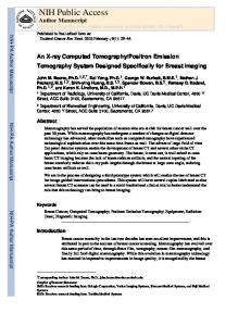
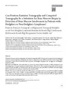
Figure 1. (a) Coronal positron emission tomography scans demonstrating a primary tumor (arrow) with intense FDG uptake in the lower mediastinal region and multiple bony metastases (arrowhead) in the vertebral column, ribs and pelvic bones. (b) Bone scintigraphy demonstrating multiple uptake of the radionuclides (arrow) in the vertebral column, ribs and pelvic bones.
bone scintigraphy were calculated using standard definitions (30). The McNemar test was used to analyze significant increases in the number of false-positive or false-negative findings (31).
Results Of the 44 patients studied, 13 had 31 bone metastases confirmed histologically or in the clinical follow-up (Figure 1). Of these 13 patients, 6 were diagnosed pretreatment and 7 had recurrence after esophagectomy surgery. Of the 31 metastases, 9 were in the vertebral column, 11 in the thoracic cage (including the ribs, clavicle, sternum and scapula), 9 in the pelvic bones and 2 in the long bones of
Kato et al: Role of PET in Evaluating Bony Metastases of Esophageal Carcinomas
Discussion
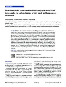
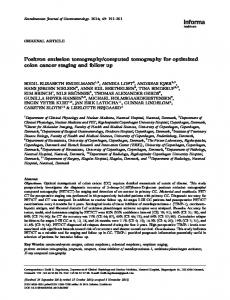
Figure 2. Coronal (a) and axial (b) images of positron emission tomography scans demonstrating intense FDG uptake in the lumbar spine (arrow), which was proved to be a false-positive lesion in a clinical follow-up.
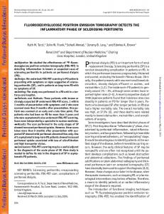
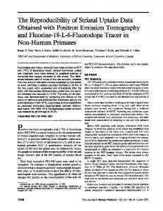
the extremities. In the detection of bony metastasis, FDGPET showed 92% sensitivity, 94% specificity and 93% accuracy, compared with 77%, 84% and 82%, respectively, for bone scintigraphy. The sensitivity, specificity and accuracy of PET were slightly higher than those of bone scintigraphy, but these differences were not statistically significant according to the McNemar’s test. A falsenegative finding with PET was found in 1 patient, because differentiation between the primary tumor and adjacent thoracic spine metastasis of the primary tumor was not possible. Two false-positive lesions occurred with PET, in the shoulder joint and lumbar spine (Figure 2), and a subcutaneous lesion in the thoracic cage was missed. Five patients with false-positive findings on their bone scans had uptake in the thoracic spine and cage and lumbar spine, and 3 patients with false-negative findings on the bone scans had positive findings with PET (Figure 3). All of these lesions were osteolytic metastases.
The current standard of practice for the detection of osseous metastatic diseases is conventional bone scans of the entire body using Tc-99m MDP. The radiotracer used is absorbed onto the bone surfaces and its uptake depends on local blood flow and osteoblastic activity (32). Although Tc-99m MDP scintigraphy is sensitive in the detection of advanced skeletal metastatic lesions, early involvement might be missed, because this technique relies on the osteoblastic reaction of the involved bone rather than actual tumor detection. In adults, the sensitivity of skeletal scintigraphy has been reported as 62% to 89% (33). Recently, PET has emerged as one of the most promising new imaging modalities for the detection of skeletal metastases. This study compared the potential role of FDG-PET compared with Tc-99m MDP in evaluating bony metastasis in esophageal carcinoma patients. Thirteen of the 44 patients studied had bone metastases. In the detection of bony metastasis, FDG-PET showed 92% sensitivity, 94% specificity and 93% accuracy, compared with 77%, 84% and 82%, respectively, for bone scintigraphy. The sensitivity, specificity and accuracy of PET were, therefore, slightly higher than those of bone scintigraphy. Three patients with false-negative findings on their bone scans had positive findings with PET; these patients had osteolytic metastases. From these results it can be suggested that PET is superior to bony scintigraphy in detecting osseous metastases. This might be because of its ability to detect the presence of tumors directly by metabolic activity, rather than indirectly by showing tumor involvement due to increased bone mineral turnover. Another reason might be its ability to detect metastatic foci earlier than bone scintigraphy. Some reports have described the high specificity, but comparable sensitivity, of FDG-PET imaging for bony metastasis (24-26, 34). Bury et al. reported that FDG-PET could detect metastatic bone involvement more accurately than bone scintigraphy in patients with non-small cell lung cancer (24). In particular, PET was more specific and had a higher positive predictive value than bone scintigraphy. Kao et al. also reported the better specificity, but lower sensitivity, of FDG-PET in detecting bone metastases (34). Cook et al. reported that FDG-PET was superior to bone scintigraphy in the detection of osteolytic breast metastases, which are associated with poor prognosis (27). In contrast, osteoblastic metastases showed lower metabolic activity and were frequently undetectable by PET. They mentioned that the cause of this difference was unclear, but it might have been attributable to the lower volumes of viable tumor tissue within osteoblastic osseous metastases, because these tend to be more acellular in nature (35, 36). Furthermore, the sensitivity of FDG-PET imaging in detecting osteoblastic bony metastases, observed especially in prostate cancer, is generally less than bone scanning using MDP
4441
ANTICANCER RESEARCH 25: 4439-4444 (2005)
Figure 3. Coronal (a) and axial (b) images of positron emission tomography scans demonstrating intense FDG uptake in the lumbar spine. A CT showed an osteolytic lesion (arrow) in the lumbar spine (c). Bone scintigraphy (d) does not demonstrate abnormal uptake, therefore this is a false-negative lesion on bone scintigraphy.
(28). In recurrent breast cancer, FDG-PET can detect significantly more lymph nodes but less bone metastases compared with conventional imaging, including bone scintigraphy (37). PET might miss, in particular, osteoblastic lesions, which are normally found by conventional bone scans (38, 39). The lesions undetectable with PET are predominantly correlated with radiologically osteoblastic or mixed osteoblastic/osteolytic lesions (37). On the other hand, there are a number of patients with positive bone lesions that have been exclusively detected by FDG-PET. Bone scintigraphy using Tc-99m MDP showed 90% sensitivity but poor specificity (61%) (23). The false-positive cases can be explained by the non-selective uptake of the radionuclide in any area of increased bone turnover (degenerative changes, inflammatory processes, mechanical stress, and so on). It was previously reported that falsenegative findings might be obtained in a small percentage of patients with purely osteolytic lesions or in patients with
4442
slow-growing lesions, where the reactive bone is not detectable (23, 40). In the present study, bony metastasis was undetected by bone scintigraphy in 3 patients with osteolytic lesions, but detected on FDG-PET. There are several advantages with PET that might lead to its superior detection of bone metastases compared with Tc-99m MDP bone scintigraphy. First, PET possesses a higher spatial resolution than bone scintigraphy (26, 40, 41). Secondly, FDG-PET and bone scintigraphy utilize different mechanisms to detect bony metastatic sites: Tc-99m MDP bone scintigraphy depends on osteoblastic bone reactions within the cancer cells, while FDG-PET gauges glucose influxes into the cancer cells (26). Therefore, FDG-PET might detect bony metastases at an earlier stage, even if cancer cells are restricted to the bone marrow (26). In conclusion, it can be suggested that FDG-PET scans are superior to bone scintigraphy in the detection of purely osteolytic diseases and in the early detection of osseous
Kato et al: Role of PET in Evaluating Bony Metastases of Esophageal Carcinomas
metastatic diseases of esophageal carcinomas. Therefore, FDG-PET is essential in the detection and follow-up of bone tumors when 99mTc-MDP bone scintigraphy produces negative results.
Acknowledgements This work was supported in part by a Grant-in-Aid for Cancer Research (13-18) from the Ministry of Health, Labor and Welfare, Japan. We would like to thank A. Nakabayashi, H. Emura, M. Ohnuma, S. Ueno, T. Ogasawara and Y. Saitoh for their excellent secretarial assistance and M. Ohno for her assistance with the data management and biostatistical analysis during the preparation of this report.
References 1 Bar-Shalom R, Valdivia AY and Blaufox MD: PET imaging in oncology. Semin Nucl Med 30: 150-185, 2000. 2 Bares R, Klever P, Hauptmann S, Hellwig D, Fass J, Cremerius U et al: F-18 fluorodeoxyglucose PET in vivo evaluation of pancreatic glucose metabolism for detection of pancreatic cancer. Radiology 192: 79-86, 1994. 3 Inoue T, Kim EE, Komaki R, Wong FC, Bassa P, Wong WH et al: Detecting recurrent or residual lung cancer with FDG-PET. J Nucl Med 36: 788-793, 1995. 4 Ahuja V, Coleman RE, Herndon J and Patz EF: The prognostic significance of fluorodeoxyglucose positron emission tomography imaging for patients with nonsmall cell lung carcinoma. Cancer 83: 918-924, 1998. 5 Coleman RE: PET in lung cancer. J Nucl Med 40: 814-820, 1999. 6 Block MI, Patterson GA, Sundaresan RS, Bailey MS, Flanagan FL, Dehdashti F, Siegel BA and Cooper JD: Improvement in staging of esophageal cancer with the addition of positron emission tomography. Ann Thorac Surg 64: 770-776, 1997. 7 Luketich JD, Schauer PR, Meltzer CC, Landreneau RJ, Urso GK, Townsend DW, Ferson PF, Keenan RJ and Belani CP: Role of positron emission tomography in staging esophageal cancer. Ann Thorac Surg 64: 765-769, 1997. 8 Flanagan FL, Dehdashti F, Siegel BA, Trask DD, Sundaresan SR, Patterson GA and Cooper JD: Staging of esophageal cancer with 18F-fluorodeoxyglucose positron emission tomography. AJR Am J Roentgenol 168: 417-424, 1997. 9 Kole AC, Plukker JT, Nieweg OE and Vaalburg W: Positron emission tomography for staging of oesophageal and gastroesophageal malignancy. Br J Cancer 78: 521-527, 1998. 10 Rankin SC, Taylor H, Cook GJ and Mason R: Computed tomography and positron emission tomography in the preoperative staging of oesophageal carcinoma. Clin Radiol 53: 659-665, 1998. 11 Kobori O, Kirihara Y, Kosaka N and Hara T: Positron emission tomography of esophageal carcinoma using (11)C-choline and (18)F-fluorodeoxyglucose: a novel method of preoperative lymph node staging. Cancer 86: 1638-1648, 1999. 12 Yeung HW, Macapinlac HA, Mazumdar M, Bains M, Finn RD and Larson SM: FDG-PET in esophageal cancer. Incremental value over computed tomography. Clin Positron Imaging 2: 255260, 1999.
13 McAteer D, Wallis F, Couper G, Norton M, Welch A, Bruce D, Park K, Nicolson M, Gilbert FJ and Sharp P: Evaluation of 18F-FDG positron emission tomography in gastric and oesophageal carcinoma. Br J Radiol 72: 525-529, 1999. 14 Skehan SJ, Brown AL, Thompson M, Young JE, Coates G and Nahmias C: Imaging features of primary and recurrent esophageal cancer at FDG-PET. Radiographics 20: 713-723, 2000. 15 Flamen P, Lerut A, Van Cutsem E, Cambier JP, Maes A, De Wever W, Peeters M, De Leyn P, Van Raemdonck D and Mortelmans L: The utility of positron emission tomography for the diagnosis and staging of recurrent esophageal cancer. J Thorac Cardiovasc Surg 120: 1085-1092, 2000. 16 Brucher BL, Weber W, Bauer M, Fink U, Avril N, Stein HJ, Werner M, Zimmerman F, Siewert JR and Schwaiger M: Neoadjuvant therapy of esophageal squamous cell carcinoma: response evaluation by positron emission tomography. Ann Surg 233: 300-309, 2001. 17 Weber WA, Ott K, Becker K, Dittler HJ, Helmberger H, Avril NE, Meisetschlager G, Busch R, Siewert JR, Schwaiger M and Fink U: Prediction of response to preoperative chemotherapy in adenocarcinomas of the esophagogastric junction by metabolic imaging. J Clin Oncol 19: 3058-3065, 2001. 18 Flamen P, Van Cutsem E, Lerut A, Cambier JP, Haustermans K, Bormans G, De Leyn P, Van Raemdonck D, De Wever W, Ectors N, Maes A and Mortelmans L: Positron emission tomography for assessment of the response to induction radiochemotherapy in locally advanced oesophageal cancer. Ann Oncol 13: 361-368, 2002. 19 Kato H, Kuwano H, Nakajima M, Miyazaki T, Yoshikawa M, Masuda N, Fukuchi M, Manda R, Tsukada K, Oriuchi N and Endo K: Usefulness of positron emission tomography for assessing the response of neoadjuvant chemoradiotherapy in patients with esophageal cancer. Am J Surg 184: 279-283, 2002. 20 Kato H, Kuwano H, Nakajima M, Miyazaki T, Yoshikawa M, Ojima H, Tsukada K, Oriuchi N, Inoue T and Endo K: Comparison between positron emission tomography and computed tomography in the use of the assessment of esophageal carcinoma. Cancer 94: 921-928, 2002. 21 Kato H, Miyazaki T, Nakajima M, Fukuchi M, Manda R and Kuwano H: Value of positron emission tomography in the diagnosis of recurrent oesophageal carcinoma. Br J Surg 94: 1004-1009, 2004. 22 Kato H: Diagnosis and treatment of oesophageal neoplasms. Jpn J Cancer Res 86: 993-1009, 1995. 23 Rybak LD and Rosenthal DI: Radiological imaging for the diagnosis of bone metastases. Q J Nucl Med 45: 53-64, 2001. 24 Bury T, Barreto A, Daenen F, Barthelemy N, Ghaye B and Rigo P: Fluorine-18 deoxyglucose positron emission tomography for the detection of bone metastases in patients with non-small cell lung cancer. Eur J Nucl Med 25: 1244-1247, 1998. 25 Gayed I, Vu T, Johnson M, Macapinlac H and Podoloff D: Comparison of bone and 2-deoxy-2-[18F]fluoro-D-glucose positron emission tomography in the evaluation of bony metastases in lung cancer. Mol Imaging Biol 5: 26-31, 2003. 26 Ohta M, Tokuda Y, Suzuki Y, Kubota M, Makuuchi H, Tajima T, Nasu S, Suzuki Y, Yasuda S and Shohtsu A: Whole body PET for the evaluation of bony metastases in patients with breast cancer: comparison with Tc99m-MDP bone scintigraphy. Nucl Med Commun 22: 875-879, 2001.
4443
ANTICANCER RESEARCH 25: 4439-4444 (2005) 27 Cook GJ, Houston S, Rubens R, Maisey MN and Fogelman I: Detection of bone metastases in breast cancer by 18FDG PET: differing metabolic activity in osteoblastic and osteolytic lesions. J Clin Oncol 16: 3375-3379, 1998. 28 Shreve PD, Grossman HB, Gross MD and Wahl RL: Metastatic prostate cancer: initial findings of PET with 2-deoxy-2-[F18]fluoro-D-glucose. Radiology 199: 751-756, 1996. 29 Inoue T, Oriuchi N, Kunio M et al: Accuracy of standardized uptake value measured by simultaneous emission and transmission scanning in PET oncology. Nucl Med Commun 20: 849-857, 1999. 30 Beck JR: Likelihood ratios. Another enhancement of sensitivity and specificity. Arch Pathol Lab Med 110: 685-686, 1986. 31 Dwyer AJ: Matchmaking and McNemar in the comparison of diagnostic modalities. Radiology 178: 328-330, 1991. 32 Malhotra P and Berman CG: Evaluation of bone metastases in lung cancer. Improved sensitivity and specificity of PET over bone scanning. Cancer Control 9: 259-260, 2002. 33 Daldrup-Link HE, Franzius C, Link TM, Laukamp D, Sciuk J, Jurgens H, Schober O and Rummeny EJ: Whole-body MR imaging for detection of bone metastases in children and young adults: comparison with skeletal scintigraphy and FDG PET. AJR Am J Roentgenol 177: 229-236, 2001. 34 Kao CH, Hsieh JF, Tsai SC, Ho YJ and Yen RF: Comparison and discrepancy of 18F-2-deoxyglucose positron emission tomography and Tc-99m MDP bone scan to detect bone metastases. Anticancer Res 20: 2189-2192, 2000. 35 Nakamoto Y, Osman M and Wahl RL: Prevalence and patterns of bone metastases detected with positron emission tomography using F-18 FDG. Clin Nucl Med 28: 302-307, 2003. 36 Peterson JJ, Kransdorf MJ and O'Connor MI: Diagnosis of occult bone metastases: positron emission tomography. Clin Orthop 415: S120-128, 2003.
4444
37 Gallowitsch HJ, Kresnik E, Gasser J, Kumnig G, Igerc I, Mikosch P and Lind P: F-18 fluorodeoxyglucose positron-emission tomography in the diagnosis of tumor recurrence and metastases in the follow-up of patients with breast carcinoma: a comparison to conventional imaging. Invest Radiol 38: 250-256, 2003. 38 Cheran SK, Herndon JE 2nd and Patz EF Jr: Comparison of whole-body FDG-PET to bone scan for detection of bone metastases in patients with a new diagnosis of lung cancer. Lung Cancer 44: 317-325, 2004. 39 Yoshioka T, Yamaguchi K, Kubota K, Saginoya T, Yamazaki T, Ido T, Yamaura G, Takahashi H, Fukuda H and Kanamaru R: Evaluation of 18F-FDG PET in patients with a metastatic or recurrent gastric cancer. J Nucl Med 44: 690-699, 2003. 40 Yang SN, Liang JA, Lin FJ, Kao CH, Lin CC and Lee CC: Comparing whole body (18)F-2-deoxyglucose positron emission tomography and technetium-99m methylene diphosphonate bone scan to detect bone metastases in patients with breast cancer. J Cancer Res Clin Oncol 128: 325-328, 2002. 41 Schirrmeister H, Guhlmann A, Elsner K, Kotzerke J, Glatting G, Rentschler M, Neumaier B, Trager H, Nussle K and Reske SN: Sensitivity in detecting osseous lesions depends on anatomic localization: planar bone scintigraphy versus 18F PET. J Nucl Med 40: 1623-1629, 1999.
Received June 14, 2005 Accepted August 25, 2005