Accepted: 17 February 2017 DOI: 10.1111/cyt.12430
ORIGINAL ARTICLE
Comparison of conventional cytology and SurePath in split thyroid fine needle aspiration materials D. Sahin1 | D. Yilmazbayhan2 | P. Firat2 | U. P. Hacisalihoglu2 | S. H. Kirimlioglu3 | E. Celenk4 | R. Arslan4 1 Department of Pathology, Acıbadem Health Group, Istanbul, Turkey 2 Department of Pathology, Istanbul Faculty of Medicine, Istanbul University, Istanbul, Turkey 3
Acıbadem University, Istanbul, Turkey
4
Department of Radiology, Istanbul Faculty of Medicine, Istanbul, Turkey Correspondence Davut Sahin, Acıbadem Saglik Grubu, 34318, Istanbul, Turkey. Email:
[email protected]
Objective: The aim of the present study was to compare the cytomorphological features and cytopathological diagnoses in thyroid aspiration materials prepared by SurePathâ (SP) and conventional cytology (CC). Materials and methods: Fine needle aspiration (FNA) materials from 180 thyroid nodules were divided into two groups to prepare three conventional smears and one SP slide. Twenty-nine cytomorphological features of thyroid lesions were compared in the CC and SP slides. The Kappa coefficiency was determined for each. The cytopathological diagnosis of CC and SP were compared. Results: The feature with the lowest Kappa coefficient was the haemorrhagic background, whereas nuclear molding had the highest Kappa coefficient. The rates of the atypical and suspicious cytopathological diagnostic categories were decreased, whereas the rates of benign and malignant categories were increased in SP. When the cytopathological diagnoses of CC and SP were compared with the histopathological diagnoses of the 31 thyroidectomy materials, the results were similar. Conclusion: The common problems seen in CC, such as an excessive number of slides, a haemorrhagic background and air drying artefact in the SP method were not encountered. Through these advantages, the rate of the indeterminate cytopathological diagnosis was low in SP. In addition to those advantages, the increased rates of non-diagnostic cases, the difficulty in evaluating the cytomorphologic features owing to tridimensional structures and the smaller size of the cells and the presence of tridimensional structures uninterpreted microscopically were the disadvantages of SP. The present results showed that SP could be used instead of CC in thyroid cytopathology. KEYWORDS
conventional cytology, fine needle aspiration, SurePath, thyroid
1 | INTRODUCTION
conventional cytology (CC) and liquid-based cytology (LBC), which are employed extensively.3–5 Problems associated with CC include
Palpable thyroid nodules occur in 4%–7% of the population,
the high incidence of non-diagnostic and indeterminate results, the
but nodules found incidentally on ultrasonography (USG) suggest a
encumbered workload for cytopathologists and the lack of reserved
prevalence of 19%–67%.1 The most common method to differentiate
material to use in cases with indeterminate diagnoses.3–6 These fac-
between neoplastic non-neoplastic and benign malignant thyroid
tors gave rise to the need to develop LBC as an alternative to CC.
nodules is fine needle aspiration (FNA) cytology.2–4 Two methods
Major reasons for non-diagnostic and indeterminate diagnosis with
are used for the preparation of thyroid aspiration materials:
CC are the hypocellularity of the aspirations, a haemorrhagic
Cytopathology. 2017;1–8.
wileyonlinelibrary.com/journal/cyt
© 2017 John Wiley & Sons Ltd
|
1
2
|
SAHIN
ET AL.
background, the thickness of the smear, inadequate fixation and
along with the other slide that was fixed with alcohol. The air-
crush artefacts. To increase the quality of the smears and establish
dried slide was stained with MGG. After the CC slides had been
standardisation in thyroid cytology, the use of FDA-approved LBC
prepared, residual material that remained in the needle and syr-
techniques, ThinPrepâ (TP) (Cytyc Corp., Marlborough, MA, USA)
inge was rinsed in the 25-cc SP CytorichTM red preservative fluid
and SurePathâ (SP) (TriPath Imaging, Inc., Burlington, NC, USA) are
in the falcon tube. The material that was placed in the SP Cytor-
increasing steadily.5,7 It has been demonstrated that the high-quality
ich solution was first homogenised via vortex, and then half of
smears and cell blocks increase the diagnostic sensitivity while
this material was extracted from the falcon tubes and placed in
decreasing the cytopathologist’s workload. LBC allows for the prepa-
special cytocentrifuge tubes to prepare SP preparate. The material
ration of new preparates before the final diagnosis in doubt cases
remaining in the falcon tubes was stored as reserve material. The
and reserves material for immunohistochemistry (IHC) or molecular
SP slides were stained with modified PAP stain in the slide pro-
pathological tests, which are the additional advantages of LBC over
cessor apparatus (Prep Stainâ Slide Processor). The SP slides were
CC.5,7,8 As a result of these advantages, LBC should be preferred
examined by a cytopathologist for specimen adequacy. From the
over CC.8,9
SP slides, 49 cases were deemed inadequate for a cytopathologi-
Most of the studies that compared CC and LBC were CC-TP
cal diagnosis, and three cases with staining artefacts were
comparisons.5,7–11 Very few studies have compared CC with SP.12–14
excluded from the study. The adequacy criteria for SP was the
One of the aims of this study was to compare the 29 cytomorpho-
same as in CC; five to six groups containing at least 10 cells were
logical features used in the diagnosis of thyroid lesions in CC and SP
deemed adequate.
preparates that have been prepared from the split materials and to
An SPSS data table were compiled comparing the Kappa coeffi-
determine the Kappa coefficient. Another aim is to answer the ques-
cient between CC and SP preparates based on 29 cytomorphological
tion of whether SP can be presented as an alternative method to
features and the CC and SP diagnoses. The SP preparates of each
CC.
case were evaluated by one senior and one junior cytopathologist (DYB, DS). Each of the 29 cytomorphological features of Kappa coefficiency that were examined was entered into the SPSS table as
2 | MATERIALS AND METHODS
absent or indistinct (0), present (1), or very prominent (2). SP cytopathological diagnoses were also noted in the table. Two inde-
This prospective study, entailing subjects who underwent FNA, was
pendent cytopathologists, one of them senior and the other junior
conducted at Istanbul University Medical Faculty’s Radiology Depart-
(PF, PH) who did not know the SP data and diagnoses, examined the
ment. Informed consent obtained from 254 patients. USG-guided
CC preparates, and entered their own scores and diagnoses into the
aspirations were performed by radiologists using 22-gauge needles
table. Five categories (other than nondiagnostic) from the Bethesda
and 10-cc syringes. Bethesda System adequacy criteria were applied.
system were entered into the diagnosis column. These diagnostic
Twenty-two cases that did not have an adequate number of cells at
categories were: 1 - Benign, 2 - Atypical, 3 - Suspicious for follicular
on-site evaluation slides, 49 cases that had an adequate number of
neoplasm (SFN), 4 - Suspicious for a malignancy (SM) and 5 - Malig-
cells for CC but not SP and three cases that had staining artefacts at
nant. After entering the data and diagnoses into the data table, the
SP slides were excluded from the study. FNA of 180 from 168
29 cytomorphological characteristics were cross-tabulated in a simi-
patients during the July–December 2013 period and the subsequent
lar Table 1. The results from the 29 cross tables were then collected
31 resections from the period between 2014 and 2015 at the same
into a common table (Table 2). Two separate tables were designed
facility were included in the study. The study was conducted with
to compare the assigned CC and SP diagnoses and the histopatho-
the FNA specimens of 180 thyroid nodules from 168 patients pos-
logical diagnoses between the CC and SP (Tables 3 and 4).
sessing an adequate number of cells for evaluation and the thyroidectomy materials of 31 patients. For the CC and SP preparates, samples composed of at least 10 cells containing five to six follicle
T A B L E 1 The cross tabulation of cellularity SP
epithelial cell groups were accepted as adequate. Re-aspiration was performed for the cases with an inadequate number of cells. No
Cellularity
more than three aspirations were performed.
CC
0
First, three CC slides were prepared from the aspiration materials from each nodule. Two of them were fixated in 95% ethyl
1
alcohol solution, and one of the slides was air-dried at room tem€ nwald-Giemsa (MGG) stain. One slide perature for the May-Gru
2
that was fixated in 95% ethyl alcohol and stained for 1 min with haematoxylin and several seconds with eosin was then subjected to on-site evaluation via a light microscope performed by a cytopathologist. The slide that was examined during onsite evaluation was destained, then restained with Papanicolaou (PAP) stain
Total
0
1
2
Total
N
2
1
0
3
%
1.1
0.6
0.0
1.7
N
1
62
7
70
%
0.6
34.4
3.9
38.9
N
0
61
46
107
%
0.0%
33.9
25.6
59.4
N
3
124
53
180
%
1.7
68.9
29.4
100.0
Kappa correlation: 0.302.
SAHIN
|
ET AL.
3
T A B L E 2 Comparison of conventional cytology and SurePath for the Kappa coefficient for 29 cytomorphological features Cytomorphological features Value n (%)
Conventional cytology 0 n (%)
Cellularity
1 n (%)
3 (1.7)
Hemorrhage
SurePath 2 n (%)
0 n (%)
70 (38.9)
107 (59.4)
3 (1.7)
1 n (%)
2 n (%)
Kappa value
124 (68.9)
53 (29.4)
0.302
57 (31.7)
78 (43.3)
45 (25.0)
170 (94.4)
9 (5.0)
1 (0.6)
0.025
Fixation artifact
5 (2.8)
101 (56.1)
74 (41.1)
9 (5.0)
73 (40.6)
98 (54.4)
0.401
Watery colloid
134 (74.4)
31 (17.2)
15 (8.3)
145 (80.6)
30 (16.7)
5 (2.8)
0.519
66 (36.7)
84 (46.7)
30 (16.7)
59 (32.8)
93 (51.7)
28 (15.6)
0.674
Dens colloid Dens cytoplasm
158 (87.8)
9 (5.0)
13 (7.2)
159 (88.3)
15 (8.3)
6 (3.3)
0.592
Honey comb
123 (58.3)
33 (18.3)
24 (13.3)
113 (62.8)
45 (25.0)
22 (12.2)
0.420
Macrofollicle
80 (44.4)
65 (36.1)
35 (19.4)
100 (55.6)
60 (33.3)
20 (11.1)
0.418
Microfollicle
121 (67.2)
47 (26.1)
12 (6.7)
131 (72.8)
40 (22.2)
9 (5.0)
0.580
Papillae
151 (83.9)
14 (7.8)
15 (8.3)
155 (86.1)
16 (8.9)
9 (5.0)
0.646
Caps
168 (93.3)
8 (4.4)
4 (2.2)
165 (91.7)
11 (6.1)
4 (2.2)
0.607
3D structures
169 (93.9)
7 (3.9)
4 (2.2)
111 (61.7)
57 (31.7)
12 (6.7)
0.045
_Isolated cells
36 (20.0)
85 (47.2)
59 (32.8)
63 (35.0)
100 (55.6)
17 (9.4)
0.372
151 (83.9)
25 (13.9)
4 (2.2)
141 (78.3)
35 (19.4)
4 (2.2)
0.683
Pleomorphism
56 (31.1)
89 (49.4)
35 (19.4)
72 (40.0)
85 (47.2)
23 (12.8)
0.613
Nucleomegaly
95 (52.8)
51 (28.3)
33 (18.3)
111 (61.7)
50 (27.8)
19 (10.6)
0.601
NCI
132 (73.3)
29 (16.1)
19 (10.6)
128 (71.1)
39 (21.7)
13 (7.2)
0.541
INSI
133 (73.9)
34 (18.9)
13 (7.2)
146 (81.1)
26 (14.4)
8 (4.4)
0.655
Monotonous atypia
Overlapping
125 (69.4)
33 (18.3)
22 (12.2)
140 (77.8)
25 (13.9)
15 (8.3)
0.646
Molding
145 (80.6)
22 (12.2)
13 (7.2)
153 (85.0)
17 (9.4)
10 (5.6)
0.778
Lymphocytes
149 (82.8)
14 (7.8)
17 (9.4)
138 (76.7)
18 (10.0)
24 (13.3)
0.549
Oncocytes
160 (88.9)
12 (6.7)
8 (4.4)
150 (83.3)
18 (10.0)
12 (6.7)
0.555
Macrophages
113 (62.8)
52 (28.9)
15 (8.3)
77 (42.8)
62 (34.4)
41 (22.8)
0.375
Swirl pattern
166 (92.2)
8 (4.4)
6 (3.3)
177 (98.3)
1 (0.6)
2 (1.1)
0.280
Giant cells
164 (91.1)
11 (6.1)
5 (2.8)
167 (92.8)
7 (3.9)
6 (3.3)
0.413
Nuclear details
23 (12.8)
90 (50.0)
67 (37.2)
22 (12.2)
80 (44.4)
78 (43.3)
0.288
137 (76.1)
30 (16.7)
13 (7.2)
150 (83.3)
23 (12.8)
7 (3.9)
0.528
Nuclear grooves
132 (73.3)
35 (19.4)
13 (7.2)
142 (78.9)
30 (16.7)
8 (4.4)
0.698
Uninterpreted
173 (96.1)
6 (3.3)
1 (0.6)
147 (81.7)
28 (15.6)
5 (2.8)
0.046
Powdrey chromatin
Caps: dome-like papillary fragments. 3D Structures: three-dimensional tissue fragments. NCI: nuclear conture irregularity. INCI: intranuclear cytoplasmic pseudoinclusion. Uninterpreted: the structures whose cytological details cannot be interpreted.
T A B L E 3 The comparison of the cytopathological diagnosis with conventional cytology (CC) and SurePath (SP) SP diagnosis
CC diagnosis
Total
Atipical
Atipical
Benign
SFN
SM
Malignant
Total
13
8
0
2
0
23
Benign
1
119
0
0
0
120
SFN
0
2
2
4
0
8
SM
2
0
1
7
2
12
Malignant
0
0
0
0
17
17
16
129
3
13
19
180
SFN, suspicious for follicular neoplasm; SM, suspicious malignancy.
4
|
SAHIN
ET AL.
T A B L E 4 Comparison of cytopathological diagnoses of conventional cytology and SurePath with histopathological diagnosis of 31 thyroidectomy materials N
CC diagnosis
SP diagnosis
Histopatholgical diagnosis
12
Malignant
Malignant
Malignant
1
Suspicious for malignancy
Malignant
Malignant
6
Suspicious for malignancy
Suspicious for malignancy
Malignant
3
Suspicious for follicular neoplasia
Suspicious for malignancy
Malignant
2
Suspicious for malignancy
Atypical
Malignant
1
Suspicious for follicular neoplasia
Benign
Benign
1
Suspicious for follicular neoplasia
Suspicious for follicular neoplasia
Malignant
3
Malignant
Suspicious for malignancy
Malignant
1
Atypical cells
Suspicious for malignancy
Malignant
1
Suspicious for follicular neoplasia
Suspicious for malignancy
Benign
3 | RESULTS
number of non-diagnostic cases in LBC in the literature is higher than that of CC.13–15 The present results did not correlate with the
This study was conducted prospectively, with 180 entailing subjects
results of the study by Jung et al., which compared CC and SP in
that underwent FNA and 31 of them entailing subjects that had sub-
split material.12 Jung et al. prepared two CC and two SP slides, and
sequent resection between January 2014 and December 2015. Ade-
subsequently stated that SP slides were more cellular than CC slides.
quate material was obtained by performing FNA once in 76.92% of
However, in the SP technique, after the vortex procedure, the cyto-
the cases, twice in 15.38% of the cases and three times in 7.69% of
logical material is divided into two parts, one of which is placed in
the cases, respectively. No more than three aspirations were per-
the cytorich solution and reserved, whereas the other part is used
formed. The average age of the patients was 49.17 years (min 15,
for the preparation of the slides. After the second centrifugation,
max 83). The time for determination of the nodules varied between
50% of the material placed in the slide processor apparatus is again
15 days and 30 years, and 78.3% of the patients did not have any
divided into two. Part of this is placed on the slides, and the rest is
clinical symptoms. The most frequent symptom was dysphagia
used in cell-block preparation. Using this technique, a maximum of
(10.6%). The average diameter of the nodules was 18.74 mm (5–
25% of the aspiration material can be found on one SP slide and
70 mm); 45% of the nodules were located in the left lobe; whereas
50% of the material on two. Because of this technical procedure, SP
43.3 were located in the right lobe; 6.1% were bilateral; and 4.4%
slides cannot be more cellular than the CC slides.
were located in the isthmus. Upon USG, 83.3% of the nodules were shown to be solid, 10% solid with a cystic component, and 6.1% cystic. USG also revealed the nodules to be 71.1% hypoechoic, 5.6%
4.2 | Benign
hyperechoic, and 23.4% isoechoic. Microcalcifications were present
In the present study, 120 (66.66%) of the cases at CC and 129
in 33.2% of the nodules. While 30.04% of the cases were being sub_ for the first time, the others had one or more aspirajected to I_IA
(71.66%) at SP were diagnosed as benign (Table 3). These results
tions performed previously. Of the samples that had prior _I_IA, 28.9%
colloid-rich background is an important feature for the diagnosis of
were reported as benign, 6.1% as insufficient, 5.6% as suspicious,
the benign nodules in thyroid aspiration materials.3 We could not
2.8% as atypical and 0.6% as malignant. The FNA results of the
find data for the comparison of colloid between SP and CC in the lit-
other subjects were unknown beforehand. The number of subjects
erature. According to the present results, SP is superior to CC in
with a history of thyroidectomy was 13.84%.
demonstrating colloid. Watery colloid is seen less frequently in SP
correlate well with the prevalent literature.16,17 The presence of a
than in CC. This may be because the liquid colloid is diluted in cytor-
4 | DISCUSSION
ich solution (Table 2). As another cytological feature of benign thyroid nodules, large-cell layers with a honeycomb appearance were
4.1 | Satisfactory and cellularity
more frequent in the CC method. The Kappa coefficiency for large
In the present study, the cellularity of CC slides was higher than that
ment. It was determined that the Kappa coefficients of lymphocytes
of SP slides. A low Kappa coefficient (fair agreement category)
and oncocytes were in the moderate-agreement category. The litera-
between CC and SP in terms of cellularity (Tables 1 and 2) was
ture states that Hashimoto thyroiditis can be more easily demon-
determined. Forty-nine of the cases (21.12%) that had adequate cells
strated in SP. In this study, the diagnosis of Hashimato thyroiditis
for CC interpretation did not have adequate cells for SP. These cases
was given to 18 (10%) cases in CC and 27 (15%) cases in SP. The
were excluded from the study. In correlation with our results, the
present results are in correlation with the literature.12 More
cell layers with a honeycomb appearance was of moderate agree-
SAHIN
|
ET AL.
lymphocytes and oncocytes in SP than in CC were determined. Differentiating lymphocytes from naked nuclei in CC is not easy.12,14
5
4.4 | Suspicious for follicular neoplasm
The nuclei and cytoplasm of lymphocytes are more easily visible in
In our study, the diagnosis of ‘suspicious for follicular neoplasm’
SP because of the clear background and good fixation. Moreover,
was given to eight (4.44%) cases in CC and three (1.66%) cases in
the fact that naked nuclei appear less in SP affords another advan-
SP (Table 3). One reason for the difference may be the misinterpre-
tage (Table 2). Although oncocytes are small, they are seen more fre-
tation of lymphocytic thyroiditis and hyperplastic benign nodules as
quently in SP than in CC. The oncocyte cytoplasms are dense, and
neoplastic lesions in CC. Because of better fixation, the differentia-
the nuclei are more hyperchromatic. One reason that the lesser diag-
tion between the reactive and neoplastic follicular epithelial cells
nosis of atypical and SFN was given at SP could be a more accurate
can be made easily in SP. The other reason can be more common
diagnosis of Hashimoto thyroiditis at SP. The Kappa coefficient was
detection of the microfollicles at CC than SP. The Kappa coefficient
found to be low in relation to demonstrating the macrophages.
for micro and macro-follicles were moderate agreement category.
There were more cases with macrophages in SP (Table 2). This was
These features were determined to be more frequent in CC
postulated to be because of the clearer demarcation of the cyto-
(Table 2).
plasm on a clean background and the maintenance of its integrity.
4.5 | Suspicious for malignancy 4.3 | Atypical (Atypia of undetermined significance or follicular lesion of undetermined significance)
The number of cases in the ‘suspicious for malignancy’ category is
In the present study, the number of cases diagnosed as atypical
because some cases that were interpreted as atypical or SFN in CC
were 23 (12.77%) at CC and 16 (8.88%) at SP. The results
were interpreted as SM in SP (Tables 3 and 4).
higher in SP (7.22%) than in CC (6.66%). This difference might be
obtained for CC and SP correlate with the previous literature (Table 3).12,13 In the literature, it has been demonstrated that, because of the fast and efficient fixation of LBC methods, drying
4.6 | Malignant
artefacts are prevented and the image quality is increased, caus-
Our results show that the numbers of cases diagnosed to be malig-
ing a decrease in the incidence of atypical and suspicious diag-
nant were 17 (9.44%) and 19 (10.55%) cases in CC and SP, respec-
noses (Figure 1A,B).12–15 The number of cases in the present
tively. This result correlates with the literature.15,16,18,19 Powdery
study with good fixation and ascribed the degree score of 2 were
chromatin is one of the nuclear features of papillary thyroid carci-
higher in SP (n=98) than in CC (n=74) (Table 2). Although pleo-
noma. The Kappa coefficient for powdery chromatin was the moder-
morphism and nucleomegaly were more frequently observed in
ate-agreement category. This feature was found more frequently
CC compared with SP, the Kappa coefficients were high for these
with CC than with SP (Table 2). The explanation of the higher pow-
features. The haemorrhagical background is a common problem in
dery chromatin detection in CC can be a misinterpretation because
CC, which makes it difficult to reach make a definite diagnosis.
of a haemorrhagic background and the pale nuclear appearance
The present results pertaining to the negative effects of a haem-
caused by a dry artefact. In terms of papillary structures and cap-like
orrhagic background are in correlation with the prevalent litera-
papillary fragments, the results of CC and SP were close, and the
ture.12–15 This study confirmed that the SP method is superior to
Kappa coefficient was a good agreement category (Table 2). Papillary
CC in terms of obtaining a non-haemorrhagical and clean back-
structures are three-dimensional in SP. Because of that difference, a
ground. Among 29 cytomorphological features that we compared,
longer time is spent determining the cytomorphological features and
we determined that the feature with the lowest Kappa coefficient
the multiple and longer duration of microfocussing. At some of the
(the poor-agreement category) was the haemorrhagical background
tridimensional papillary structures, nuclear details of the cells cannot
(Table 2). Because of the haemorrhagical background in CC prepa-
be visualised (Figure 1C,D). It was determined that isolated cells are
rations, some cells with enlarged nuclei, cyst epithelium, macro-
seen more frequently in CC than in SP and that the Kappa coeffi-
phages and endothelial cells might not be recognised and
cient was low. Isolated cells appear on the CC slides because the
misinterpreted as atypical follicle epithelial cells (Figure 1A,B). As
cells become separated from the mechanical effect created by
described in the literature, differentiation between the follicle
spreading the material on the slide. In the present study, isolated
epithelial cells and the other cells was difficult, and this may be
epithelial cells did not appear on SP slides of benign thyroid lesions,
the reason for
increased atypical and suspicious diagnoses
but this feature was seen in papillary and medullary carcinomas. This
(Table 3).15,18,19 In SP, the number of cases with benign and
is because malignant cells lose their cohesion properties. It is stated
malignant diagnoses is high because in CC, the morphology of
that nuclear membrane irregularity is seen more frequently in LBC
the cells are disrupted because of the haemorrhagical background,
than CC (Table 2). The results of this study are correlated with the
The advantages of the
literature.12,18 In SP, some of the nuclei of some papillary carcinoma
SP method are erythrolysis and good fixation; the artefacts caus-
cells revealed nuclear membrane irregularities, which can be
artefacts of air drying and spreading.
11,12
ing atypical appearance are reduced. These advantages are effi-
described as ‘the broken-stone appearance’. The nuclei with the bro-
cient in decreasing the atypical diagnosis in SP.
ken-stone
appearance
and
a
half-missing
appearance
were
6
|
(A)
SAHIN
ET AL.
(B)
(C)
(D)
(E)
(F)
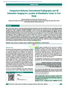
F I G U R E 1 Benign cyst lining cells. (a) Conventional cytology. PAP 9 400. (b) Cyst lining cells with fine chromatin and prominent nucleoli. SurePath. PAP 9 400. Papillary carcinoma. (c) Papillary structures. Conventional cytology. PAP 9 200. (d) Because there are disadvantages to this technique, the cytologic details cannot be identified at the three-dimensional papillary structures. SurePath. PAP 9 200. Papillary carcinoma. (e). Intranuclear cytoplasmic pseudoinclusions. MGG 9 1000. (f) Intranuclear cytoplasmic pseudoinclusions, nuclear grooves, nuclear membrane irregularity, and pleomorphism in a threedimensional cell layer. At the threedimensional cell layers, cytologic details are hardly fixed. The size of the cells in SP are smaller than that of CC at the same highpower fields. SurePath. PAP 9 1000.
demonstrated at different levels with microfocussing. The Kappa
demonstrated than CC (Figure 1A,B). It can be said that, in the
coefficient were in the good agreement category for intranuclear
demonstration of nuclear details, the SP method is harder but also
cytoplasmic pseudo-inclusions and nuclear grooves. In SP, along with
more reliable than the CC method.
the shrinking nuclei, the inclusions also show concomitant shrinkage.
Three-dimensional structures were not seen in CC, whereas they
Because of this shrinkage, it is more difficult to see nuclear inclu-
were seen in 38.4% of the SP cases (Figure 1C,D). Similar results
sions and grooves with SP than it is with CC (Figure 1E,F). The fact
have been stated in the literature.12 The reason for the increased
that overlapping, molding and nuclear grooving demonstrate a high
frequency of the three-dimensional tissue structures in the SP
Kappa coefficient shows that these three characteristics are reliable
method is that, in this technique, aspiration material is not spreading.
indicators of neoplastic proliferation (Table 2). The swirl pattern can
Another cytomorphological feature that was found to have the low-
be demonstrated in 14%–17% of the cases with papillary carci-
est Kappa coefficient (fair-agreement category) after a haemorrhagi-
noma.18 The Kappa coefficient was low for swirl patterns that were
cal background and tridimensional figures was determined to be
demonstrated more commonly in CC than SP. The Kappa coefficient
tissue fragments, whose cytomorphological details cannot be clearly
was found to be low for chromatin structure and nucleoli-like
identified. This feature was seen in 18.4% of SP (Table 2). When
nuclear details. These characteristics were demonstrated more com-
preparing SP samples, the material taken from the centrifuge and
monly with SP. In SP, there is shrinkage of the cell and nucleus, and
applied to the slides contains tridimensional tissue fragments and cell
one must perform more careful and detailed evaluations to demon-
layers. As some of these structures are very thick and stain deeply,
strate chromatin structure, inclusions, nuclear grooves and nuclear
adequate evaluation is not possible because the cytological details
membranes. Although the nuclei are smaller in SP, morphological
are not visualised with microfocussing. Another problem caused by
details, such as nucleoli and chromatin structure, can be better
tridimensional tissue fragments is that they cause covering of
SAHIN
|
ET AL.
T A B L E 5 Comparison of conventional cytology and SurePath in thyroid cytology Parameters
Conventional cytology
7
concomitant risk for SFN category to be 15%-30%.2–4 The present results coincide with the literature.
SurePath
5 | CONCLUSIONS
Background
Hemorrhagic
Clear
Fixation quality
_Inadequate
High
Cell type intepretation
Difficult
Easy
The problems of thyroid cytology in CC were determined, such as a
Rate of indeterminate diagnosis
High
Low
haemorrhagic
Cellularity
High
Low
SP. Those properties of SP that are superior to CC afford fewer
Nondiagnostic rate
Low
High
diagnoses of undetermined, and more diagnoses of benign and
Uninterpreted tissue fragments
No
Yes
malignant. However, the hypocellularity of the preparates, as well as
Three dimensional cell layers
No
Yes
Artifacts
Crush and air drying
Staining and covering
cussing. Although this study was conducted with only a few cases,
Number of slides
Much
Less
method to CC, which could be used in thyroid cytopathology.
€nwald-Giemsa May-Gru stainable
Yes
No
Reserved material and additional tests
No
Yes
Cost effectivty
Yes
background,
air-drying
and
crush
artefacts,
an
increased workload owing to high numbers of slides were solved in
the high number of non-diagnostic cases and structures that cannot be interpreted, are problems in SP. An disadvantage of SP, tri-dimentional structures and smaller cells need more attention and microfothe present results (Table 5) showed that SP was an alternative
REFERENCES
High cost
artefacts that may preclude microscopic evaluation (Table 5). Twenty-six (14%) SP slides covered by an automatic apparatus containing covering artefacts that precluded examination were uncovered and recovered manually. According to previous literature, some of the cytomorphological features found in LBC preparates but not in CC are tridimensional structures which cannot be interpreted microscopically, hobnail pattern of cells, naked capillaries, eosinophilic nucleoli and perinuclear halo, which are seen in papillary carcinoma.12,15,18–20. Other features are cell shrinkage, a ball shape of the follicles caused by the invisible follicular lumens, the dense basophilic debris on the background in the haemorrhagic aspirations in which inadequate Cytorich solution is used and a tridimensional appearance of the nuclei in papillary carcinomas, which makes some nuclear parts invisible (broken-stone appearance). When the cytopathological diagnoses of CC and SP with the histopathologic diagnoses of 31 thyroidectomy cases were compared, incompatibility for only three cases that affect the therapeutic approach was determined (Table 4). Of the two cases with histopathological diagnoses of malignancy, one had the diagnosis of atypical in CC, and the other had the same diagnosis in SP. For a case in which the SP and histological diagnoses were benign (in lymphocytic thyroiditis), it was noted that CC assigned a diagnosis of ‘suspicious for follicular neoplasia’. According to these results, both methods were inefficient in determining one of 30 thyroid carcinoma. Using the CC method, one benign lesion had the overdiagnosis of ‘suspicious for follicular neoplasia’. In the current literature, the rates of cases assigned with atypical diagnoses have a 5%-15% chance of being found to be malignant upon resection and the
1. Welker MJ, Orlov D. Thyroid nodules. Am Fam Physician. 2003;67:559-566. 2. Ali SZ, Cibas ES. The Bethesda System for Reporting Thyroid Cytopathology: Definition, Criteria and Explanatory Notes. Newyork: Springer Press; 2010:5-128. 3. DeMay R. The Art and Science of Cythopathology, 2th edn. Chicago: ASP. 2012;830-942. 4. Cibas ES, Ducatman BS. Cytology: Diagnostic Principles and Clinical Corralates, 4th edn. Philadelphia: Saunders Elsevier; 2014:255-282. 5. Afify AM, Liu J, Al-Khafaji BM. Cytologic artifacts and pitfalls of thyroid fine-needle aspiration using ThinPrep: a comparative retrospective review. Cancer. 2001;93:179-186. 6. Kocjan G, Feichter G, Hagmar B, et al. Fine needle aspiration cytology: a survey of current European practice. Cytopathology. 2006;17:219-226. 7. Cavaliere A, Colella R, Puxeddu E, et al. Fine needle aspiration cytology of thyroid nodules: conventional vs thin layer technique. J Endocrinol Invest. 2008;31:303-308. 8. Poller DN, Stelow EB, Yiangou C. Thyroid FNAC cytology: can we do it better? Cytopathology. 2008;19:4-10. 9. Fadda G, Rossi ED. Liquid-based cytology in fine-needle aspiration biopsies of the thyroid gland. Acta Cytol. 2011;55:389-400. 10. Cochand-Priollet B, Prat JJ, Polivka M, et al. Thyroid fine needle aspiration: the morphological features on ThinPrep slide preparations. Eighty cases with histological control. Cytopathology. 2003;14:343-349. 11. Saleh H, Bassily N, Hammoud MJ. Utility of a liquid-based, monolayer preparation in the evaluation of thyroid lesions by fine needle aspiration biopsy: comparison with the conventional smear method. Acta Cytol. 2009;53:130-136. 12. Jung CK, Lee A, Jung ES, Choi YJ, Jung SL, Lee KY. Split sample comparison of a liquid-based method and conventional smears in thyroid fine needle aspiration. Acta Cytol. 2008;52:313-319. 13. Nagarajan N, Schneider EB, Ali SZ, Zeiger MA, Olson MT. How do liquid-based preparations of thyroid fine-needle aspiration compare with conventional smears? An analysis of 5475 specimens. Thyroid. 2015;25:308-313. 14. Geers C, Bourgain C. Liquid-based FNAC of the thyroid: a 4-year survey with SurePath. Cancer Cytopathol. 2011;119:58-67.
8
|
15. Rossi ED, Morassi F, Santeusanio G, Zannoni GF, Fadda G. Thyroid fine needle aspiration cytology processed by ThinPrep: an additional slide decreased the number of inadequate results. Eur J Endocrinol. 2013;168:853-859. 16. Ravinsky E, Safneck JR. Fine needle aspirates of follicular lesions of the thyroid gland. The intermediate-type smear. Acta Cytol. 1990;34:813-820. 17. Policarpio-Nicolas ML, Sirohi D. Macrofollicular variant of papillary carcinoma, a potential diagnostic pitfall: a report of two cases including a review of literature. Cytojournal. 2013;30:16. 18. Lee JS, Choi HS, Park IA, Ryu HS. Liquid-based fine needle aspiration biopsy of papillary thyroid carcinoma: logistic regression analysis with conventional and new cytomorphologic features. Acta Cytol. 2013;57:233-240. 19. Chang H, Lee E, Lee H, et al. Comparison of diagnostic values of thyroid aspiration samples using liquid-based preparation and
SAHIN
ET AL.
conventional smear: one-year experience in a single institution. APMIS. 2013;121:139-145. 20. Suzuki A, Hirokawa M, Higuchi M, et al. Cytological characteristics of papillary thyroid carcinoma on LBC specimens, compared with conventional specimens. Diagn Cytopathol. 2015;43:108-113.
How to cite this article: Sahin D, Yilmazbayhan D, Firat P, et al. Comparison of conventional cytology and SurePath in split thyroid fine needle aspiration materials. Cytopathology. 2017;00:1-8. https://doi.org/10.1111/cyt.12430