support initiation (15,16). Similar results were obtained when other mitogens, such ethylene dibromide (EDB), nafenopine (BAF), and cyproterone acetate (CPA), ...
Enzvironimental Health Perspectives 101(Suppl 5): 163-168 (1993)
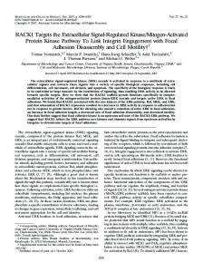
Compensatory Regeneration, MitogenInduced Liver Growth, and Multistage Chemical Carcinogenesis by Giovanna Maria Ledda-Columbano,1 Pierpaolo Coni,1 Gabriella Simbula,1 Ignazio Zedda,1 and Amedeo Columbano Liver cell proliferation has often been implicated to play a major role during different steps of the carcinogenic process. Most of the experimental studies indicating a close association between cell proliferation and liver cancer development have made use of a compensatory type of proliferative stimulus. However, liver growth may also be caused by direct hyperplasia after administration of primary mitogens. Our recent studies examined the possible differences between these two types of cell proliferation. Our studies indicate that a) increased expression of proto-oncogenes such as c-fos, c-jun, and c-myc is not necessary for entry into the cell cycle during mitogen-induced liver growth; b) mitogen-induced liver growth does not support initiation of chemical hepatocarcinogenesis; c) repeated proliferative stimuli induced by primary mitogens do not stimulate the growth of initiated cells to a focal and/or nodular stage; and d) mitogen-induced liver growth, unlike compensatory regeneration, is followed by a particular mode of cell death, namely, apoptosis. This type of cell death may be responsible for the elimination of carcinogen-initiated cells.
Introduction Cell proliferation appears to be intimately involved in several phases of the carcinogenic process. Although the detailed mechanism by which cell proliferation participates in initiation, promotion, and progression phases is far from understood, several epidemiological as well as experimental studies have recently led many investigators to hypothesize that any condition characterized by enhanced cell division results in an accumulation of genetic errors that may ultimately lead to neoplasia (1-3). Interestingly, most of the information about the role of cell proliferation in experimental carcinogenesis, especially data indicating a positive correlation between cell proliferation and the initiation step of liver chemical carcinogenesis, comes from experi'Istituto di Patologia Sperimentale, Universita di Cagliari, Via Porcell 4, 09124 Cagliari, Italy. Address reprints request to A. Columbano, Istituto di Patologia Sperimentale, Universita di Cagliari, Via Porcell 4, 09124 Cagliari, Italy. This paper was presented at the Symposium on Cell Proliferation and Chemical Carcinogenesis that was held January 14-16, 1992, in Research Triangle Park, NC.
mental protocols wherein a compensatory type of proliferative stimulus was used. In this article we summarize our recent research presenting evidence that there are at least two different types of proliferative stimuli and that these two different stimuli may exert different effects on initiation and promotion of hepatochemical carcinogenesis.
Compensatory Regeneration Cell proliferation can be induced in the liver after surgical removal of cells [partial hepatectomy; PH (4)]. This regenerative growth seems to be under control of a growth regulatory mechanism, and it stops once the organ has reached its original mass. A similar regenerative growth can be observed in the liver after treatment with several chemicals, including many carcinogens, that induce cell necrosis. This type of cell proliferation, called compensatory regeneration, is positively correlated with the initiation phase of chemical hepatocarcinogenesis (5-8). In recent years, this type of proliferative stimulus, especially after PH, has been extensively characterized in terms of expression of cellular proto-oncogenes production of growth factors such as HGF (hepatocyte growth factor) and trans-
164
LEDDA-COLUMBANO ET AL.
forming growth factor TGFa, as well as of mitoinhibitory signals such as TGF3 [for reviews see Fausto and Mead (9) and Michalopoulos (10)].
so+
-GST-P + 40 -
Mitogen-Induced Liver Growth Compensatory regeneration is not the only mode of cell proliferation. Certain chemicals may induce growth in organs such as the liver and kidney without causing prior cell loss. These agents, called primary mitogens, induce cell proliferation as the primary event, thus increasing the mass of the organ due to an increase in DNA content. After cessation of the mitogenic stimulus, there is a rapid regression of the hyperplasia until the organ reaches its original mass. The regression of the original hyperplasia appears to be due to a particular mode of cell death, namely apoptosis (11). The fact that apoptotic bodies do not occur until liver or renal cells have completed their replicative cycle and the fact that apoptotic bodies are not detected once the organs have regressed to their original mass (12-14) support the hypothesis that apoptosis is involved in the regulation of organ size by playing a complementary but opposite role to mitosis. Apoptosis under these conditions may be referred to as "compensatory cell death," in the same way we define "compensatory regeneration" as the mitotic response of a tissue after cell removal. It is also important to stress that mitogen-induced liver growth occurs without any previous cell loss, which seems to suggest that mitogens can interfere with the regulatory mechanisms normally operating in the target organ. The interference is only transient. In fact, refractoriness of liver cells to proliferation is observed after a few days, even in the presence of the mitogenic stimulus. Based on the differences exhibited by these two types of proliferative stimulus, we designed experiments to study their effects in different steps of liver carcinogenesis.
Initiation of Liver Carcinogenesis As already mentioned, several studies have shown that proliferative stimuli of the compensatory type applied before or immediately after administration of a non-necrogenic dose of carcinogens support the initiation of chemical hepatocarcinogenesis (5-8). To study whether different proliferative stimuli could exert a different effect on the initiation phase, experimental protocols were adopted wherein the same non-necrogenic dose of the carcinogen N-methyl-N-nitrosourea (MNU) was administered during compensatory regeneration induced by PH or CCl4 and during liver growth induced by the primary mitogen lead nitrate (LN). The results obtained indicate that, unlike compensatory regeneration, mitogen-induced liver growth did not support initiation (15,16). Similar results were obtained when other mitogens, such ethylene dibromide (EDB), nafenopine (BAF), and cyproterone acetate (CPA), or
30 -
E .,,
U 0
U-
MNU
MNU+PH
MNU+CC14 MNU+LN MNU+EDBMNU+CPAMNU+NAF
FIGURE 1. Effect of proliferative stimuli of different types on initiation of chemical carcinogenesis. Male Wistar rats (200 g) were given N-methyl-N-nitrosourea (60 mg/kg) during S phase after induction of compensatory regeneration by partial hepatectomy and CC14 (2 mL/kg) or mitogen-induced liver growth by lead nitrate (100 gmole/kg), ethylene dibromide (100 mg/kg), cyproterone acetate (60 mg/kg), and nafenopin (200 mg/kg). The initiated hepatocytes were assayed as GST-P+ (placental glutathione-Stransferase) foci after promotion with the resistant hepatocyte model.
other carcinogens, such as benzo(a)pyrene (BaP) and diethylnitrosamine (DEN), were used (Fig. 1). Similar findings were obtained whether the initiated hepatocytes were monitored as enzyme-altered foci using the resistant hepatocyte model [RH (17)] or using the phenobarbital (PB) (18,19) or the orotic acid (OA) model (20). Thus, it appears that only compensatory liver cell proliferation, but not direct hyperplasia, supports carcinogen-induced initiation.
Development of Foci and Nodules Cell proliferation is involved not only in the initiation step of chemical carcinogenesis but also in the promotion phase. A number of experimental findings suggest that proliferative stimuli such as those exerted by PH or necrogenic doses of CCl4 promote the appearance of nodules and hepatocellular carcinomas after initiation with different carcinogens (21-23). In contrast, conflicting information comes from studies where mitogens were used as promoting agents in long-term regimens. Although long-term feeding with mitogenic compounds exerts a promoting effect on the growth of preneoplastic and neoplastic lesions (24-27), it has been reported that compounds with mitogenic activity paradoxically accelerate the regression of enzymealtered foci (28-30). A precise assessment of the role of cell proliferation during promotion of liver carcinogenesis is further complicated by the fact that, with few exceptions (24), the mitogenic compounds elicit a very transient proliferative response of the liver (usually the proliferative response is almost completely abolished in a matter of days). Thus, under these condi-
CELL PROLIFERATION AND LIVER CARCINOGENESIS
tions it is difficult to conceive of an association between the promoting ability of these chemicals (promotion requires a long time) and their capacity to induce cell proliferation in the target organ, which is limited to a few days. A specific effect of these compounds on initiated cells rather than a generalized proliferative stimulation in the target organ may better justify the promoting ability of these chemicals. To learn more about the relation between cell proliferation and promotion, an experimental protocol was adopted wherein rat liver was initiated with DEN and then stimulated to proliferate by repeated administration of the necrogenic compound CC14 (compensatory regeneration) or by repeated treatment with LN (mitogen-induced liver growth). Under these experimental conditions, it is possible to determine whether a mitotic response of liver cells occurs any time the proliferative stimulus is applied and to associate the proliferative capacity of the two different proliferative stimuli with their ability to promote the growth of carcinogen-initiated cells. The results obtained indicate that y-glutamyltranspeptidase-(GGT)-positive foci and/or nodules were observed only when cell proliferation was induced by CC14 (compensatory regeneration) but not when the proliferative stimulus was represented by a primary mitogen (Fig. 2); this finding indicates that liver growth induced by the mitogen LN, unlike compensatory regeneration, does not possess any promoting ability. Interestingly, the extent of cell proliferation observed after the last proliferative stimulus was essentially similar in both the experimental groups, indicating that DNA synthesis per se is not a sufficient condition for the growth of preneoplastic lesions (31).
Development of Enzyme-Altered Foci during Promotion It is possible that in lead-nitrate-induced hyperplasia there is no difference between initiated and normal cells (lack of "differential"). To answer this question, we designed an experimental protocol wherein the "differential" was generated by the exposure of initiated liver cells to several promoting regimens. It is known that in several models of rat liver tumor promotion, compensatory regeneration is either a necessary component (resistant hepatocyte model) or it potentiates the promoting ability of promoting agents such as OA and PB (20,32,33). Therefore, we investigated whether induction of mitogen-induced liver growth in the presence of a promoting environment could now enhance the growth of initiated cells to a focal or nodular stage. The results, which are shown in Table 1, indicate that mitogenic stimulation induced by LN in rats previously initiated with DEN to promoting regimens such as OA or PB, unlike that exerted by PH, is unable to potentiate the promoting ability of these agents. Analysis of DNA synthesis in the various conditions did not show any significant difference, again suggest-
u
165
-5|
20
10-
117~~~~~~~~~~~~0
51
DENA
CCI4
DENA+CCI4
DENA+LN
FIGURE 2. Incidence of y-glutamyltranspeptidase-positive foci versus hepatic cell proliferation after compensatory regeneration or direct hyperplasia. Rats were treated with an initiating dose of diethylnitrosamine (100 mg/kg). After 15 days, cell proliferation was induced for 8 times, once every 20 days, by a compensatory type of proliferative stimulus (CCI4) or by direct hyperplasia (lead nitrate). Incorporation of tritiated thymidine was measured at the time of sacrifice after the last injection of CCI4 or lead nitrate.
ing a dramatic difference between these two types of proliferative stimuli despite the fact that they both stimulate a similar degree of liver cell proliferation.
Conclusions The following hypotheses may be considered to explain the different effect of the two types of liver cell proliferative stimuli on initiation and promotion of chemical hepatocarcinogenesis: a) mitogen-induced hyperplasia is not conducive for the growth of initiated cells; b) mitogen-induced hyperplasia stimulates the growth of initiated cells, but initiated cells are elimiTable 1. Effect of different proliferative stimuli given during promotion with OA on (3H)thymidine incorporation into hepatic DNA and on the incidence of GGT+ foci.a
[ H]Thymidine Proliferative incorporation GGT+ Treatment stimulus foci/cm2 (cpm/4g DNA) DEN+ OA None 18 + 3 0.7 ± 0.2 DEN+ OA PH 90 ± 30 12.0 ± 1.0 DEN+OA LN 102 ± 7 1.2 ± 0.3 DEN+ BD PH ND 1.3 ± 0.6 DEN+ BD LN ND 0.7 ± 0.2 Abbreviations: ND, not determined; OA, orotic acid; GGT, y-glutamyltranspeptidase; DEN, diethylnitrosamine; BD, basal diet; PH, partial hepatectomy; LN, lead nitrate. aDEN was given at a dose of 100 mg/kg IP. Two weeks after DEN administration, rats were fed a diet containing 1% OA or BD. Liver cell proliferation was induced 2 weeks later by PH or LN (100 micromole/kg). For determination of thymidine incorporation, osmotic minipumps containing [3 H]thymidine releasing a constant flow rate of 1 iCi/hr were implanted IP, and the rats were killed 3 days afterwards. For determination of the enzyme-altered foci, rats were sacrificed 2 weeks after application of the proliferative stimuli and the foci were monitored as GGT+.
166
LEDDA-COLUMBANO ET AL.
nated by apoptosis during the regression phase. The former hypothesis argues that the signal transduction pathways for these two types of growth stimuli are different. It is possible that although the normal hepatocytes may respond to both the proliferative stimuli, initiated hepatocytes can respond to growth signals induced by compensatory cell proliferation but not to those induced by mitogen-induced direct hyperplasia. In this respect, it is of interest that the signal transduction pathways induced by these different growth stimuli appear to be different. In fact, while a transient and sequential expression of c-fos, c-jun, and c-myc is induced in rat liver after CC14 and PH, hyperplasia induced by the mitogen CPA is not accompanied by any increase in the expression of these proto-oncogenes (34). Preliminary studies indicate that a similar pattern can be observed during hepatic cell growth induced by NAF. Furthermore, no significant increase in the expression of c-fos was observed during hyperplasia induced by two other mitogens, LN and EDB. The differences observed between the various proliferative stimuli seem to concern immediate early genes. In fact, an increased expression of genes associated with S phase such as H-ras, K-ras, and thymidilate synthase (TS) was observed in all proliferative conditions (Table 2). Whether these differences are a reflection of different pathways of signal transduction has to be investigated. It is conceivable that primary mitogens use different mechanisms leading to the entry into the cell cycle. For example, the mitogens CPA (a synthetic steroid) and NAF (a hypolipidemic drug) could exert their effect by binding to a superfamily of intracellular steroid hormone receptors (35). In this way these chemicals could directly modulate specific gene transcription. In contrast, another type of cell growth stimulus related to membrane receptors exerts this function through different transcription-activating factors [AP1 family (36)]. Recent studies suggesting that steroid receptors and AP1 factors are the primary regulators in two separate signal transduction pathways (37) may possibly explain why CPA-induced hyperplasia is not accompanied by an increased expression of Table 2. Schematic representation of the pattern of expression of some cell-cycle-related genes during cell proliferation induced by different proliferative stimuli.
Gene
Compensatory regeneration PH CC14
T
Hyperplasia CPA
LN
EDB
T T T
t
T
1
c-fos c-jun c-myc
T
T T
T
1
T T
c-Ha-ras TS
T T
T T
T T
Abbreviations: PH, partial hepatectomy, LN, lead nitrate; EDB, ethylene dibromide; CPA, cyproterone acetate; TS, thymidilate synthase.
c-fos, c-jun, and c-myc mRNA, while an increased expression of these proto-oncogenes is present during cell proliferation induced by stimuli of a different nature. Another possibility is that mitogens induce growth factors that are different from those induced during compensatory regeneration. It will be of interest to determine whether mitogen-induced liver growth is associated with an increase in growth factors such as HGF or TGFa, as observed during CC14, galactosamine, or PH-induced compensatory regeneration (38,39). It will also be of interest to determine whether a difference exists in terms of modulation of positive and negative growth-regulating factors between the two types of proliferative stimuli. As mentioned above, another possible explanation for the absence of enzyme-altered hepatic lesions in mitogen-treated rat liver is that initiated cells divide after the mitogenic stimulus, but they are eliminated by apoptosis during the regression of the initial hyperplasia (16). The possibility that this type of cell death might be responsible for the lack of foci formation implies that initiated cells are susceptible to apoptosis as much or even more than normal hepatocytes. This possibility is supported by studies based on mathematical models that indicated an extensive loss of initiated cells shortly after their formation (40). In this respect, it is also interesting to note that preneoplastic liver cells induced by three different promoting procedures, the RH model, the CD model, and the OA model, show a high apoptotic index at the early stage of the process (41). In this study as well as in other reports, the incidence of apoptotic bodies was found to be much higher in preneoplastic hepatocytes than in surrounding normal cells (12,41,42), suggesting that carcinogen-initiated liver cells (that are quite resistant to necrosis) are somehow more sensitive than normal cells to apoptosis. The possibility that carcinogen-altered cells may be preferentially eliminated by apoptosis is very intriguing. In fact, it may be hypothesized that induction of apoptosis at a time when initiated cells have not yet undergone the changes responsible for their neoplastic development may selectively kill these cells without severe loss of normal hepatocytes (43). On the other hand, carcinogen-altered hepatocytes are generally believed to be more resistant than normal cells to another type of cell death, necrosis. It is known that administration of compounds such as CCl4 and dimethylnitrosamine at doses that induce severe necrosis in normal liver do not elicit necrosis of hepatocytes of preneoplastic lesions (44). The possibility that initiated hepatocytes may be resistant to one type of cell death (necrosis) but not to the other (apoptosis) deserves consideration. Although at present we do not understand the exact significance of apoptosis in the carcinogenic process, an in-depth study of the mechanisms underlying the differences between this type of cell death and cell necrosis may help to clarify the mechanism(s) responsible for the inability of mitogen-
CELL PROLIFERATION AND LIVER CARCINOGENESIS
induced cell proliferation to support the growth of carcinogen-initiated cells. This work was supported by funds from Ministero Universita e Pubblica Istruzione (60%), Associazione Italiana Ricerca sul Canczo, and Consiglio Nazionale della Ricezche (Progetto Finalizzato Oncologia), Italy. REFERENCES 1. Preston-Martin, S., Pike, M. C., Ross, R. K., Jones, P. A., and Henderson, B. E. Increased cell division as a cause of human cancer. Cancer Res. 50: 7415-7421 (1990). 2. Ames, B. N., and Gold, L. S. Chemical carcinogenesis: too many rodent carcinogens. Proc. Natl. Acad. Sci. U.S.A. 87: 7772-7776 (1990). 3. Cohen, S. M.,. and Ellwein, L. B. Cell proliferation in carcinogenesis. Science 249: 972-975 (1990). 4. Higgins, G. M., and Anderson, R. M. Experimental pathology of the liver. 1. Restoration of the liver of the white rat following partial surgical removal. Arch. Pathol. 12: 186-202 (1931). 5. Cayama, E., Tsuda, H., Sarma, D. S. R., and Farber, E. Initiation of chemical carcinogenesis requires cell proliferation. Nature 275: 60-61 (1978). 6. Columbano, A., Rajalakshmi, S., and Sarma, D. S. R. Requirement of cell proliferation for the initiation of liver carcinogenesis as assayed by three different procedures. Cancer Res. 41: 2079-2083 (1981). 7. Kaufmann, W. K., Rahija, R. J., MacKenzie, S. A., and Kaufman, D. G. Cell cycle dependent initiation of hepatocarcinogenesis in rats by (7r, 8t-dihydroxy-9t, 10t-epoxy)-7, 8, 9, 10-tetrahydrobenzo(a)pyrene. Cancer Res. 47: 3771-3775 (1987). 8. Rabes, H. M., Muller, L., Hartmann, A., Kerler, R., and Schuster, C. Cell cycle dependent initiation of adenosine triphosphatase-deficient populations in adult rat liver by a single dose of N-methyl-N-nitrosourea. Cancer Res. 46: 645-650 (1986). 9. Fausto, N., and Mead, J. E. Biology of Disease. Regulation of liver growth: protooncogenes and transforming growth factors. Lab Invest. 60:4-13 (1989). 10. Michalopoulos, G. K. Liver regeneration: molecular mechanisms of growth control. FASEB J. 4: 240-249 (1990). 11. Wyllie, A. H., Kerr, J. F. R., and Currie, A. R. Cell death: the significance of apoptosis. Int. Rev. Cytol. 68:251-306 (1980). 12. Bursch, W., Lauer, B., Timmermann-Trosiener, I., Barthel, G., Schuppler, J., and Schulte-Hermann, R. Controlled cell death (apoptosis) of normal and putative preneoplastic cells in rat liver following withdrawal of tumor promoters. Carcinogenesis 52:
453-458(1984). 13. Columbano, A., Ledda-Columbano, G. M., Coni, P., Faa, G., Liguori, C., Santacruz, G., and Pani, P. Occurrence of cell death (apoptosis) during the involution of liver hyperplasia. Lab. Invest. 52: 670-675 (1985). 14. Ledda-Columbano, G. M., Columbano, A., Coni, P., Faa, G., and Pani, P. Cell deletion by apoptosis during regression of renal hyperplasia. Am. J. Pathol. 135: 657-662 (1989). 15. Columbano, A., Ledda-Columbano, G. M., Coni, P., and Pani, P. Failure of mitogen-induced cell proliferation to achieve initiation of rat liver carcinogenesis. Carcinogenesis 8: 345-347 (1987). 16. Columbano, A., Ledda-Columbano, G. M., Lee, G., Rajalakshmi, S., and Sarma, D. S. R. Inability of mitogen-induced liver hyperplasia to support the induction of enzyme-altered islands induced by liver carcinogens. Cancer Res. 47: 5557-5559 (1987). 17. Solt, D. B., and Farber, E. New principle for the analysis of chemical carcinogenesis. Nature 263: 701-703 (1976). 18. Peraino, C., Fry, R. J. M., and Staffeldt, E. Reduction and enhancement by phenobarbital of hepatocarcinogenesis induced in the rat by 2-acetylaminofluorene. Cancer Res. 31: 1506-1512 (1971). 19. Pitot, H. C., Barsness, L. Goldsworthy, T., and Kitagawa, T. Biochemical characterization of stages of hepatocarcinogenesis
167
after a single dose of diethylnitrosamine. Nature 275: 60-61 (1978). 20. Columbano, A., Ledda, G. M., Rao, P. M., Rajalakshmi, S., and Sarma, D. S. R. Dietary orotic acid, a new selective growth stimulus for carcinogen altered hepatocytes in rat. Cancer Lett. 16: 191-196 (1982). 21. Pound, A. W., and McGuire, L. J. Repeated partial hepatectomies as a promoting stimulus for carcinogenic response of liver to nitrosamines in rats. Br. J. Cancer 37: 585-594 (1978). 22. Dragani, T. A., Maneti, G., and Della Porta, G. Enhancing effect of carbon tetrachloride in mouse hepatocarcinogenesis. Cancer Lett. 31: 171-179 (1986). 23. Pound, W. Carcinogenesis and cell proliferation. N. Z. Med. J. 67: 88-99 (1968). 24. Marsman, D. S., Cattley, R. C., Conway, J. G., and Popp, J. A. Relationship of hepatic peroxisome proliferation and replicative DNA synthesis to the hepatocarcinogenicity of the peroxisome proliferators di (2-ethylhexyl)-phthalate, and (4-chloro-6-(2,3xylidini)-2-pyrimidinylthio) acetic acid (Wy-14,643) in rats. Cancer Res. 48: 6739-6744 (1988). 25. Yager, J. D., Roebuck, B. D., Paluszcyc, T. L., and Memoli, V. A. Effects of ethynil estradiol and tamoxifen on liver DNA turnover and new synthesis and appearance of gamma glutamyl transpeptidase-positive foci in female rats. Carcinogenesis 7: 2007-2014 (1986). 26. Rao, M. S., and Reddy, J. K. Peroxisome proliferation and hepatocarcinogenesis. Carcinogenesis 8: 631-636 (1987). 27. Kraupp-Grasl, B., Bursch, W., Gerbrancht, U., SchulteHermann, R. Tumor promotion by the peroxisome inducer nafenopin in rat liver may involve a new subtype of phenotypically altered foci. Cancer Res. 50: 3701-3708 (1990). 28. Staubli, W., Bentley, P., Bieri, F., Frolich, E., and Waechter, F. Inhibitory effect of nafenopin upon the development of diethylnitrosamine-induced enzyme altered foci within the rat liver. Carcinogenesis 5:4146 (1984). 29. De Angelo, A. B., Garret, C. T., and Queral, A. E. Inhibition of phenobarbital and dietary choline deficiency promoted preneoplastic lesions in rat liver by environmental contaminant di (2ethylhexyl)-phthalate. Cancer Lett. 13: 323-328 (1984). 30. Perera, M. I. R., and Shinozuka, H. Accelerated regression of carcinogen-induced preneoplastic hepatocyte foci by peroxisome proliferators BR931, 4-chloro-6-(2,3-xylidino)-2-pyrimidinylthio (N-B-hydroxyethyl) acetamide and di (2-ethylhexyl) phthalate. Carcinogenesis 5: 1193-1198 (1984). 31. Columbano, A., Ledda-Columbano, G. M., Curto, M., Ennas, M. G., Chelo, A., and Pani, P. Cell proliferation and promotion of rat liver carcinogenesis: different effect of hepatic regeneration and mitogen-induced hyperplasia on the development of enzymealtered foci. Carcinogenesis 11: 771-776 (1990). 32. Ford, J. O., and Pereira, M. A. Short term in vivo initiation/promotion bioassay for hepatocarcinogens. J. Environ. Pathol. Toxicol. 4: 3946 (1980). 33. Tatematsu, M., Nakanishi, K., Murasaki, G., Miyata, Y., Hirose, M., and Ito, N. Enhancing effect of inducers of liver microsomal enzymes on induction of hyperplastic liver nodules by N-2-fluorenylacetamide in rats. J. Natl. Cancer Inst. 63: 1411-1416
(1979).
34. Coni, P., Pichiri-Coni, G., Ledda-Columbano, G. M., Rao, P. M., Rajalakshmi, S., Sarma, D. S. R., and Columbano, A. Liver hyperplasia is not necessarily associated with increased expression of c-fos and c-myc mRNA. Carcinogenesis 11: 835-840 (1990). 35. Issemann, I., and Green, S. Activation of a member of the steroid hormone receptor superfamily by peroxisome proliferators. Nature 347: 645-650 (1990). 36. Chiu, E., Boyle, W. J., Meek, J., Smeal, T., Hunter, T., and Karin, M. The c-fos protein interacts with c-jun/AP1 to stimulate transcription of AP1 responsive genes. Cell 54: 541-552 (1988). 37. Miner, J. N., Diamond, M. I., and Yamamoto, K. R. Joints in the regulatory lattice: composite regulation by steroid receptor-AP1 complexes. Cell Growth Differen. 2:525-530 (1991).
168
LEDDA-COLUMBANO ET AL.
38. Lindroos, P., Zarnegar, R., and Michalopoulos, G. K. Hepatocyte growth factor (hepatopoietin A) rapidly increases in plasma before DNA synthesis and liver regeneration stimulated by partial hepatectomy and carbon tetrachloride administration. Hepatology 13: 743-750 (1991). 39. Kinoshita, T., Tashiro, K., and Nakamura, T. Marked increase of HGF mRNA in non-parenchymal liver cells of rats treated with hepatotoxins. Biochem. Biophys. Res. Commun. 165: 1229-1234 (1989). 40. Moolgavkar, S. H., Luebeck, G., and DeGunst, M. Two mutation model for carcinogenesis: relative roles of somatic mutations and cell proliferation in determining risk. In: Scientific Issues in Quantitative Cancer Risk Assessment (S. H. Moolgavkar, Ed.), Birkhauser, Boston, MA, 1990, pp. 136-152. 41. Columbano, A., Ledda-Columbano, G. M., Rao, P. M., Rajalak-
shmi, S., and Sarma, D. S. R. Occurrence of cell death (apoptosis) in preneoplastic and neoplastic liver cells. Am. J. Pathol. 116: 441-446(1984). 42. Rotstein, J., Sarma, D. S. R., and Farber, E. Sequential alterations in growth control and cell dynamics of rat hepatocytes in early precancerous steps in hepatocarcinogenesis. Cancer Res. 46: 2377-2385 (1986). 43. Ledda-Columbano, G. M., and Columbano, A. Apoptosis and hepatocarcinogenesis. In: Apoptosis: The Molecular Basis of Cell Death (L. Tomei and F. 0. Cope, Eds.), Cold Spring Harbor Laboratory Press, Cold Spring Harbor, NY, 1991, pp. 101-120. 44. Farber, E., Parker, S., and Gruenstein, M. The resistance of putative premalignant liver cell populations hyperplastic nodules to the acute cytotoxic effect of some hepatocarcinogen. Cancer Res. 36: 3879-3887 (1976).