proximal segment, called the ''top of the basilar''syndrome, is known for its wide variety of clinical manifestations (1). Bilateral complete ophthalmoplegia has, ...
ORIGINAL CONTRIBUTION
Complete Bilateral Ophthalmoplegia Resistant to Caloric Stimulation in Bilateral Paramedian Midbrain-Thalamic Infarction Miguel Angel Tola-Arribas, MD, Alejandro Vara-Castrodeza, MD, and Juan Ernesto Alonso-Santor, MD Abstract: A 79-year-old woman who developed bilateral paramedian midbrain-thalamic infarction manifested complete bilateral ophthalmoplegia resistant to caloric stimulation, indicating impairment of the vestibulo-ocular reflex (VOR). Previous reports have mentioned this phenomenon but have not explicitly reported the results of caloric testing. Why a lesion apparently confined to the upper brainstem should produce impairment of the horizontal VOR remains unexplained. (J Neuro-Ophthalmol 2009;29:284–285)
O
cclusion of the midbrain and thalamic perforating branches of the posterior cerebral arteries in their proximal segment, called the ‘‘top of the basilar’’syndrome, is known for its wide variety of clinical manifestations (1). Bilateral complete ophthalmoplegia has, however, been rarely described (2–7). None of the reports includes information about eye movement responses to caloric testing or passive head movement. We describe a patient with acute complete bilateral ophthalmoplegia due to a bilateral paramedian midbrain-thalamic infarction in whom such testing produced no improvement in eye movements, indicating that the vestibulo-ocular reflex (VOR) pathway was impaired. Why a lesion apparently limited to the upper brainstem should impair the horizontal VOR pathway remains unexplained.
CASE REPORT A 79-year-old woman with a history of hypertension and bilateral carotid endarterectomy suddenly became Department of Neurology (MAT-A), Department of Radiology (AV-C), and Department of Internal Medicine (JEA-S), Hospital Universitario Rı´o Hortega, Valladolid, Spain. Address correspondence to Miguel A. Tola-Arribas, MD, c/o Martı´n Santos Romero, 9, 1° A, 47016-Valladolid, Spain; E-mail: mtolaa@ seneurologia.org
284
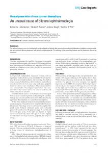
unresponsive to noxious stimuli. She also had complete bilateral ptosis. Her eyes were in the primary position, and there were no spontaneous eye movements. Pupils measured 8 mm in dim illumination and did not constrict to direct light. Caloric testing with cold water or with passive head movement failed to elicit any eye movement. Corneal and gag reflexes were normal. There were bilateral spontaneous movements of all four extremities and normal deep tendon reflexes. There was no Babinski reflex. Based on these findings, a toxic process was initially suspected. Results of electrocardiography were normal. Brain CT showed mild signs of small vessel ischemic white matter disease. Brain MRI performed 2 days after symptom onset demonstrated acute bilateral paramedian midbrainthalamic infarction (Fig. 1A–D). MRA images showed complete basilar artery occlusion (Fig. 1E–F). Over the following days, she remained unresponsive, and her bilateral ophthalmoplegia and mydriasis remained unchanged. She died of pneumonia a few weeks after presentation. No autopsy was performed.
DISCUSSION This patient had occlusion of the arterial supply of the paramedian thalamus and midbrain, an infrequent and grave variant of the top of the basilar syndrome that manifests clinically with a wide variety of ocular motor, sleep-wake cycle, alertness, behavioral, and motor abnormalities (8,9). Despite imaging evidence of basilar artery thrombosis, no pontine clinical or imaging abnormalities were observed. However, based on caloric testing, the VOR pathway to stimulate abduction, which does not course through the midbrain or thalamus, was absent. This phenomenon is unexplained. Patients with complete ophthalmoplegia in this setting have generally died within days of onset. Survivors have demonstrated improvement of abduction within a few days (2). J Neuro-Ophthalmol, Vol. 29, No. 4, 2009
Midbrain-Thalamic Infarction
J Neuro-Ophthalmol, Vol. 29, No. 4, 2009
FIG. 1. Diffusion-weighted (A), fluid-attenuated inversion recovery (B), T2 coronal (C), and T2 axial (D) MRI sequences show acute bilateral paramedian midbrain-thalamic infarction. Coronal (E) and sagittal (F) MRA time-of-flight images show occlusion of the middle and distal basilar arteries (arrowheads).
REFERENCES 1. Caplan LR. ‘‘Top of the basilar’’ syndrome. Neurology 1980;30: 72–79. 2. Thurtell MJ, Halmagyi GM. Complete ophthalmoplegia. An unusual sign of bilateral paramedian midbrain-thalamic infarction. Stroke 2008;39:1355–1357. 3. Finocchi C, Del Sette M, Croce R, et al. Bilateral ophthalmoplegia: an unusual sign of the top of the basilar artery syndrome. Ital J Neurol Sci 1996;17:301–304. 4. Biller J, Sand JJ, Corbett JJ, et al. Syndrome of the paramedian thalamic arteries: clinical and neuroimaging correlation. J Clin Neuroophthalmol 1985;5:217–223.
5. De Mendonca A, Pimentel J, Morgado F, et al. Mesencephalic haematoma: case report with autopsy study. J Neurol 1990;237:55–58. 6. Tomecek FJ, Morgan JK. Ophthalmoplegia with bilateral ptosis secondary to midbrain hemorrhage: a case with clinical and radiologic correlation. Surg Neurol 1994;41:131–136. 7. Worthington JM, Halmagyi GM. Bilateral total ophthalmoplegia due to midbrain hematoma. Neurology 1996;46:1176–1177. 8. Waterston JA, Stark RJ, Guilligan BS. Paramedian thalamic and midbrain infarction: the ‘‘mesencephalothalamic syndrome.’’ Clin Exp Neurol 1987;24:45–53. 9. Castaigne P, Lhermitte F, Buge A, et al. Paramedian thalamic and midbrain infarct: clinical and neuropathological study. Ann Neurol 1981;10:127–148.
285