Composite Tumor Associating TB and SK
Ann Dermatol Vol. 27, No. 5, 2015
http://dx.doi.org/10.5021/ad.2015.27.5.601
CASE REPORT
Composite Tumor Associating Trichoblastoma and Seborrheic Keratosis Seung-Hee Loh, Bark-Lynn Lew, Woo-Young Sim Department of Dermatology, Kyung Hee University College of Medicine, Seoul, Korea
Seborrheic keratosis is a common benign epidermal tumor histologically composed of basaloid and squamous cells. It mainly occurs on the face, scalp, and trunk, and presents clinically as a well-circumscribed, brownish to black papule, nodule, or plaque. Trichoblastoma is a relatively rare benign, slow-growing tumor showing differentiation toward the primitive hair follicle. It clinically manifests as a solitary, skin to erythematous colored, well-circumscribed dermal nodule located predominantly on the head and neck with a predilection for the scalp. Histologically, a well-demarcated mass of follicular germinative cells that show various degrees of differentiation, arranged in lobules, sheets, and nests, is located in the dermis or subcutaneous fat layer. We report the case of a 28-year-old female patient with a solitary, 2.0×4.0 cm black plaque with a 0.7-cm skin-colored nodule on the scalp. Histologically, the entire black plaque had prominent hyperkeratosis, acanthosis, and papillomatosis with horn cysts. The central nodule showed well-circumscribed, various-sized dermal tumor lobules without a connection to the overlying epidermis. The lobular aggregation was composed of numerous basaloid epithelial nests and multiple primitive papillary structures with distinct peripheral palisading of nuclei. According to these findings, the scalp lesion was diagnosed as a composite tumor associating trichoblastoma and seborrheic keratosis. (Ann Dermatol 27(5) 601∼604, 2015)
Received February 25, 2014, Revised February 5, 2015, Accepted for publication February 18, 2015 Corresponding author: Bark-Lynn Lew, Department of Dermatology, Kyung Hee University Hospital at Gangdong, 892 Dongnam-ro, Gangdong-gu, Seoul 05278, Korea. Tel: 82-2-440-7329, Fax: 82-2440-7336, E-mail:
[email protected] This is an Open Access article distributed under the terms of the Creative Commons Attribution Non-Commercial License (http:// creativecommons.org/licenses/by-nc/4.0) which permits unrestricted non-commercial use, distribution, and reproduction in any medium, provided the original work is properly cited.
-KeywordsComposite tumor, Seborrheic keratosis, Trichoblastoma
INTRODUCTION Trichoblastoma (TB) was first introduced by Headington1 in 1970, and was described as a benign and well-circumscribed tumor with follicular differentiation. Ackerman et al.2 further defined this tumor and classified TBs into five subtypes according to the histological growth pattern: large nodular, small nodular, cribriform, racemiform, and retiform. TBs are characterized by a symmetrical growth, smooth borders, and a sharp circumscription. The most common locations of TB are the face, scalp, and pelvic girdle2,3. TB occurs sporadically in most cases. One case of sporadic TB arising within an apocrine poroma has been reported4. Other cases of TBs are associated with seborrheic keratosis (SK), inverted follicular keratosis, or verruca vulgaris-like inverted follicular keratosis5. We report herein a rare case of composite tumor associating TB and SK.
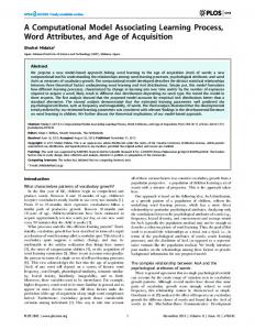
CASE REPORT A 28-year-old healthy woman presented with a skin-colored nodule within a darkly pigmented plaque on the occipital scalp. She identified the lesion 2 months before her first visit to our department. The patient had no personal or family history of cutaneous or internal malignancies. Physical examination of the scalp revealed a 2.0×4.0-cm darkly pigmented, verrucous plaque. A distinct 0.7-cm skin-colored, hairless nodule appeared within the plaque (Fig. 1). A biopsy specimen was taken from the margin of the central skin-colored nodule. Histopathologic findings of the central nodule showed well-circumscribed, various-sized dermal tumor lobules. The lobular aggregation Vol. 27, No. 5, 2015
601
SH Loh, et al
was composed of numerous basaloid epithelial nests and multiple primitive papillary structures with a distinct peripheral palisading of nuclei (Fig. 2A, B). Prominent hyperkeratosis, acanthosis, papillomatosis, and horn cysts were found adjacent to this nodule (Fig. 2A, C, D). Because the patient desired that the entire lesion be removed, complete excision was performed. To investigate the characteristics of each tumor and the connection be-
Fig. 1. A 2.0×4.0-cm dark pigmented, verrucous plaque on the scalp. Centrally, a distinct 0.7-cm skin-colored, hairless nodule was observed.
tween these two tumor types, various immunohistochemical staining procedures were also performed. The excised specimen showed centrally well-demarcated dermal tumor lobules spanning the entire dermis without a connection to the overlying epidermis. Tumor cells were arranged in nodular nests of palisading basaloid cells and multiple primitive papillary structures. Various degrees of hair follicle differentiation were observed. There was neither an area of necrosis en masse, nor a cleft between the epithelium and stroma. Therefore, this tumor can be diagnosed as a TB. Prominent hyperkeratosis, acanthosis, papillomatosis, and horn cysts, consistent with typical SK, were found around the tumor on the entire plaque beside the central nodule. The entire TB tumor lobules were stained against cytokeratin 19 (CK19) monoclonal antibody (Fig. 3A). Focal tumor cells showed weak CK17 expression. CK17 expression in SK is limited to a collarette of small keratinocytes around the TB nodules, depicting a transitional zone between the CK17-positive TB and the CK17-negative SK (Fig. 3B). Some of the tumor cells were weakly immunolabeled with Bcl-2 and CD10, and the stroma around the tumor lobules were Bcl-2 and CD10 positive. None of the tumor lobules showed immunolabeling against the CD34 monoclonal antibody (Fig. 3C).
Fig. 2. (A) Scanning view of the excision specimen (H&E, scanning view). (B) Biopsy specimen showing well-circumscribed, various-sized dermal tumor lobules. Prominent hyperkeratosis, acanthosis, papillomatosis, and pseudohorn cysts were seen adjacent to the tumor nodule (H&E, ×20). (C) The lobular aggregation was composed of numerous basaloid epithelial nests and multiple primitive papillary structures with distinct peripheral palisading of nuclei (H&E, ×200). (D) Marked hyperkeratosis, acanthosis, papillomatosis, several horn cysts, and mild basal hyperpigmentation were detected in the epidermis (H&E, ×100).
602 Ann Dermatol
Composite Tumor Associating TB and SK
Fig. 3. (A) The entire trichoblastoma tumor lobules were stained against cytokeratin 19 (CK19) monoclonal antibody, showing a mild peripheral palisading of nuclei (×100). (B) Focal tumor cells showed weak CK17 expression (×100). (C) None of the tumor lobules showed immunolabeling against CD34 monoclonal antibody (×100).
On the basis of the clinical and histological findings, we diagnosed this case as a composite tumor associating TB and SK. After surgical excision of the entire lesion, the patient is currently well, and has not experienced recurrence even after a year.
DISCUSSION TB is a rare benign tumor composed of germinative cells, from which a folliculosebaceous unit arises. TB most commonly occurs on the scalp and face. Clinically, it presents as small exophytic and endophytic, skin-colored nodules without ulceration. Basaloid proliferation is an essential histologic finding of TB, in which the tumor cells are arranged in cords, sheets, or discrete clusters surrounded by fibrous stroma1. In our study, the TB was positive for CK17. However, the expression was rather focal and weaker than that in previously reported cases. CK17 was expressed in the inner cell layers of the outer root sheath in normal adult hair follicles and in hyperproliferative conditions of the epidermis, such as psoriasis and wound healing6. Moreover, CK19 was expressed in outer cell layers of the outer root sheath. Coexpression of CK17 and CK19 in neoplastic cells of TB seems to be related to an outer root sheath phenotype7. The differential diagnosis between TB and basal cell carcinoma may be difficult in some cases. Histologically, the absence of epitheloid necrosis en masse, myxoid stromal induction, and peritumoral cleft, as well as an evidence of follicular papilla differentiation in the stroma surrounding the basaloid nodule, lead to the diagnosis of TB. Moreover, relatively symmetrical and smooth tumor borders can suggest TB8. In this study, CD34 was immunolabeled in the stroma between tumor lobules. CD34 expression has originally been shown in normal endothelium and the perifollicular area9. The stroma of TB showed diffuse CD34
expression, whereas basal cell carcinoma stroma did not show CD34 expression10. Moreover, Bcl-2 and CD10 immunohistochemical studies were conducted to distinguish TB from basal cell carcinoma and showed similar stromal expression patterns to CD34. Other studies about TB revealed similar immunohistochemical staining patterns similar to our study7. Composite tumors associating sporadic TB (i.e., not occurring in the context of a preexisting nevus sebaceous or of Brooke-Spiegler syndrome) with another epithelial neoplasm have been described in two reports on seven cases: one was within an apocrine poroma and others were associated with melanocytic nevus, SK, verruca vulgaris, angiokeratoma, and basal cell carcinoma4,5. A composite tumor associating TB and SK was reported in only two cases5. In one case, some tumor lobules of TB were connected to the SK. However, in another case, there was no connection between TB and SK. In our case, no direct connection between TB nodules and SK was observed. The collision of such tumors does not seem coincidental. TB might induce hyperproliferation of the overlying epidermis during self-proliferation and differentiation. SK is present in almost all healthy adults older than 60 years. Previous reports have documented associated benign or malignant tumors occurring either adjacent to or contiguous with SK11,12. The rates of coexistence of such tumors with SK have been reported with rates varying between 0.14% and 9% of SK examined histologically12,13. Lesions associated with SK are of various types: basal cell or squamous cell carcinoma, melanoma, melanocytic nevus, keratoacanthoma, trichilemmoma, eccrine porocarcinoma, and hemangioma11-13. TB is suggested as another possible SK-associated neoplasm. We report herein a rare and interesting case of a composite tumor associating TB and SK.
Vol. 27, No. 5, 2015
603
SH Loh, et al
REFERENCES 1. Headington JT. Differentiating neoplasms of hair germ. J Clin Pathol 1970;23:464-471. 2. Ackerman AB, DeViragh PA, Chongchitnant N. Neoplasms with follicular differentiation. Philadelphia: Lea and Febiger, 1993:401. 3. Lee HJ, Choe TJ, Park JG, Ha SJ, Kang SJ, Kim JW. A case of trichoblastoma. Ann Dermatol 1999;11:286-288. 4. Santos-Briz A, Rodríguez-Peralto JL, Miguélez A, López-Ríos F. Trichoblastoma arising within an apocrine poroma. Am J Dermatopathol 2002;24:59-62. 5. Battistella M, Peltre B, Cribier B. Composite tumors associating trichoblastoma and benign epidermal/follicular neoplasm: another proof of the follicular nature of inverted follicular keratosis. J Cutan Pathol 2010;37:1057-1063. 6. Wilson CL, Dean D, Lane EB, Dawber RP, Leigh IM. Keratinocyte differentiation in psoriatic scalp: morphology and expression of epithelial keratins. Br J Dermatol 1994;131: 191-200. 7. Yamamoto O, Asahi M. Cytokeratin expression in trichoblastic fibroma (small nodular type trichoblastoma), tricho-
604 Ann Dermatol
8.
9. 10.
11.
12.
13.
epithelioma and basal cell carcinoma. Br J Dermatol 1999; 140:8-16. Johnson WC. Metastatic carcinoma of the skin. In: Elder D, Elenitas R, Jaworsky C, Johnson B Jr, editors. Lever's histopathology of the skin. 10th ed. Philadelphia: Lippincott, 2009:1155-1156. Kim HJ, Min HG, Lee ES. Alopecia neoplastica in a patient with gastric carcinoma. Br J Dermatol 1999;141:1122-1124. Kirchmann TT, Prieto VG, Smoller BR. CD34 staining pattern distinguishes basal cell carcinoma from trichoepithelioma. Arch Dermatol 1994;130:589-592. Conner KB, Cohen PR. Cutaneous metastasis of breast carcinoma presenting as alopecia neoplastica. South Med J 2009;102:385-389. Kohno A, Saruta T, Kimura H. A case of alopecia neoplastica due to cutaneous metastasis from stomach. Rhinsho Derm 1983;25:334-335. Yuen YF, Lewis EJ, Larson JT, Wilke MS, Rest EB, Zachary CB. Scalp metastases mimicking alopecia areata. First case report of placental site trophoblastic tumor presenting as cutaneous metastasis. Dermatol Surg 1998;24:587-591.