coenzyme A (Sigma), 0.9 mu (60 mg/liter) imidazole. (Sigma), 5% fetal calf ..... cholinergic (Phi%, 1970; DeFeudis, 1974); however; syn- aptic connections have ...
The Journal of Neuroscience Vol. 2, No. 5, pp. 623-632 May 1982
0270~6474/82/0205-0623$02.00/O
Copyright 0 Society for Neuroscience Printed in U.S.A.
CONDITIONED MEDIUM FROM CULTURES OF EMBRYONIC NEURONS CONTAINS A HIGH MOLECULAR WEIGHT FACTOR WHICH INDUCES ACETYLCHOLINE RECEPTOR AGGREGATION CULTURED MYOTUBES ANNE
E. SCHAFFNER2~~~ Laboratory
MATHEW
ON
P. DANIELS
of Biochemical Genetics, National Heart, Lung, and Blood Institute, National Institutes of Health,
Bethesda, Maryland 20205
Received
June
1, 1981; Revised
September
8, 1981; Accepted
December
28, 1981
Abstract The developmental mechanisms involved in the formation of stable arrays of postsynaptic neurotransmitter receptors near sites of neurotransmitter release are essentially unknown. However, several recent studies have shown that cells or tissues of neural origin produce macromolecular factors which induce an increase in the number of acetylcholine (ACh) receptors or the number of receptor aggregates on cultured embryonic myotubes. We have tested primary cultures of embryonic neurons and other cell types for the release of an ACh receptor aggregation factor. Conditioned medium from the cultures was applied to cultures of embryonic rat myotubes for 1 day; ACh receptors on the myotubes were stained with tetramethylrhodamine-labeled a-bungarotoxin and ACh receptor aggregation activity, defined as the change in the number of receptor aggregates per myotube, was assayed. Aggregation activity with a molecular weight I 50,000 was released by cultures of neurons from sympathetic ganglia, dorsal root ganglia, spinal cord, and cerebellum. Little or no activity was released by glial or other non-neuronal cultures. Release of aggregation activity by different neuronal cell types varied by up to an order of magnitude; however, this variation was not well correlated with the differences in ACh synthesis. The factor(s) in neuronal cell conditioned medium induced a rearrangement of pre-existing receptors at the cell surface, and its action was not dependent on new protein synthesis. The results of this study are consistent with the idea that one or more receptor aggregation factors secreted by neurons are involved in the organization of neurotransmitter receptors during synapse formation in vivo.
Embryonic skeletal muscle fibers at early stages of tors (Chang and Huang, 1975; Berg and Hall, 1975; development are sensitive to acetylcholine (ACh) along Devreotes and Fambrough, 1975; Burden, 1977b; Michler their entire length and exhibit an essentially uniform and Sakmann, 1980) as well as, in some species, a change distribution of ACh receptors in their plasma mem- in the ion channel properties of the receptors (Michler and Sakmann, 1980). Synaptic localization of ACh senbranes. With further development, both ACh sensitivity and the distribution of receptors become restricted to the sitivity also has been demonstrated in parasympathetic area of the neuromuscular junction (Diamond and Mi- neurons of the frog interatrial septum (Harris et al., ledi, 1962; Bevan and Steinbach, 1977; Burden, 1977a). 1971). This spatial restriction of receptors is followed by a Receptor localization to regions of nerve contact has marked increase in the metabolic stability of the recep- been described in developing systems in vitro. Acetylcholine receptor clusters have been identified at nervemuscle synapses in co-cultures of embryonic chick spinal ’ We are grateful to Scott Dubit and Lisa Chang for technical cord and muscle (Cohen and Fischbach, 1977; Frank and assistance and to Marty Green for manuscript preparation. A. E. S. was Fischbach, 1979). Rat skeletal muscle cells from the the recipient of a Muscular Dystrophy Association Postdoctoral Reclonal line L6 exhibit increased acetylcholine sensitivity search Fellowship. on areas of the membrane where they are in contact with ’ To whom correspondence should be addressed. Present address: nerve cells from the mouse neuroblastoma clone N18 Laboratory of Neurophysiology, National Institute of Neurological and (Steinbach et al., 1973). In co-cultures of Xenopus emCommunicative Disorders and Stroke, National Institutes of Health, Bethesda, MD 20205. bryonic spinal cord or neural tube cells and muscle, ACh
624
Schaffner and Daniels
receptors are redistributed so as to be localized along points of contact between the two cell types (Anderson and Cohen, 1977; Anderson et al., 1977; Cohen and Weldon, 1980). The mechanisms whereby innervation leads to receptor localization are unknown. It is possible that receptors are synthesized preferentially and inserted at the endplate region in response to nerve contact or to soluble factors released by the nerve. Another possibility, not exclusive of the first, may involve a redistribution of receptors already present in the plasma membrane in response to the influence of the nerve. Soluble factors which increase the number of ACh receptors and the number of receptor aggregates on cultured muscle cells have been found in cell-free extracts of fetal rat and chick nervous tissue (Podleski et al., 1978; Jessell et al., 1979). Christian et al. (1978) identified a high molecular weight ACh receptor aggregation factor in the medium of cultured neuroblastoma x glioma hybrid cells. That such a factor may be neural specific was suggested by the finding that the aggregation factor was released by the parent neuroblastoma but not the parent glioma cell line. The activity of this factor was independent of new receptor synthesis. If such a factor is operating in vivo, it should be released by embryonic neurons during the period of synaptogenesis. In this study, we present evidence that several types of rat embryonic neurons in cell culture produce a high molecular weight factor which causes the aggregation of ACh receptors in the plasma membrane of cultured myotubes. This activity appears to be specific to neurons and occurs in the absence of protein synthesis. Materials and Methods Muscle cultures. Rat myotube cultures were prepared by a modification of the methods of Nelson et al. (1976). Minced muscle from the hindlimbs of 20-day-old Sprague-Dawley rat embryos (Taconic Farms, Germantown, NY) was dissociated for 30 min at 37°C in 0.2% trypsin (three times crystallized and lyophilized; Millipore Corp., Bedford, MA), 0.01% DNase (Type I, Sigma, St. Louis, MO), and 1% glucose in calcium- and magnesium-free Dulbecco’s phosphate-buffered saline (CMFPBS). Cells were resuspended in 80% Dulbecco’s minimal essential medium (DMEM; Gibco, Grand Island, NY), 10% fetal calf serum (North American Biologicals, Inc., Miami, FL), and 10% horse serum. The cell suspension was passed through a Nitex filter (pore size, 120 pm2) to remove large pieces of tissue, incubated for 20 min at 37°C in lOO-mm plastic Petri dishes to allow for the preferential adhesion of fibroblasts, and plated in 1.5 ml of the same medium in 35-mm collagen-coated plastic tissue culture dishes (Falcon, Becton-Dickinson, Cockeysville, MD). After 3 to 4 days, the medium was replaced with 90% DMEM, 10% horse serum, and 10 PM cytosine arabinoside (ara-C, Sigma) to kill dividing cells. Cytosine arabinoside was removed after 2 to 3 days and the cultures were fed twice weekly. Cultures were incubated in a humidified atmosphere with 10% COZ and 90% air at 36°C. Sympathetic neuron cultures. Superior cervical ganglia (SCG) were removed from 18- to 20-day-old SpragueDawley rat embryos, cut in pieces, and dissociated for 30
Vol. 2, No. 5, May 1982
min at 37°C in CMF-PBS containing 0.1% trypsin, 0.01% DNase, and 1% glucose. Cells were resuspended in SCG medium (see below). Clumps were removed by low speed centrifugation or by passing the suspension through Nitex (pore size, 120 pm’). Cells were plated in 0.5 ml of SCG medium into collagen-coated 16-mm plastic tissue culture wells (Costar, Cambridge, MA) at a density of 8 to 10 x lo4 cells/well. Methocel (hydroxypropyl methylcellulose, Dow Chemical Co., Midland, MI) was added to increase the viscosity of the medium and enhance the attachment of neurons to the substrate (Bray, 1970). Medium was replaced every 2 days. “Adrenergic” cultures (SCG-AIR) were grown in SCG medium according to a modification of the method of Mains and Patterson (1973). The SCG medium consisted of L15 (Leibowitz medium, Microbiological Associates, Bethesda, MD) supplemented with 28 mM (5 gm/liter) glucose, 36 PM (5 mg/ liter) p-aminobenzoic acid (Sigma), 0.5 PM (0.4 mg/liter) coenzyme A (Sigma), 0.9 mu (60 mg/liter) imidazole (Sigma), 5% fetal calf serum, 5% rat serum (Microbiological Associates), 0.4 to 0.6% Methocel, and 100 rig/ml of nerve growth factor (NGF; kindly supplied by Dr. Gordon Guroff, National Institutes of Health, Bethesda, MD). Cultures were maintained in a humidified air atmosphere at 36°C. Although non-neuronal cells did not proliferate extensively in medium without sodium bicarbonate, it was sometimes necessary to add 10 PM ara-C for 1 to 2 days after the cells had been in culture for 1 week. “Cholinergic” cultures (SCG-C02) were prepared in a manner similar to that described for adrenergic cultures with the following exceptions. SCG medium minus NGF was supplemented with 26 mM (2.2 gm/liter) sodium bicarbonate and conditioned by incubation with rat myotube cultures. Conditioned medium (CM) then was diluted with fresh medium to give a final concentration of 60% CM and supplemented with 100 rig/ml of NGF. SCG cells were plated in this medium with Methocel to yield cholinergic cultures (Patterson and Chun, 1977). Cytosine arabinoside was added to the cultures 4 to 5 days after plating and removed after 2 days. Cultures prepared in this manner were essentially free of non-neuronal cells (Fig. la). SCG glial cultures were obtained by omitting Methocel and NGF from SCG medium. Muscle cultures used for CM, cholinergic neuronal cultures, and glial cultures were maintained in a humidified atmosphere with 5% CO2 and 95% air at 36°C. Co-culture of rat myotubes and SCG neurons. Muscle cells were prepared as described above. A cell suspension of 7.5 x lo5 SCG neurons in 90% DMEM, 5% horse serum, 5% fetal calf serum, 100 rig/ml of NGF, and 10 PM ara-C was added to lo-day muscle cultures and cocultured for a 4-day period. Dorsal root ganglion cultures. Dorsal root ganglia (DRG) were removed from 16-day-old rat embryos. Cultures were prepared in a manner identical to cholinergic SCG neurons except that they were grown in nonconditioned medium. Cytosine arabinoside was added 4 to 5 days after plating to obtain cultures free of non-neuronal cells (Fig. Id). Cerebellar cultures. Cerebella were removed from 12to 15-day-old Sprague-Dawley rat embryos and dissociated by vigorous trituration followed by passage
The Journal of Neuroscience
Acetylcholine
Receptor
Aggregation
Factor from Neurons
625
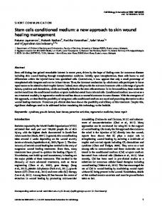
Figure 1. Cultures of dissociated primary neurons from (a) superior cervical ganglia, (b) cerebellum, (c) spinal cord, and (d) dorsal root ganglia, typical of the cultures used as sources of neuronal CM. In a, b, and d, very few non-neuronal cells are present. These are phase contrast photomicrographs. Bar, 100 pm.
through a Nitex falter (pore size, 120 pm’). Approximately 5 x lo5 cells were plated per collagen-coated 16-mm plastic tissue culture well (Costar or Linbro, Linbro Division, Flow Laboratories, Inc., Hamden, CT) in 0.5 ml of 80% DMEM, 10% fetal calf serum, and 10% horse serum. Medium was supplemented with an additional 31 mu (5.5 gm/liter) glucose (final glucose concentration, 56 mu (10 gm/liter)) and 80 units/liter of insulin (crystalline, bovine pancreas; Sigma) according to Sotelo et al. (1980). After 5 days, the medium was replaced with DMEM containing 10% horse serum and 10 PM ara-C. After 4 to 5 days, cultures were fed the same medium without araC. Cultures were maintained in a humidified atmosphere with 10% COP and 90% air at 36°C. A typical culture is shown in Figure 1 b. Spinal cord cultures. Spinal cords were removed from lZday-old Sprague-Dawley rat embryos, stripped of dorsal root ganglia and meninges, and dissociated in CMFPBS containing 0.02% trypsin and 0.01% DNase. Cells were resuspended in 80% DMEM, 10% horse serum, and 10% fetal calf serum. The medium was supplemented with 28 mM (5 gm/liter) glucose and 18 mM (1.5 gm/liter)
sodium bicarbonate (Ransom et al., 1977). Cells were plated in 1.5 ml of medium into 35-mm collagen-coated dishes at a density of 1.2 x lo6 cells/dish. After 4 to 5 days, the medium was replaced with 90% DMEM, 10% horse serum, 10 pM fluorodeoxyuridine (Sigma), and 100 PM uridine (Sigma). The fluorodeoxyuridine was removed after 2 days. A typical culture is shown in Figure lc. Spinal cord glial cultures were obtained by incubating lweek-old cultures with AzBs, a neuron-specific cytotoxic antibody (Eisenbarth et al., 1979) in the presence of guinea pig complement (Cappel Laboratories, Inc., Cochranville, PA). Antibody was added under saturating conditions at a final concentration of 7 pg/ml. Glial cultures were grown in 90% DMEM and 10% horse serum supplemented as above. Spinal cord cultures enriched for small neurons and containing very low levels of choline acetyltransferase activity were prepared as described elsewhere (Schnaar and Schaffner, 1981) and grown in a manner identical to whole dissociated spinal cord (nonseparated) neuronal cultures. All spinal cord cultures were grown in a humidified atmosphere with 10% CO2 and 90% air at 36°C.
626
Schaffner
Other non-neuronal cell cultures. Conditioned medium was obtained from cultures of adult rat skin fibroblasts and adult peritoneum courtesy of Dr. George Eisenbarth, Duke University Medical Center, Durham, NC. Conditioned medium. Conditioned medium (CM) from neuronal and non-neuronal cell cultures was removed sterilely every 2 days, centrifuged at 1,200 x g for 5 min, decanted, and frozen at -20°C. Before application to muscle cultures, CM was thawed, recentrifuged, and pooled. Conditioned medium to be concentrated was pooled and placed in a 50-ml Amicon filtration cell (Amicon Corp., Lexington, MA) fitted with an XM50 membrane (nominal molecular weight cutoff, 50,000). The medium was concentrated at 4°C and this process required 2 to 4 hr for each sample. Both the retentates and their respective ultrafiltrates were passed through a 0.45~pm Millex filter wetted with medium (Millipore Corp.) and refrigerated until use, usually within 16 hr. Serial dilutions of concentrated CM were made by the addition of appropriate volumes of fresh medium. Bioassay. Conditioned medium was applied to 12-dayold muscle cultures and removed after 24 hr. Cultures were rinsed twice for 5 min in DMEM and 10% fetal calf serum and stained for 1 hr at 36°C with tetramethyhhodamine-labeled a-bungarotoxin (TMR-aBTx; kindly supplied by Dr. Zvi Vogel, Weizmann Institute of Science, Rehovot, Israel) in the same medium. Cultures were rinsed for 10 min in DMEM and 10% fetal calf serum and twice for 10 min in DMEM buffered with 25 mM HEPES (4-(2-hydroxyethyl)-1-piperazine-ethanesulfonic acid, Gibco), pH 7.4, in the absence of serum. Then cultures were fixed for 30 min in 2% formaldehyde, 0.1 M sodium phosphate buffer, pH 7.4, at 4”C, rinsed twice for 30 min in 0.1 M sodium phosphate buffer at 4”C, and placed in 80% ethanol at -20°C. Cultures were examined with a Zeiss Photomicroscope II equipped with epi-illumination. Light from an HBO W/4 mercury arc lamp was passed through BP546/9 and KP600 excitation filters and a Kodak 23A barrier filter. The appearance of the myotubes under these conditions is shown in Figure 2. To quantify the effects of CM, each concentration or dilution of CM was added to duplicate cultures. Twenty-five fields (at least 100 myotube segments) in each culture were viewed and the number of fluorescent ACh receptor aggregates (A) was divided by the number of myotube segments (M). A myotube segment was defined as any continuous length of myotube visible in the field. The length of myotube segments did not change noticeably under the various conditions and cultures were always viewed at the same final magnification. The average A/M of duplicate cultures exposed to CM was divided by the average A/M of control cultures exposed to fresh medium to give an A/M ratio. In almost all cases, duplicate plates gave A/M values which differed by