Communicative & Integrative Biology 5:5, 440-447; September/October 2012; ©2012 Landes Bioscience
Conserved role of dopamine in the modulation of behavior Andrés G. Vidal-Gadea and Jonathan T. Pierce-Shimomura* Section of Neurobiology; The University of Texas at Austin; Austin, TX USA
D
opamine is an ancient signaling molecule. It is responsible for maintaining the adaptability of behavioral outputs and is found across taxa. The following is a summary of the role of dopamine and the mechanisms of its function and dysfunction. We discuss our recent findings on dopaminergic control of behaviors in C. elegans and discuss its potential implications for work in the fields of C. elegans and Parkinson research. A Ubiquitous Signaling Molecule
Keywords: Caenorhabditis elegans, Parkinson’s disease, gait transitions Submitted: 04/24/12 Revised: 06/01/12 Accepted: 06/01/12 http://dx.doi.org/10.4161/cib.20978 Correspondence to: Jonathan T. Pierce-Shimomura; Email:
[email protected]
440
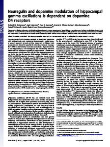
The monoamine dopamine is a small signaling molecule that can be synthesized from the amino acid tyrosine by the enzyme tyrosine hydroxylase. In the absence of tyrosine hydroxylase, dopamine can be made inefficiently by tyrosinase (Fig. 1).1,2 The widespread use of dopamine by different taxa hints at its ancient origin. Some photosynthesizing protists have daily vertical migrations in the water column triggered by the presence of daylight. An antagonist dopamine-acetylcholine system has been shown to control this activity by affecting light sensitivity: with dopamine decreasing it.3,4 Dopamine is also found in fruits and vegetables where its oxidation results in the familiar brown spots on ripe bananas.5,6 Its role in plants appears to be as a strong antioxidant, providing protection from lipid peroxidative damage caused by the intense heat and sunlight of the tropics.7 In bacteria, fungi, protozoans, cnidarians, nematodes, arthropods, mollusks, annelids and vertebrates, dopamine seems present wherever it is sought (Fig. 2).8-17 Although the main role of dopamine is in intraorganismal signaling, opportunistic organisms sometimes exploit dopamine
Communicative & Integrative Biology
signaling for inter-species interactions. For example, mammals release dopamine as part of their systemic response to infection; pathological organisms use this signal in an attempt to survive the immune response. Gram-negative bacteria respond to this dopamine signal by accelerating their division rate often overwhelming the host’s defenses.18 Pathogenic fungi respond to this signal by synthesizing melanin, making them resistant to ionic oxidants released by the host’s macrophages.19 Since many fungi use dopamine as a precursor for melanin synthesis, some fungi selectively invade dopamine producing areas of the brain, causing meningitis.20 Other animals have cracked the dopamine code of their prey. Some wasps for example inject dopamine into the cockroach nervous system forcing them to passively host their larvae.21,22 A wealth of specialized receptors has allowed the use of dopamine to be widespread across taxa as well as within organisms where it can modulate diverse processes.23,24 This diversity may have risen through processes of gene duplication and horizontal gene transfers beginning with bacteria.25 In mammals, five serpentine dopamine receptors have been described in two pharmacologically distinct classes. The D1-like receptors (DOP1 and DOP5) act postsynaptically to increase cyclic adenosine monophosphate (cAMP) levels, while D2-like receptors (DOP2, DOP3 and DOP4) act both pre- and postsynaptically to reduce cAMP levels.25-27 In addition to D1- and D2-like receptors, some invertebrates also have ionotropic dopamine receptors.28 The competing regulation of cAMP levels by the different D1-like and D2-like receptor types allows the use of dopamine in the
Volume 5 Issue 5
Perspective
Figure 1. Synthesis pathway for dopamine. Dopamine is primarily synthesized by tyrosine hydroxylase (A) but can also be made in its absence (B). 2
fine control of behavior. This is particularly effective when rapidly changing environmental forces require the modification of ongoing behavioral patterns, such as during locomotion or during risk-reward evaluations.23 In animals, one of the main roles of dopamine is to act as a behavioral switch in the transition from faster to slower motor patterns. This has been best demonstrated in well-controlled electrophysiological studies of fictive forms of rhythmic locomotion in reduced (semi-intact) preparations. For instance, dopamine slows down locomotion in snails.29 Both in sea slugs and in leeches, dopamine inhibits swimming and induces crawling, while in lamprey, zebrafish and crabs it slows down locomotor rhythms.14-17,30 However, the role of dopamine in controlling locomotion in these systems has not been demonstrated in freely-behaving animals. Dopamine Signaling in C. elegans Dopaminergic signaling has been intensely studied in the nematode worm Caenorhabditis elegans. Hermaphrodite worms have eight (mechanosensory) dopaminergic neurons (the male has additional neurons but won’t be discussed here). Although developmentally distinct, these neurons are divided into three classes on the basis of their morphology: four CEP neurons innervate the tip of the nose, two ADE neurons innervate the head cuticle,
www.landesbioscience.com
and two posterior PDE neurons innervate the posterior cuticle.13 As in other animals, worm dopaminergic neurons express genes encoding tyrosine hydroxylase (TH/cat-2), dopa decarboxylase (bas-1), vesicular monoamine transporter (cat-1), dopamine transporter (dat-1), as well as autoreceptors (dop-2). The coordinated expression of each of these genes and others that typify differentiated dopaminergic neurons is initiated and maintained throughout life by a terminal selector (EST transcription factor) that binds a 10 base-pair promoter element that is conserved in mammals.31 The eight dopamine neurons act both through classic synapses (429 synapses in total) as well as extrasynapticly by releasing dopamine into the worm’s body cavity.32,33 All worm dopamine neurons also express the mechanotransduction channel TRP-4 and are thought to be mechanoreceptive.34-37 C. elegans has D1-like receptor genes (dop-1 and dop-4) and D2-like receptor genes (dop-2 and dop-3) similar to the ones found in mammals (in addition to some unique nematode receptors).25,28,38-40 Depending on the type, dopamine receptors are expressed exclusively in neurons or also in non-neuronal cells.33 D1-like receptors are expressed in neuronal and non-neuronal targets alike (e.g., muscle, glia); D2-like receptors are restricted to neurons which is in keeping with a neuroregulatory role.25 Notably, dopaminergic neurons express the autoreceptor
Communicative & Integrative Biology
DOP-2 that operates in negative feedback loops.25 The parallel between the dopaminergic systems of C. elegans and mammals extends to the role of dopamine in modulation of behavioral patterns by environmental cues, and to processes of learning and memory. A classical example of environmental modulation of behavior via D1- and D2-like receptor interactions comes from studies of DOP-3 and DOP-1 receptors coexpressed in mechanosensory and motor neurons and that antagonistically mediate the decrease in locomotion by well-fed worms as they enter a patch of food.33 Evaluation of cost vs. benefit for environmental cues is a crucial process. In mammals this process is tightly associated with balance between D1- and D2-like pathways whose chemical disruption can lead to addiction.41 C. elegans also uses dopamine to compare the quality of its environment against its internal physiological state and re-evaluates its responses accordingly. For example, in the presence of food worms will habituate their escape response more rapidly; a behavior presumably mediated by their dopaminergic mechanosensory neurons that detect the texture of the food.42 In C. elegans, this kind of learning can be in response to both gustatory and odor information.43-46 While some forms or learning (e.g., habituation to mechanical stimuli) take place through the D1-like pathway, others (e.g., associative learning) are facilitated by presynaptic changes in dopaminergic neurons.47,48 Dopaminergic signaling appears to be influenced in C. elegans by compounds that are addictive in humans such as ethanol and cocaine.49-51 In addition to learning and memory, dopamine also modulates many behavioral patterns in C. elegans, inhibiting many behaviors that are promoted by serotonin.52,53 Serotonin promotes a number of behaviors in C. elegans: including egglaying, through its action on command and motor neurons; pharyngeal pumping (feeding), through its action on motor neurons and muscles; and more recently swimming, through yet unspecified targets.52,53 Age-related locomotor changes have been correlated with changes in the dopamine/serotonin balance in older worms.54
441
Figure 2. Evolutionary tree showing the presence of dopamine across different taxa as well as its use. Dopamine is generally involved in the production of slower gaits often associated with feeding.
Our lab has recently discovered a new role for dopamine in transitioning between motor patterns on land and in water. In C. elegans, crawling on land consists of slow (~0.5 HZ) dorsoventral body bends of high angular amplitude that result in a persistent “S-like” body shape. By contrast, swimming in water is characterized by alternating dorsoventral body bends of high frequency (~1.5 HZ) and low angular amplitude that result in worms sequentially alternating between “C-like” shapes. While both behaviors propel the animal forward, the spatial patterns of bending forces have been shown to be the result of differential patterns of muscular activity produced by the animal to locomote in environments with distinct drag coefficient ratios.55,56 When the speed of the worm is constrained by a range of viscous solutions, C. elegans displays discontinuous bouts of crawl- or swim-like kinematics rather than a simple continuous modulation of locomotory kinematics. This result, together with additional experiments that reveal bimodal switching of locomotory patterns, demonstrates that crawling and swimming represent mutually exclusive forms of locomotion, also commonly known as distinct “locomotory gaits.” Of the three classes of dopaminergic neurons described above, we found that
442
only ADE and PDE were required for worms to transition from swimming to crawling.57 This is consistent with how ADE and PDE neurons have mechanoreceptive endings on the sides of the worm that could detect firm contact with the ground, while the CEP neurons instead innervate the tip of the nose which the worm typically keeps off the ground. Our finding that both anterior (ADEs) and posterior (PDEs) dopaminergic mechanoreceptor neurons are required during gait transitions could seem surprising—diffusion is thought to be very rapid in such small organisms. In fact, we found that injection of dopamine into the anterior and posterior regions of the worm’s body produced distinct results; with anterior injections alone being able to induce gait transitions.57 This suggests that (at least while swimming) worms can effectively compartmentalize their pseudocoelomic space. Both ADE and PDE neurons send processes to the anterior half of the body where they would presumptively release dopamine locally and effect a swim-tocrawl transition. Downstream from the dopamine neurons we found that worms transitioned from distinct swimming to crawling gaits through the D1-like dopaminergic pathway.57 Elimination of dopamine or
Communicative & Integrative Biology
D1-like receptor genes (dop-1 or dop-4) caused worms to collapse upon exit from a puddle. While many worms stopped moving altogether for up to 45 min, other worms displayed unproductive crawling often by propagating a single dorsoventral bend from head to tail before stopping or initiating backward locomotion. Gently prodding these worms induced crawling. Conversely, raising dopamine levels caused worms to inappropriately switch from swimming to crawling in water (an effect also dependent on D1-like receptors).57 In keeping with other behaviors, the transition to crawling depended not only on the balance between D1- and D2-like receptors (as mutants lacking both receptor types showed no phenotype, Fig. 3) but also on the balance between dopamine and serotonin. Altering the balance between these amines by means of exogenous drug application, endogenous photo-uncaging of the amines, or stimulating their release via optogenetic stimulation (of the producing neurons), induced gait transitions between swimming and crawling. Our results suggest that as worms emerge from a puddle, ground contact is sensed through a subset of mechanosensitive dopaminergic neurons. These in turn release dopamine into the anterior body cavity and trigger
Volume 5 Issue 5
Figure 3. The dopaminergic system is required for swim to crawl transitions in C. elegans. The crawling frequency before and after swimming was compared for worms with impairments in their aminergic systems. Only worms deficient in dopamine production, or in the D1-like dopamine receptor pathway showed a significantly marked deficit transitioning from swimming to crawling. The plit shows the ratio of crawling head bends following swimming to that before swimming. The assay includes all available bioaminergic pathway C. elegans mutants. Bars report means and SEMs.
crawling through the D1-like pathway. Conversely, immersion in water stimulates release of serotonin and inhibits dopamine release (caused by the removal from the substrate). This last finding is consistent with previous work on the leech where endogenous levels of serotonin were found in association with the induction of swimming behavior.58 It should be noted that in our experiments we altered the balance between serotonin and dopamine without removing the underlying environmental context. Therefore worms swimming in water, once induced to crawl (by dopamine release), remained nevertheless immersed in water (therefore receiving conflicting sensory information). This could account for the episodic nature of the behavioral inductions, where worms could be seen alternating between swimming and crawling bouts as a result of the conflicting sensory inputs represented by the applied amine and the actual physical environment. Furthermore, under certain experimental conditions crawling waves induced in water failed to propagate all the way to the tail, suggesting that the full production of the behavior likely involves additional control systems as those proposed by other groups.32,59-61 These findings suggest a combinatorial system for behavior selection where synergies between different dopaminergic pathways interact with those between
www.landesbioscience.com
dopamine and other amines. For example, swimming behavior seems to occur by the combined effects of a decrease in dopamine release (brought about by loss of ground contact), and an increase in serotonin release (brought about by entrance into an aquatic medium). It is perhaps through these interactions that dopamine seems to not just trigger one behavioral transition, but rather a whole host of behaviors associated with crawling on land; like foraging, feeding and defecation.62 One interesting method for studying how the balance within dopaminergic signaling pathways and between dopamine and other neurotransmitter signaling systems is maintained is in a comparison between land-grown and liquid-grown C. elegans. As mentioned above, worms entering a liquid environment experience a decrease in dopamine and an increase in serotonin that triggers transition into swimming and the inhibition of crawling as well as many other behaviors like feeding, defecation and egglaying which are crucial for survival. However, in order to survive and reproduce in a liquid environment, worms would need to overcome this aquatically-induced shift toward serotonin (by increasing dopamine production, decreasing serotonin production, or a combination of the two) long enough to carry on the afore-mentioned vital functions. In their original description of the
Communicative & Integrative Biology
dopaminergic system of C. elegans, Sulston et al. reported no less than a 62-fold increase in levels of L-dopa (the precursor of dopamine) for worms grown in liquid culture.13 Thus, it seems that culture of C. elegans in liquid is accompanied by a chronic—and significant—upregulation of dopamine production that may enable temporary reversal of the serotonin-dopamine balance and allow animals to engage in feeding and other vital functions while immersed in liquid. The dopaminergic effects described above primarily involve feeding and reductions in locomotory rates. These are consistent with other known roles of dopamine. For instance, when worms enter a patch of food (bacteria) dopamine is responsible for a couple of well-characterized behaviors that facilitate food ingestion, namely “basal slowing” and “area-restricted search.” During basal slowing, dopaminergic neurons in non-starved worms are thought to mechanically sense surrounding bacteria and decrease crawling velocity (termed as basal slowing).37 At the same time dopamine is proposed to also increase the turning rate resulting in worms thus remaining in the vicinity of a food source (termed Area Restricted Search).63 Faced with adverse environmental conditions, C. elegans larva enter an alternative developmental stage known as dauer, dopamine has also been implicated in reducing
443
locomotor output and causing the characteristic quiescent states of these animals.64 Studies of mutant animals have revealed motor phenotypes for many mutants deficient in dopaminergic signaling.37 For example, mutations in the gene encoding the dopamine reuptake transporter dat-1 lead to a buildup of extrasynaptic dopamine resulting in the swim-induced paralysis phenotype (SWIP).65,66 Our model of swim induction as the combined effect of a decrease in dopamine release and an increase in serotonin release is consistent with the SWIP phenotype. Here a buildup of endogenously released dopamine in swimming dat-1 mutants would eventually lead to a transition to crawling, termination of swimming and finally paralysis (SWIP). These findings hint to dopamine as a possible master switch, capable of altering the behavioral state of an animal by inhibiting and promoting sets of motor outputs in a context-dependent way. The great degree of conservation in molecular and functional pathways seems to span across taxa to mammals where the dopaminergic system has been intensely studied due to its role in Parkinson disease. Parkinson Disease In 1817 James Parkinson described a disease (later renamed by Charcot in his honor) affecting the motor system with tremors, rigidity, bradykinesia and imbalance.67,68 Parkinson disease (PD) etiology involves the death of dopaminergic neurons in the substantia nigra pars compacta of the brain. Environmentally, death of dopaminergic neurons can be induced by acute exposure to manganese, by 6-hydroxidopamine (6OHDA), MPTP, or by other toxic species that dopaminergic neurons selectively uptake.69-71 Alternatively, mutations in genes involved in dopaminergic signaling can also lead to PD.72 Examples of mutations known to cause dopaminergic neuron loss include those affecting the LRRK2, PARK2 and SNCA genes.73-75 The presence of different alleles for these genes, and the endogenous synthesis of 6OHDA from dopamine may all contribute to the development of PD.74,76,77 During the development of the disease, it is possible that the balance between
444
the D1- and the D2-like dopaminergic pathways becomes compromised as the gradual reduction of dopamine levels is mitigated by postsynaptic compensatory changes.78-81 Herein may lie one of the greatest challenges of PD treatment, as merely replacing lost dopamine in the brain (or its precursor L-dopa) does not repair potential receptor imbalances created by the disease. Therefore, L-dopa alone is unable to prevent the eventual onset of secondary motor deficits.82 Further complicating treatment, differences in dopamine receptor polymorphisms have been shown to result in differential responses to standard treatments.83 For example, treatment with Levodopa has been shown to upregulate the expression of D1-like (but not D2-like) receptors and is associated with the development of a disorder known as Levodopainduced dyskinesias (LID).84,85 Treatment with both Levodopa and D2-like agonist (derived from non-mammalian systems) seem to mitigate LID supporting the idea that preserving the balance between these two pathways is crucial for recovery.86,87 In light of this bleak picture, it is evident that studies in model systems permitting fast genetic and molecular evaluation of pathway dynamics and interactions are necessary to complement ongoing efforts in classical mammalian systems. In a recent manuscript we introduced a fast, facile behavioral assay capable of assaying potential drugs (or mutations) affecting some of the motor deficiencies associated with loss of dopaminergic signaling during PD. We found that (paralleling findings with Parkinson patients) animals with impaired D1- to D2-like receptor balance exhibited deficits in transitioning between different behaviors.57 Besides showing remarkable conservation of function from worms to human, our findings suggest that the use of C. elegans in Parkinson research could expand to include behavioral approaches, besides the genetic and molecular ones already in use. The Future of C. elegans in Parkinson Research The genetic amenability of C. elegans, coupled with the level of conservation in dopaminergic pathway and function has
Communicative & Integrative Biology
already led to its profitable use in the field of PD research.88,89 Many studies have sought to establish C. elegans as a valid model system in this field by showing parallels between its dopaminergic system and that of mammals. Overexpression of the human gene encoding α-synuclein (a protein associated with a familiar form of PD) leads to dopamine neuron degeneration in the worm.90-93 Work on C. elegans has shown that 6OHDA causes degeneration of dopaminergic neurons through DAT-1, and that the chaperone molecule TorsinA can protect against this effect.94,95 C. elegans has also been used to study the roles of mitochondrial toxicity in the development of PD, MPTP toxicity, or of calorie restriction in protection against neurodegeneration.96,97 Automated systems using C. elegans can now quickly screen thousands of putative drugs. Fast and economical high-throughput screens have already began yielding drugs and gene targets that can serve as potential therapeutic approaches for the treatment of human afflictions such as PD.98-104 The remarkable conservation in the dopaminergic pathway and function across phyla strongly hints to the importance of its role in the survival of organisms. In evolutionary terms, no sooner is an organism capable of performing two incompatible tasks than it becomes subservient of the need to determine which one is adaptive in which situation. For Parkinson research, this conservation means that there is much useful information to be gained from studying extramammalian dopaminergic systems. The diversity of motor outputs found in the animal kingdom, when compared with the conservation of the systems modulating them evidences how forgiving natural selection can be to one process and how strict to another. References 1. Elsworth JD, Roth RH. Dopamine synthesis, uptake, metabolism and receptors: relevance to gene therapy of Parkinson’s disease. Exp Neurol 1997; 144:49; PMID:9126143; http://dx.doi.org/10.1006/ exnr.1996.6379. 2. Rios M, Habecker B, Sasaoka T, Eisenhofer G, Tian H, Landis S, et al. Catecholamine synthesis is mediated by tyrosinase in the absence of tyrosine hydroxylase. J Neurosci 1999; 19:3519-26; PMID:10212311. 3. Forward RB Jr. Effects of neurochemicals upon a dinoflagellate photoresponse. J Protozool 1977; 24:401-5; PMID:21286.
Volume 5 Issue 5
4. Kiefer DA, Lasker R. Two blooms of Gymnodinium splendens, an unarmored dinoflagelate. Fish Bull US 1975; 73:675-8. 5. Udenfriend S, Lovenberg W, Sjoerdsma A. Physiologically active amines in common fruits and vegetables. Arch Biochem Biophys 1959; 85:487-90; PMID:13840159; http://dx.doi.org/10.1016/00039861(59)90516-8. 6. Waalkes TP, Sjoerdsma A, Creveling CR, Weissbach H, Udenfriend S. Serotonin, norepinephrine and related compounds in bananas. Science 1958; 127:648-50; PMID:17808884; http://dx.doi. org/10.1126/science.127.3299.648. 7. Kanazawa K, Sakakibara H. High content of dopamine, a strong antioxidant, in Cavendish banana. J Agric Food Chem 2000; 48:844-8; PMID:10725161; http://dx.doi.org/10.1021/jf9909860. 8. Murooka Y, Doi N, Harada T. Distribution of membrane-bound monoamine oxidase in bacteria. Appl Environ Microbiol 1979; 38:565-9; PMID:120132. 9. Calne DB. Role of ergot derivatives in the treatment of parkinsonism. Fed Proc 1978; 37:2207-9; PMID:658459. 10. Ness JC, Morse DE. Regulation of galactokinase gene expression in Tetrahymena thermophila. II. Identification of 3,4-dihydroxyphenylalanine as a primary effector of adrenergic control of galactokinase expression. J Biol Chem 1985; 260:10013-8; PMID:2991271. 11. Carlberg M, Anctil M. Biogenic amines in coelenterates. Comp Biochem Physiol C 1993; 106:1-9; PMID:7903605; http://dx.doi.org/10.1016/07428413(93)90250-O. 12. Ostroumova TV, Markova LN. The effects of dopamine synthesis inhibitors and dopamine antagonists on regeneration in the hydra Hydra attenuata. Neurosci Behav Physiol 2002; 32:293-8; PMID:12135343; http://dx.doi.org/10.1023/A:1015066424928. 13. Sulston J, Dew M, Brenner S. Dopaminergic neurons in the nematode Caenorhabditis elegans. J Comp Neurol 1975; 163:215-26; PMID:240872; http:// dx.doi.org/10.1002/cne.901630207. 14. Martinez EA, Murray M, Leung MK, Stefano GB. Evidence for dopaminergic and opiod involvement in the regulation of locomotor activity in the land crab Gecarcinus lateralis. Comp Biochem Phys Part C 1988; 90:89-93; http://dx.doi.org/10.1016/07428413(88)90103-X. 15. McClellan AD, Brown GD, Getting PA. Modulation of swimming in Tritonia: excitatory and inhibitory effects of serotonin. J Comp Physiol A 1994; 174:25766; PMID:7908336; http://dx.doi.org/10.1007/ BF00193792. 16. Crisp KM, Mesce KA. A cephalic projection neuron involved in locomotion is dye coupled to the dopaminergic neural network in the medicinal leech. J Exp Biol 2004; 207:4535-42; PMID:15579549; http:// dx.doi.org/10.1242/jeb.01315. 17. McPherson DR, Kemnitz CP. Modulation of lamprey fictive swimming and motoneuron physiology by dopamine, and its immunocytochemical localization in the spinal cord. Neurosci Lett 1994; 166:23-6; PMID:8190353; http://dx.doi.org/10.1016/03043940(94)90831-1. 18. Lyte M, Ernst S. Catecholamine induced growth of gram negative bacteria. Life Sci 1992; 50:203-12; PMID:1731173; http://dx.doi.org/10.1016/00243205(92)90273-R. 19. Gómez BL, Nosanchuk JD. Melanin and fungi. Curr Opin Infect Dis 2003; 16:91-6; PMID:12734441; http://dx.doi.org/10.1097/00001432-20030400000005. 20. Williamson PR, Wakamatsu K, Ito S. Melanin biosynthesis in Cryptococcus neoformans. J Bacteriol 1998; 180:1570-2; PMID:9515929.
www.landesbioscience.com
21. Weisel-Eichler A, Haspel G, Libersat F. Venom of a parasitoid wasp induces prolonged grooming in the cockroach. J Exp Biol 1999; 202:957-64; PMID:10085268. 22. Weisel-Eichler A, Libersat F. Venom effects on monoaminergic systems. J Comp Physiol A Neuroethol Sens Neural Behav Physiol 2004; 190:683-90; PMID :15160282 ; http://dx.doi.org/10.1007/ s00359-004-0526-3. 23. Jorgensen EM. Dopamine: should I stay or should I go now? Nat Neurosci 2004; 7:101921; PMID:15452567; http://dx.doi.org/10.1038/ nn1004-19. 24. Iyer LM, Aravind L, Coon SL, Klein DC, Koonin EV. Evolution of cell-cell signaling in animals: did late horizontal gene transfer from bacteria have a role? Trends Genet 2004; 20:292-9; PMID:15219393; http://dx.doi.org/10.1016/j.tig.2004.05.007. 25. Suo S, Sasagawa N, Ishiura S. Cloning and characterization of a Caenorhabditis elegans D2-like dopamine receptor. J Neurochem 2003; 86:869-78; PMID:12887685; http://dx.doi.org/10.1046/j.14714159.2003.01896.x. 26. Le Crom S, Kapsimali M, Barôme PO, Vernier P. Dopamine receptors for every species: gene duplications and functional diversification in Craniates. J Struct Funct Genomics 2003; 3:161-76 ; PMID:12836695; http://dx.doi. org/10.1023/A:1022686622752. 27. Nass R, Blakely RD. The Caenorhabditis elegans dopaminergic system: opportunities for insights into dopamine transport and neurodegeneration. Annu Rev Pharmacol Toxicol 2003; 43:521-44; PMID :12415122 ; http://dx.doi.org/10.1146/ annurev.pharmtox.43.100901.135934. 28. Ringstad N, Abe N, Horvitz HR. Ligand-gated chloride channels are receptors for biogenic amines in C. elegans. Science 2009; 325:96-100; PMID:19574391; http://dx.doi.org/10.1126/science.1169243. 29. Pavlova GA. Effects of serotonin, dopamine and ergometrine on locomotion in the pulmonate mollusc Helix lucorum. J Exp Biol 2001; 204:1625-33; PMID:11398751. 30. Souza BR, Romano-Silva MA, Tropepe V. Dopamine D2 receptor activity modulates Akt signaling and alters GABAergic neuron development and motor behavior in zebrafish larvae. J Neurosci 2011; 31:5512-25; PMID:21471388; http://dx.doi. org/10.1523/JNEUROSCI.5548-10.2011. 31. Flames N, Hobert O. Gene regulatory logic of dopamine neuron differentiation. Nature 2009; 458:8859; PMID:19287374; http://dx.doi.org/10.1038/ nature07929. 32. White JG, Southgate E, Thomson JN, Brenner S. The structure of the nervous system of the nematode Caenorhabditis elegans. Philos Trans R Soc Lond B Biol Sci 1986; 314:1-340; PMID:22462104; http:// dx.doi.org/10.1098/rstb.1986.0056. 33. Chase DL, Pepper JS, Koelle MR. Mechanism of extrasynaptic dopamine signaling in Caenorhabditis elegans. Nat Neurosci 2004; 7:1096-103; PMID :15378064 ; http://dx.doi.org/10.1038/ nn1316. 34. Li W, Feng Z, Sternberg PW, Xu XZ. A C. elegans stretch receptor neuron revealed by a mechanosensitive TRP channel homologue. Nature 2006; 440 : 684-7; PMID:16572173; http://dx.doi. org/10.1038/nature04538. 35. Li W, Kang L, Piggott BJ, Feng Z, Xu XZ. The neural circuits and sensory channels mediating harsh touch sensation in Caenorhabditis elegans. Nat Commun 2011; 2:315; PMID:21587232; http:// dx.doi.org/10.1038/ncomms1308. 36. Stephens GJ, Johnson-Kerner B, Bialek W, Ryu WS. From modes to movement in the behavior of Caenorhabditis elegans. PLoS One 2010; 5:13914; PMID:21103370; http://dx.doi.org/10.1371/journal. pone.0013914.
Communicative & Integrative Biology
37. Sawin ER, Ranganathan R, Horvitz HR. C. elegans locomotory rate is modulated by the environment through a dopaminergic pathway and by experience through a serotonergic pathway. Neuron 2000; 26:619-31; PMID:10896158; http://dx.doi. org/10.1016/S0896-6273(00)81199-X. 38. Sugiura M, Fuke S, Suo S, Sasagawa N, Van Tol HHM, Ishiura S. Characterization of a novel D2-like dopamine receptor with a truncated splice variant and a D1-like dopamine receptor unique to invertebrates from Caenorhabditis elegans. J Neurochem 2005; 94:1146-57; PMID:16001968; http://dx.doi. org/10.1111/j.1471-4159.2005.03268.x. 39. Suo S, Sasagawa N, Ishiura S. Identification of a dopamine receptor from Caenorhabditis elegans. Neurosci Lett 2002; 319:13-6; PMID:11814642; http://dx.doi.org/10.1016/S0304-3940(01)02477-6. 40. Tsalik EL, Niacaris T, Wenick AS, Pau K, Avery L, Hobert O. LIM homeobox gene-dependent expression of biogenic amine receptors in restricted regions of the C. elegans nervous system. Dev Biol 2003; 263:81-102; PMID:14568548; http://dx.doi. org/10.1016/S0012-1606(03)00447-0. 41. Self DW, Barnhart WJ, Lehman DA, Nestler EJ. Opposite modulation of cocaine-seeking behavior by D1- and D2-like dopamine receptor agonists. Science 1996; 271:1586-9; PMID:8599115; http://dx.doi. org/10.1126/science.271.5255.1586. 42. Kindt KS, Quast KB, Giles AC, De S, Hendrey D, Nicastro I, et al. Dopamine mediates context-dependent modulation of sensory plasticity in C. elegans. Neuron 2007; 55:662-76; PMID:17698017; http:// dx.doi.org/10.1016/j.neuron.2007.07.023. 43. Ezcurra M, Tanizawa Y, Swoboda P, Schafer WR. Food sensitizes C. elegans avoidance behaviours through acute dopamine signalling. EMBO J 2011; 30:1110-22; PMID:21304491; http://dx.doi. org/10.1038/emboj.2011.22. 44. Hukema RK, Rademakers S, Jansen G. Gustatory plasticity in C. elegans involves integration of negative cues and NaCl taste mediated by serotonin, dopamine and glutamate. Learn Mem 2008; 15:82936; PMID:18984564; http://dx.doi.org/10.1101/ lm.994408. 45. Ezak MJ, Ferkey DM. The C. elegans D2-like dopamine receptor DOP-3 decreases behavioral sensitivity to the olfactory stimulus 1-octanol. PLoS One 2010; 5:9487; PMID:20209143; http://dx.doi. org/10.1371/journal.pone.0009487. 46. Kimura KD, Fujita K, Katsura I. Enhancement of odor avoidance regulated by dopamine signaling in Caenorhabditis elegans. J Neurosci 2010; 30:1636575; PMID:21123582; http://dx.doi.org/10.1523/ JNEUROSCI.6023-09.2010. 47. Voglis G, Tavernarakis N. A synaptic DEG/ENaC ion channel mediates learning in C. elegans by facilitating dopamine signalling. EMBO J 2008; 27:328899; PMID:19037257; http://dx.doi.org/10.1038/ emboj.2008.252. 48. Sanyal S, Wintle RF, Kindt KS, Nuttley WM, Arvan R, Fitzmaurice P, et al. Dopamine modulates the plasticity of mechanosensory responses in Caenorhabditis elegans. EMBO J 2004; 23:47382; PMID:14739932; http://dx.doi.org/10.1038/ sj.emboj.7600057. 49. Ward A, Walker VJ, Feng Z, Xu XZ. Cocaine modulates locomotion behavior in C. elegans. PLoS One 2009; 4:5946; PMID:19536276; http://dx.doi. org/10.1371/journal.pone.0005946. 50. Lee J, Jee C, McIntire SL. Ethanol preference in C. elegans. Genes Brain Behav 2009; 8:578-85; PMID:19614755; http://dx.doi.org/10.1111/j.1601183X.2009.00513.x. 51. Bettinger JC, McIntire SL. State-dependency in C. elegans. Genes Brain Behav 2004; 3:266-72; PMID:15344920; http://dx.doi.org/10.1111/j.1601183X.2004.00080.x.
445
52. Horvitz HR, Chalfie M, Trent C, Sulston JE, Evans PD. Serotonin and octopamine in the nematode Caenorhabditis elegans. Science 1982; 216:1012-4; PMID:6805073; http://dx.doi.org/10.1126/science.6805073. 53. Weinshenker D, Garriga G, Thomas JH. Genetic and pharmacological analysis of neurotransmitters controlling egg laying in C. elegans. J Neurosci 1995; 15:6975-85; PMID:7472454. 54. Murakami H, Bessinger K, Hellmann J, Murakami S. Manipulation of serotonin signal suppresses early phase of behavioral aging in Caenorhabditis elegans. Neurobiol Aging 2008; 29:1093-100; PMID:17336425; http://dx.doi.org/10.1016/j.neurobiolaging.2007.01.013. 55. Pierce-Shimomura JT, Chen BL, Mun JJ, Ho R, Sarkis R, McIntire SL. Genetic analysis of crawling and swimming locomotory patterns in C. elegans. Proc Natl Acad Sci USA 2008; 105:209827; PMID:19074276; http://dx.doi.org/10.1073/ pnas.0810359105. 56. Shen XN, Sznitman J, Krajacic P, Lamitina T, Arratia PE. Undulatory locomotion of C. elegans on wet surfaces. arXiv:1112.1118v1 [physics.flu-dyn]. 57. Vidal-Gadea AG, Topper S, Young L, Crisp A, Kressin L, Elbel E, et al. Caenorhabditis elegans selects distinct crawling and swimming gaits via dopamine and serotonin. Proc Natl Acad Sci USA 2011; 108:17504-9; PMID:21969584; http://dx.doi. org/10.1073/pnas.1108673108. 58. Willard AL. Effects of serotonin on the generation of the motor program for swimming by the medicinal leech. J Neurosci 1981; 1:936-44; PMID:7288474. 59. Korta J, Clark DA, Gabel CV, Mahadevan L, Samuel ADT. Mechanosensation and mechanical load modulate the locomotory gait of swimming C. elegans. J Exp Biol 2007; 210:2383-9; PMID:17575043; http://dx.doi.org/10.1242/jeb.004572. 60. Berri S, Boyle JH, Tassieri M, Hope IA, Cohen N. Forward locomotion of the nematode C. elegans is achieved through modulation of a single gait. HFSP J 2009; 3:186-93; PMID:19639043; http://dx.doi. org/10.2976/1.3082260. 61. Fang-Yen C, Wyart M, Xie J, Kawai R, Kodger T, Chen S, et al. Biomechanical analysis of gait adaptation in the nematode Caenorhabditis elegans. Proc Natl Acad Sci USA 2010; 107:20323-8; PMID:21048086; http://dx.doi.org/10.1073/pnas.1003016107. 62. Vidal-Gadea AG, Davis S, Becker L, PierceShimomura JT. Coordination of behavioral hierarchies during environmental transitions in Caenorhabditis elegans. Worm 2012; 1:1-7; http:// dx.doi.org/10.4161/worm.19148. 63. Hills T, Brockie PJ, Maricq AV. Dopamine and glutamate control area-restricted search behavior in Caenorhabditis elegans. J Neurosci 2004; 24:121725; PMID:14762140; http://dx.doi.org/10.1523/ JNEUROSCI.1569-03.2004. 64. Gaglia MM, Kenyon CJ. Stimulation of movement in a quiescent, hibernation-like form of Caenorhabditis elegans by dopamine signaling. J Neurosci 2009; 29:7302-14; PMID:19494152; http://dx.doi. org/10.1523/JNEUROSCI.3429-08.2009. 65. Carvelli L, Matthies DS, Galli A. Molecular mechanisms of amphetamine actions in Caenorhabditis elegans. Mol Pharmacol 2010 ; 78:151-6 ; PMID:20410438; http://dx.doi.org/10.1124/ mol.109.062703. 66. McDonald PW, Hardie SL, Jessen TN, Carvelli L, Matthies DS, Blakely RD. Vigorous motor activity in Caenorhabditis elegans requires efficient clearance of dopamine mediated by synaptic localization of the dopamine transporter DAT-1. J Neurosci 2007; 27:14216-27; PMID:18094261; http://dx.doi. org/10.1523/JNEUROSCI.2992-07.2007. 67. Charcot JM. Leçons Du Mardi À La Salpêtrière. In: Lecrosnier E, Babé, Eds., Progrès Médical. I. Paris: Aux Bureaux de Progrès Médical 1889; 1-579.
446
68. Parkinson J. An essay on the shaking palsy. London: Whittingham and Rowland 1817; 1-66. 69. Bouchard M, Mergler D, Baldwin ME, Panisset M. Manganese cumulative exposure and symptoms: a follow-up study of alloy workers. Neurotoxicology 2008; 29:577-83; PMID:18562007; http://dx.doi. org/10.1016/j.neuro.2008.04.013. 70. Breese GR, Traylor TD. Depletion of brain noradrenaline and dopamine by 6-hydroxydopamine. Br J Pharmacol 1971; 42:88-99; PMID:5580702. 71. Langston JW, Palfreman J. The case of the frozen addicts. New York: Pantheon Books 1995; 1-309. 72. Pramstaller PP, Schlossmacher MG, Jacques TS, Scaravilli F, Eskelson C, Pepivani I, et al. Lewy body Parkinson’s disease in a large pedigree with 77 Parkin mutation carriers. Ann Neurol 2005; 58:41122; PMID:16130111; http://dx.doi.org/10.1002/ ana.20587. 73. Whaley NR, Uitti RJ, Dickson DW, Farrer MJ, Wszolek ZK. Clinical and pathologic features of families with LRRK2-associated Parkinson’s disease. J Neural Transm Suppl 2006; 70:221-9; PMID:17017533; http://dx.doi.org/10.1007/978-3211-45295-0_34. 74. Farrer M, Chan P, Chen R, Tan L, Lincoln S, Hernandez D, et al. Lewy bodies and parkinsonism in families with parkin mutations. Ann Neurol 2001; 50:293-300; PMID:11558785; http://dx.doi. org/10.1002/ana.1132. 75. Perez RG, Waymire JC, Lin E, Liu JJ, Guo F, Zigmond MJ. A role for α-synuclein in the regulation of dopamine biosynthesis. J Neurosci 2002; 22:3090-9; PMID:11943812. 76. Senoh S, Witkop B, Creveling CR, Udenfriend S. 2,4,5-trihydroxyphenethylamine, a new metabolite of 3,4-dihydroxyphenethylamine. J Am Chem Soc 1959; 81:1768-9; http://dx.doi.org/10.1021/ ja01516a065. 77. Senoh S, Creveling CR, Udenfriend S, Witkop B. Chemical, Enzymatic and Metabolic Studies on the Mechanism of Oxidation of Dopamine. J Am Chem Soc 1959; 81:6236-40; http://dx.doi.org/10.1021/ ja01532a030. 78. Gerfen CR. D1 dopamine receptor supersensitivity in the dopamine-depleted striatum animal model of Parkinson’s disease. Neuroscientist 2003; 9:455-62; PMID:14678578; http://dx.doi. org/10.1177/1073858403255839. 79. Gerfen CR, Engber TM, Mahan LC, Susel Z, Chase TN, Monsma FJ Jr, et al. D1 and D2 dopamine receptor-regulated gene expression of striatonigral and striatopallidal neurons. Science 1990; 250:142932; PMID:2147780; http://dx.doi.org/10.1126/science.2147780. 80. Paul ML, Graybiel AM, David JC, Robertson HA. D1-like and D2-like dopamine receptors synergistically activate rotation and c-fos expression in the dopamine-depleted striatum in a rat model of Parkinson’s disease. J Neurosci 1992; 12:3729-42; PMID:1357113. 81. Gerfen CR, Miyachi S, Paletzki R, Brown P. D1 dopamine receptor supersensitivity in the dopamine-depleted striatum results from a switch in the regulation of ERK1/2/MAP kinase. J Neurosci 2002; 22:5042-54; PMID:12077200. 82. Nadjar A, Gerfen CR, Bezard E. Priming for l-dopainduced dyskinesia in Parkinson’s disease: a feature inherent to the treatment or the disease? Prog Neurobiol 2009; 87:1-9; PMID:18938208; http:// dx.doi.org/10.1016/j.pneurobio.2008.09.013. 83. Asghari V, Sanyal S, Buchwaldt S, Paterson A, Jovanovic V, Van Tol HH. Modulation of intracellular cyclic AMP levels by different human dopamine D4 receptor variants. J Neurochem 1995; 65:115765; PMID:7643093; http://dx.doi.org/10.1046/ j.1471-4159.1995.65031157.x.
Communicative & Integrative Biology
84. Guigoni C, Aubert I, Li Q, Gurevich VV, Benovic JL, Ferry S, et al. Pathogenesis of levodopa-induced dyskinesia: focus on D1 and D3 dopamine receptors. Parkinsonism Relat Disord 2005; 11:25-9; PMID:15885624; http://dx.doi.org/10.1016/j. parkreldis.2004.11.005. 85. Fabbrini G, Brotchie JM, Grandas F, Nomoto M, Goetz CG. Levodopa-induced dyskinesias. Mov Disord 2007; 22:1379-89; PMID:17427940; http:// dx.doi.org/10.1002/mds.21475. 86. Brooks DJ. Dopamine agonists: their role in the treatment of Parkinson’s disease. J Neurol Neurosurg Psychiatry 2000; 68:685-9; PMID:10811688; http://dx.doi.org/10.1136/jnnp.68.6.685. 87. Kalda A, Herm L, Rinken A, Zharkovsky A, Chen JF. Co-administration of the partial dopamine D2 agonist terguride with L-dopa attenuates L-dopa-induced locomotor sensitization in hemiparkinsonian mice. Behav Brain Res 2009; 202:232-7; PMID:19463706; http://dx.doi.org/10.1016/j.bbr.2009.03.037. 88. Nass R, Miller DM, Blakely RD. C. elegans: a novel pharmacogenetic model to study Parkinson’s disease. Parkinsonism Relat Disord 2001; 7:185-91; PMID:11331185; http://dx.doi.org/10.1016/S13538020(00)00056-0. 89. Wintle RF, Van Tol HHM. Dopamine signaling in Caenorhabditis elegans-potential for parkinsonism research. Parkinsonism Relat Disord 2001; 7:177-83; PMID:11331184; http://dx.doi.org/10.1016/S13538020(00)00055-9. 90. Kuwahara T, Koyama A, Gengyo-Ando K, Masuda M, Kowa H, Tsunoda M, et al. Familial Parkinson mutant α-synuclein causes dopamine neuron dysfunction in transgenic Caenorhabditis elegans. J Biol Chem 2006; 281:334-40; PMID:16260788; http:// dx.doi.org/10.1074/jbc.M504860200. 91. Lakso M, Vartiainen S, Moilanen AM, Sirviö J, Thomas JH, Nass R, et al. Dopaminergic neuronal loss and motor deficits in Caenorhabditis elegans overexpressing human alpha-synuclein. J Neurochem 2003; 86:165-72; PMID:12807436; http://dx.doi. org/10.1046/j.1471-4159.2003.01809.x. 92. Hamamichi S, Rivas RN, Knight AL, Cao S, Caldwell KA, Caldwell GA. Hypothesis-based RNAi screening identifies neuroprotective genes in a Parkinson’s disease model. Proc Natl Acad Sci USA 2008; 105:728-33; PMID:18182484; http://dx.doi. org/10.1073/pnas.0711018105. 93. Dexter PM, Caldwell KA, Caldwell GA. A predictable worm: application of Caenorhabditis elegans for mechanistic investigation of movement disorders. Neurotherapeutics 2012; 9:393-404; PMID:22403010; http://dx.doi.org/10.1007/s13311012-0109-x. 94. Nass R, Hall DH, Miller DM, 3rd, Blakely RD. Neurotoxin-induced degeneration of dopamine neurons in Caenorhabditis elegans. Proc Natl Acad Sci USA 2002; 99:3264-9; PMID:11867711; http:// dx.doi.org/10.1073/pnas.042497999. 95. Cao S, Gelwix CC, Caldwell KA, Caldwell GA. Torsin-mediated protection from cellular stress in the dopaminergic neurons of Caenorhabditis elegans. J Neurosci 2005; 25:3801-12; PMID:15829632; http:// dx.doi.org/10.1523/JNEUROSCI.5157-04.2005. 96. Ved R, Saha S, Westlund B, Perier C, Burnam L, Sluder A, et al. Similar patterns of mitochondrial vulnerability and rescue induced by genetic modification of α-synuclein, parkin, and DJ-1 in Caenorhabditis elegans. J Biol Chem 2005; 280:42655-68; PMID:16239214; http://dx.doi.org/10.1074/jbc. M505910200. 97. Braungart E, Gerlach M, Riederer P, Baumeister R, Hoener MC. Caenorhabditis elegans MPP+ model of Parkinson’s disease for high-throughput drug screenings. Neurodegener Dis 2004; 1:175-83; PMID:16908987; http://dx.doi. org/10.1159/000080983.
Volume 5 Issue 5
98. Jadiya P, Chatterjee M, Sammi SR, Kaur S, Palit G, Nazir A. Sir-2.1 modulates ‘calorie-restrictionmediated’ prevention of neurodegeneration in Caenorhabditis elegans: implications for Parkinson’s disease. Biochem Biophys Res Commun 2011; 413:306-10; PMID:21889494; http://dx.doi. org/10.1016/j.bbrc.2011.08.092. 99. Rohde CB, Gilleland C, Samara C, Norton S, Haggarty S, et al. Microfluidic in vivo screen identifies compounds enhancing neuronal regeneration. Ann Int Conf IEEE EMBS 2009; 5950-2. 100.Kwok TC, Ricker N, Fraser R, Chan AW, Burns A, Stanley EF, et al. A small-molecule screen in C. elegans yields a new calcium channel antagonist. Nature 2006; 441:91-5; PMID:16672971; http:// dx.doi.org/10.1038/nature04657.
www.landesbioscience.com
101. L emieux GA, Liu J, Mayer N, Bainton RJ, Ashrafi K, Werb Z. A whole-organism screen identifies new regulators of fat storage. Nat Chem Biol 2011; 7:20613; PMID:21390037; http://dx.doi.org/10.1038/ nchembio.534. 102.Doitsidou M, Flames N, Lee AC, Boyanov A, Hobert O. Automated screening for mutants affecting dopaminergic-neuron specification in C. elegans. Nat Methods 2008; 5:869-72; PMID:18758453; http:// dx.doi.org/10.1038/nmeth.1250. 103.Flames N, Hobert O. Gene regulatory logic of dopamine neuron differentiation. Nature 2009; 458:8859; PMID:19287374; http://dx.doi.org/10.1038/ nature07929.
Communicative & Integrative Biology
104.Ruan Q, Harrington AJ, Caldwell KA, Caldwell GA, Standaert DG. VPS41, a protein involved in lysosomal trafficking, is protective in Caenorhabditis elegans and mammalian cellular models of Parkinson’s disease. Neurobiol Dis 2010; 37:330-8; PMID:19850127; http://dx.doi.org/10.1016/j.nbd.2009.10.011.
447