Clin Kidney J (2012) 5: 456–458 doi: 10.1093/ckj/sfs102
Clinical Report
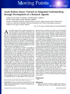
Contrast-induced acute kidney injury following iodine opacification other than by intravascular injection Tilman Perrin1, Ould Maouloud Hemett1, Markus Menth2 and Eric Descombes1 1
Internal Medicine and Nephrology Department, Hospital Fribourg, Fribourg, Switzerland and 2Surgery Department, Hospital Fribourg, Fribourg, Switzerland Correspondence and offprint requests to: Eric Descombes; E-mail:
[email protected]
Abstract Contrast-induced acute kidney injury (CI-AKI) classically occurs following the intravascular administration of iodinated contrast medium (CM). However, some cases of iodine-induced nephrotoxicity have been reported in patients who did not receive intravascular CM, as a consequence of iodine absorption through mucosae, burned skin or interstitial tissues. Recently, we observed the first case of CI-AKI occurring after an enteroclysis without any direct intravascular injection of CM. Here, we report this case, and review other clinical situations in which renal toxicity has been reported following the non-intravascular use of iodinated compounds. Keywords: acute kidney injury; acute renal failure; contrast-induced acute kidney injury; enteroclysis; iodine toxicity
Introduction
Case report
Contrast-induced acute kidney injury (CI-AKI) remains a common cause of in-hospital acute renal failure. Its pathophysiology is multifactorial and has not yet been understood completely, involving iodine-induced renal ischaemia and tubular toxicity. Risk factors for CI-AKI include pre-existing renal failure, hypotension, heart failure, older age, anaemia, diabetes mellitus and concomitant nephrotoxic medications [1, 2]. In clinical practice, CI-AKI is defined as a relative (≥25%) or absolute (≥0.5 mg/dL = 44 µmol/L) rise in serum creatinine level over baseline within 3 days of the administration of an iodinated compound, in the absence of any other explanation for the acute kidney injury. In the case of CI-AKI, serum creatinine typically reaches a peak value on Day 3–7 but usually returns to, or close to, baseline within 1–3 weeks [1, 2]. Classically, CI-AKI complicates the intravascular injection of iodinated contrast medium (CM), but we report here a case of CI-AKI that occurred after an intraenteric radiocontrast administration without any direct intravascular injection of CM. To our knowledge, this is the first reported case of CI-AKI after enteroclysis, but in the literature some other cases of iodine-induced nephrotoxicity following non-intravascular use of iodinated compounds have been reported. In the following, the different clinical situations in which this complication occurred as well as the mechanisms involved will be discussed.
A 57-year-old woman having two stomies due to a long history of intestinal resections was admitted to the intensive care unit (ICU) because of an acute rise in serum creatinine level (from 67 to 134 µmol/L in 24 h) with oliguria and drowsiness. Three days before, she developed fever with C-reactive protein elevation attributed to a digestive bacterial translocation and was treated by Imipenem 500 mg t.i.d without any other change in her current medication. On the same day, looking for a peritoneal fistula, a trans-jejunostomy enteroclysis was performed with 300 mL of Iopamiro 300 (low-osmolar CM; 90 g iodine load), which showed no intestinal leak. Since ICU admission, the patient remained haemodynamically stable and after rehydration a normal diuresis resumed. To note, the urine sediment was normal, in particular without leukocyturia nor eosinophiluria, while the urinary βNAG and lysozyme were markedly increased. Thereafter, the serum creatinine level continued to increase rapidly up to 481 µmol/L on Day 6 after enteroclysis and then slowly returned to baseline within a month (Figure 1). In the attempt to find the reason for this acute renal failure, it turned out that the radiologist had noticed the presence of CM in the urinary tracts already during the enteroclysis. For this reason, he had performed a native abdominal computed tomography (CT)-scan 1 h later, which confirmed the unexpected presence of CM in both renal pelvis and ureters, without evidence for any peritoneal resorption of CM (Figure 2). Therefore, although no
© The Author 2012. Published by Oxford University Press on behalf of ERA-EDTA. This is an Open Access article distributed under the terms of the Creative Commons Attribution Non-Commercial License (http://creativecommons.org/ licenses/by-nc/3.0/), which permits non-commercial re-use, distribution, and reproduction in any medium, provided the original work is properly cited. For commercial re-use, please contact
[email protected]
CI-AKI after enteroclysis
457
intravascular-iodinated CM had been administered to this patient, we considered the diagnosis of CI-AKI on the basis of the presence of CM in both urinary tracts associated with a typical clinical evolution. As no leak of CM into the peritoneal space was detected on the CT-scan, we assumed that the rapid absorption of CM occurred through the intestinal mucosa, the permeability of which was apparently increased by the local inflammatory
Fig. 1. Evolution of blood creatinine level after enteroclysis (arrow).
Fig. 2. Abdominal native CT-scan made 1 h after enteroclysis showing contrast media in the enterostomy (A), intestine (B) and renal pelvis bilaterally (C).
state. Moreover, in our patient, the transmucosal resorption of CM could have been sustained, as suggested by the persistence of CM in the caecum on a native CT-scan made 7 days after the enteroclysis.
Discussion To our knowledge, this is the first case of CI-AKI after enteroclysis reported in the literature. However, on review of the PubMed database, we found other cases of CI-AKI in patients who did not receive any intravascular injection of CM. In fact, these patients either underwent diagnostic hepatobiliary [3–8] and gynaecological [9–11] procedures with iodinated CM, or had topical treatments using iodinated antimicrobials [12–18]. Some clinical data of the 33 cases reported in the literature, including the one here presented, are summarized in Table 1. In most of them, the diagnosis of CI-AKI was suggested either by the detection of CM in urinary tracts on radiological exams, or by highly supra-physiological serum iodine concentrations. The assumed mechanism for toxicity was to be iodine absorption through mucosae or burned skin or interstitial tissues. It is important to note that since the first cases of nephrotoxicity reported after oral cholecystography in the 1960s [3, 8], it appeared that Iopanoic acid was a common cause of AKI, causing even irreversible renal damage. Accordingly, this extremely hazardous workup examination has been replaced by more accurate and safe diagnostic tests [19]. Similarly, because of several reported cases of systemic toxicity, it is clear that the use of povidone-iodine as CM for gynaecological diagnostic procedures should also be avoided [9, 11]. In patients with cutaneous burns, one previous study showed that the iodine absorption through the burned skin is proportional to the surface of the injury [20], and therefore, repeated topical antimicrobial treatment of large-area burns with povidone-iodine should be discouraged [17]. Similarly, a rapid interstitial absorption of iodine during experimental mediastinal irrigation with povidone-iodine has been documented in dogs [21]. According to the results of this latter study and to the cases of CI-AKI reported in patients with mediastinitis treated by mediastinal irrigation with povidone-iodine, this antiseptic treatment should obviously be undertaken only with great caution [14, 21]. In summary, the present case report as well as the aforementioned data highlight that CI-AKI sometimes
Table 1. Reported cases of CI-AKI following non-intravascular iodinated compounds use Use of iodinated product
Procedure
Type of iodinated product
Contrast agent
Percutaneous cholangiography
Diatrizoate meglumine sodium [4, 6] Unknown [5, 7] Iopanoic acid [3] Bunamiodyl sodium [8] Povidone-iodine [9–11] Iopamidol (presented case) Povidone-iodine [12, 17]
Transmucosal Transmucosal Transcutaneous (burned skin)
Povidone-iodine [13–15, 18]
Interstitial
4
Povidone-iodine [16]
Interstitial
1
Oral cholecystography
Antimicrobial
Gynaecological tract opacification Enteroclysis Topical disinfection of large burned areas Continuous mediatinal irrigation (mediastinitis) Local repetitive irrigation (cellulitis)
Suspected mechanism of resorption
Number of patients with CI-AKI
Transmucosal
7
Local trauma Transmucosal
6 3 1 11
458
occurs in patients who did not receive intravascular injection of iodinated CM and in whom iodine causes systemic toxicity after absorption through mucosae, burned skin or interstitial tissues. Therefore, the diagnosis of CI-AKI should not be excluded solely by the absence of intravascular injection of CM, and clinicians should always consider the diagnosis of CI-AKI when an acute kidney injury develops soon after the use of iodine-based products. Conflict of interest statement. None declared.
References 1. Lameire N. Contrast-induced nephropathy—prevention and risk reduction. Nephrol Dial Transplant 2006; 21: 11–23 2. Solomon R. Contrast media nephropathy—how to diagnose and how to prevent? Nephrol Dial Transplant 2007; 22: 1812–1815 3. Canales CO, Smith GH, Robinson JC et al. Brief recordings. Acute renal failure after the administration of iopanoic acid as a cholecystographic agent. N Engl J Med 1969; 281: 89–91 4. Gregory MC. Acute renal failure after percutaneous cholangiography. Arch Intern Med 1984; 144: 1288–1289 5. Kabza R, Dyktynski P. 2 cases of acute renal failure following cholangiography through the Kehr’s drain. Wiad Lek 1969; 22: 1319–1321 6. Kone BC, Watson AJ, Gimenez LF et al. Acute renal failure following percutaneous transhepatic cholangiography. A retrospective study. Arch Intern Med 1986; 146: 1405–1407 7. Mohan A, Paturi A, Schlein A et al. Acute renal failure associated with use of contrast in the biliary tree in an 82-year-old woman. J Am Geriatr Soc 2010; 58: 1806–1807 8. Wennberg JE, Okun R, Hinman EJ et al. Renal toxicity of oral cholecystographic media. Bumamiodyl sodium and iopanoic acid. J Am Med Assoc 1963; 186: 461–467 9. Beji S, Kaaroud H, Ben Moussa F et al. Acute renal failure following mucosal administration of povidone-iodine. Presse Med 2006; 35: 61–63
T. Perrin et al. 10. Lakhal K, Faidherbe J, Choukhi R et al. Povidone-iodine: features of critical systemic absorption. Ann Fr Anesth Reanim 2011; 30: 1–3 11. Moudden MK, Labaye J, Sarret D et al. Acute renal failure following internal administration of povidone-iodine: a case report. Rev Med Intern 2007; 28: 556–558 12. Aiba M, Ninomiya J, Furuya K et al. Induction of a critical elevation of povidone-iodine absorption in the treatment of a burn patient: report of a case. Surg Today 1999; 29: 157–159 13. Campistol JM, Abad C, Nogue S et al. Acute renal failure in a patient treated by continuous povidone-iodine mediastinal irrigation. J Cardiovasc Surg 1988; 29: 410–412 14. Glick PL, Guglielmo BJ, Tranbaugh RF et al. Iodine toxicity in a patient treated by continuous povidone-iodine mediastinal irrigation. Ann Thorac Surg 1985; 39: 478–480 15. Kanakiriya S, De Chazal I, Nath KA et al. Iodine toxicity treated with hemodialysis and continuous venovenous hemodiafiltration. Am J Kidney Dis 2003; 41: 702–708 16. Labbe G, Mahul P, Morel J et al. Iodine intoxication after subcutaneous irrigations of povidone-iodine. Ann Fr Anesth Reanim 2003; 22: 58–60 17. Pietsch J, Meakins JL. Complications of povidone-iodine absorption in topically treated burn patients. Lancet 1976; 1: 280–282 18. Ryan M, Al-Sammak Z, Phelan D. Povidone-iodine mediastinal irrigation: a cause of acute renal failure. J Cardiothorac Vasc Anesth 1999; 13: 729–731 19. Tse F, Barkun JS, Romagnuolo J et al. Nonoperative imaging techniques in suspected biliary tract obstruction. HPB 2006; 8: 409–425 20. Hunt JL, Sato R, Heck EL et al. A critical evaluation of povidone-iodine absorption in thermally injured patients. J Trauma 1980; 20: 127–129 21. Glick PL, Guglielmo BJ, Winter ME et al. Iodine toxicity secondary to continuous povidone-iodine mediastinal irrigation in dogs. J Surg Res 1990; 49: 428–434 Received for publication: 23.3.12; Accepted in revised form: 16.7.12