Aug 16, 2005 - and the pantropical tramp millipede R. purpureus. Two of these ant species, A. grandidieri and T. electrum, were also found in Mantella.
Convergent evolution of chemical defense in poison frogs and arthropod prey between Madagascar and the Neotropics Valerie C. Clark*†‡, Christopher J. Raxworthy†, Vale´rie Rakotomalala§, Petra Sierwald¶, and Brian L. Fisher储 *Department of Chemistry, Columbia University, New York, NY 10027; †Department of Herpetology, American Museum of Natural History, New York, NY 10024; §Department of Animal Biology, Universite´ d’Antananarivo, Antananarivo, 1001, Madagascar; ¶Department of Zoology, Field Museum of Natural History, Chicago, IL 60605; and 储Department of Entomology, California Academy of Sciences, San Francisco, CA 94103
alkaloid occurrence 兩 dietary sequestration 兩 nicotine
A
mphibians use a variety of skin chemicals for protection against predators, and anurans that store lipophilic basic alkaloids in granular glands are collectively termed ‘‘poison frogs’’ (1–3). Poison frogs have been recognized in four families: South American Melanophyrniscus (Bufonidae); Australian Pseudophryne (Myobatrachidae); Central and South American Dendrobates, Epipedobates, and Phyllobates (Dendrobatidae); and Malagasy (Madagascan) Mantella (Mantellidae) (1, 4–8). Additionally, trace amounts of such alkaloids have been detected in a Thai ranid frog, Limnonectes kuhli (9). More than 500 frog skin alkaloids belonging to 25 structural classes have so far been categorized, with each coded by a boldface number representing the nominal mass and a letter to distinguish among alkaloids of the same molecular weight (MW code) (1). Although wild poison frogs retain skin alkaloids for several years in captivity (10–12), they do not seem to produce alkaloids; rather, it seems that they sequester and accumulate such toxins from dietary arthropods by using an as-yet-uncharacterized alkaloid uptake system (11–13). Alkaloids are absent in dendrobatid and Mantella frogs raised in captivity on a diet of Drosophila, but these individuals will readily accumulate alkaloids added to their diet (10–15). Some anurans also have the ability to modify ingested www.pnas.org兾cgi兾doi兾10.1073兾pnas.0503502102
alkaloids: pumiliotoxin 307A was metabolized by one species of the genus Pseudophyrne (8), and pumiliotoxin (⫹)-251D was efficiently and stereoselectively hydroxylated by Dendrobates spp. into allopumiliotoxin (⫹)-267A, which is five times more toxic (14). The only known example of direct alkaloid production is in the Australian Pseudophryne frogs, which seem to be capable of synthesizing indolic pseudophrynamines (8). Recently, the putative dietary sources of representative alkaloids of several structural classes of ‘‘poison frog alkaloids’’ were reported; these dietary arthropods include beetles, ants, and millipedes. Dendrobatid and bufonid frogs and coccinellid beetles all share precoccinelline (193C), suggesting that these beetles represent a dietary source of this coccinelline-like tricyclic alkaloid and others like it (1, 13, 16, 17–21). Poison-dart frogs, genus Phyllobates of the Neotropics, contain highly toxic steroidal alkaloids, the batrachotoxins (22), as do passerine birds (23–24) and putative dietary melyrid beetles of Papua New Guinea (25); all are brightly colored. Regarding ants as sources of dietary alkaloids, pumiliotoxins were recently detected in formicine genera Brachymyrmex and Paratrechina, where they occurred microsympatrically with the poison frog Dendrobates pumilio in Panama (26). Poison frogs and Neotropical ants of the subfamily Myrmicinae share several classes of alkaloids, including 2,5-disubstituted (ds) pyrrolidines, 2,6-ds piperidines, 3,5-ds pyrrolizidines, 3,5-ds indolizidines, 4,6-ds quinolizidines, and 2,5-ds decahydroquinolines (1, 13, 16, 27). Reports of alkaloids from African ants are limited to the subfamily Myrmicinae: 2,5-dialkylpyrrolidines and 1-pyrrolines were detected in Monomorium in South Africa and Kenya (28), and substituted pyrazine alkaloids detected in Eutetramorium mocquerysi, a genus endemic to Madagascar (29). Two other Malagasy myrmicine ants of the genus Metapone use methyl pyrrole-2-carboxylate as a trail pheromone and also contain pyrazines (30). Seven alkaloids of the spiropyrrolizidine (SpiroP) class have been detected in poison frogs; three of these alkaloids have also been reported from two millipede species: (i) polyzonamine (151B) and nitropolyzonamine (238) from Petaserpes cryptocephalum (McNeill 1887) [Polyzoniidae: Polyzoniida, often misidentified as Polyzonium rosalbum (Cope 1879)] (31; see p. 32 of ref. 32) of Ithaca, New York (33–34) and (ii) the 238 and SpiroP O-methyloxime 236 from the widespread Rhinotus purpureus (Pocock 1894) (Siphonotidae: Polyzoniida) that occurs sympatrically in Panama with the poison frog D. pumilio (35). Other alkaloids detected in millipedes include glomerins (quinazolinones) from the Glomeridae (36–37) and the This paper was submitted directly (Track II) to the PNAS office. Abbreviations: ds, disubstituted; SpiroP, spiropyrrolizidine; Saha, Sahavondrona; Vato, Vatoharanana; GCT, GC-TOF mass spectrometer; TAS, transcutaneous amphibian stimulator. ‡To
whom correspondence should be sent at the present address: Department of Chemistry and Chemical Biology, Cornell University, Ithaca, NY 14853. E-mail: vcc4@ cornell.edu.
© 2005 by The National Academy of Sciences of the USA
PNAS 兩 August 16, 2005 兩 vol. 102 兩 no. 33 兩 11617–11622
CHEMISTRY
With few exceptions, aposematically colored poison frogs sequester defensive alkaloids, unchanged, from dietary arthropods. In the Neotropics, myrmicine and formicine ants and the siphonotid millipede Rhinotus purpureus are dietary sources for alkaloids in dendrobatid poison frogs, yet the arthropod sources for Mantella poison frogs in Madagascar remained unknown. We report GC-MS analyses of extracts of arthropods and microsympatric Malagasy poison frogs (Mantella) collected from Ranomafana, Madagascar. Arthropod sources for 11 ‘‘poison frog’’ alkaloids were discovered, 7 of which were also detected in microsympatric Mantella. These arthropod sources include three endemic Malagasy ants, Tetramorium electrum, Anochetus grandidieri, and Paratrechina amblyops (subfamilies Myrmicinae, Ponerinae, and Formicinae, respectively), and the pantropical tramp millipede R. purpureus. Two of these ant species, A. grandidieri and T. electrum, were also found in Mantella stomachs, and ants represented the dominant prey type (67.3% of 609 identified stomach arthropods). To our knowledge, detection of 5,8-disubstituted (ds) indolizidine iso-217B in T. electrum represents the first izidine having a branch point in its carbon skeleton to be identified from ants, and detection of 3,5-ds pyrrolizidine 251O in A. grandidieri represents the first ponerine ant proposed as a dietary source of poison frog alkaloids. Endemic Malagasy ants with defensive alkaloids (with the exception of Paratrechina) are not closely related to any Neotropical species sharing similar chemical defenses. Our results suggest convergent evolution for the acquisition of defensive alkaloids in these dietary ants, which may have been the critical prerequisite for subsequent convergence in poison frogs between Madagascar and the Neotropics.
ECOLOGY
Edited by Jerrold Meinwald, Cornell University, Ithaca, NY, and approved July 1, 2005 (received for review April 27, 2005)
terpenoid alkaloid buzonamine from the Polyzoniidae, genus Buzonium (38). More than 100 alkaloids of 12 classes have been detected in skin of Mantella (1, 4, 6, 39–40), and many also occur in Neotropical poison frogs. In addition to their ability to sequester and accumulate alkaloids from diet, Mantella and some dendrobatid frogs also share the following features: terrestrial eggs, small body size (⬍50-mm snout-vent length), toothless jaws, a specialist diet composed largely of ants, active diurnal foraging behaviors, and aposematic coloration; all features are considered to have been produced by convergent evolution (4, 41–45). In both groups, sequestered defensive chemicals appear to be closely associated with (i) the evolution of aposematism, as a visual warning of their toxicity to potential predators, and (ii) active diurnal foraging, a behavior that is generally rare in frogs (46). Although the foraging behavior and diet of Mantella is not yet well documented relative to dendrobatids (46–52), ants are known to dominate the diet of Mantella (42–43). A study of 774 prey items taken from the stomachs of 15 Mantella specimens of four species found that ants represented 74% of the total prey and that all prey items were ⬍5 mm in length (43). However, to date there had been no studies concerning arthropods as potential sources of frog skin alkaloids in Madagascar, and the potential convergence of alkaloid defenses in arthropod groups between the Neotropics and Madagascar had not yet been investigated. Here, we report results of our alkaloid survey conducted in and around Ranomafana National Park (in Fianarantsoa Province, southeast Madagascar) that targeted both Mantella poison frogs and potential dietary microsympatric leaf-litter arthropods. Materials and Methods Field Collections. Mantellid frogs and arthropods were collected within and around Ranomafana National Park. Four collecting sites (all within 15 m of 2- to 4-m-wide streams) were surveyed: Vohiparara, 21°13.587⬘ S, 47°22.193⬘ E; Sahavondrona (Saha), 21°15.450⬘ S, 47°21.609⬘ E; Vatoharanana (Vato), 21°17.444⬘ S, 47°25.569⬘ E; and Ampasimpotsy, 21°28.796⬘ S, 47°33.424⬘ E, with sampling conducted during the latter part of the rainy season (March 13 to May 1, 2003). Mantella were photographed, killed with chloroform, and skinned into 100% methanol, and their bodies were fixed in 10% formalin within 1 h of capture to preserve stomach contents. In addition, a transcutaneous amphibian stimulator (TAS) (53) was used to obtain skin exudates from live frogs, which were then photographed and released. All frog voucher specimens have been deposited at the Department of Animal Biology at University of Antananarivo and at the American Museum of Natural History; frog stomach contents were removed for subsequent identification. All Mantella capture sites were also surveyed for leaf-litter arthropods to produce samples for alkaloid analyses. Forcepsmediated collections were made by searching through leaf-litter on white cloth and removing arthropods with entomological forceps. Arthropod specimens were put in methanol in taxon-specific tubes, and forceps were wiped clean with methanol between samples. Collecting efforts focused primarily on arthropods ⬍5 mm in length (considered the maximum prey size for Mantella); these samples largely comprised ants, beetles, and millipedes. Mixed arthropod collections were made by using 10 mini-Winkler extractors per locality (methods are described in refs. 54–56) to provide additional arthropod reference collections for identifications. All arthropods were identified by B.L.F., P.S., and V.R. and deposited at the Department of Animal Biology at University of Antananarivo, the American Museum of Natural History, the Field Museum of Natural History, and the California Academy of Sciences. Alkaloid Analyses. Methanol extracts of arthropods and frogs were
analyzed for alkaloids by GC-MS on a GC-TOF mass spectrometer (GCT) (Micromass, Manchester, U.K.) in electron-impact and chemical-ionization (with NH3) modes. A 30-m ⫻ 0.25-mm i.d. 11618 兩 www.pnas.org兾cgi兾doi兾10.1073兾pnas.0503502102
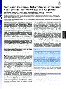
Fig. 1. Mantella poison frogs and their putative arthropod prey that contain the same defensive alkaloids. (a) M. madagascariensis (shown actual size). (b) M. bernhardi. (c) M. baroni. (d) R. purpureus. (e) T. electrum. ( f) A. grandidieri. (Scale bars, 1 mm.)
Supelco Equity 5 column with 0.25-m film thickness was used for all GCT injections, and the oven temperature of the GCT was increased at 10°C兾min from 100°C to 280°C. GCT calibration samples supplied by John Daly (National Institutes of Health, Bethesda) for Mantella baroni (Saha, January 1993; Vato, December 1989) provided reference retention times for known alkaloids (4). Methanolic frog skin alkaloid fractions were prepared for individual frogs following the methodology of ref. 11. A volume of 1 l, corresponding to 1 mg of wet weight frog skin, was injected for alkaloid fractions from Mantella skins, whereas 4 l was injected for each TAS frog extract. Arthropod extracts were injected without work-up into alkaloid fractions. Alkaloids were identified by using the MS library of ref. 1. Results Three species of Mantella poison frog were recorded during this survey of Ranomafana: M. baroni, Mantella bernhardi, and Mantella madagascariensis (Fig. 1). GC-MS analyses of the methanol extracts obtained from 22 individual Mantella representing these three species (18 skins and 4 TAS extracts) recovered 80 coded alkaloids (1). Additionally, nicotine (which we code as 162), previously undetected in any frog species, was detected in M. baroni of Saha. The occurrence of these 81 alkaloids in Mantella are given in Table 3, which is published as supporting information on the PNAS web site, and the 33 alkaloids that were previously undetected in Mantella are highlighted; 9 of these 33 Mantella alkaloids are isomers not previously reported in frogs. Eleven of these 81 alkaloids are now known from a specific Malagasy arthropod source. TAS extracts yielded detectable alkaloids similar in diversity compared with skin extracts. The distributions of arthropod source alkaloids in individual Mantella frogs are presented by locality in Table 1. Analyses of the 154 extracts of arthropod morphospecies samples (65 ants and 89 others) detected 11 known poison frog alkaloids Clark et al.
Table 1. Distribution of selected alkaloids in individual Mantella frogs at four survey sites in the Ranomafana region of Madagascar Number of frogs containing alkaloid M. mad.
M. bernhardi
Saha Vato Vohi
Vohi
Ampasimpotsy
— — 2 — —
—* — —* —* —*
1 — 1 1 —
1 1 1 1 1
— — — — —
— — 4
—* — 3*
3 3 8
— 1 1
— 5 6
—
—
3
—
—
4 — 1
3 — —
8 — —
1 — —
5 5* —
—
1
—
—
—
— 3 1 4
— 3 0 3
— 5 3 8
— 1 0 1
1 6 0 6
Previously Undetected Alkaloids in Mantella. To our knowledge, our
—, not detected; Vohi, Vohiparara; M. mad., M. madagascariensis. *This alkaloid was also found at this site in arthropods sampled by the forceps method (see Table 2). †Data are combined for three additional isomers of 223H with earlier retention times in Mantella than in the ant cis-223H, which has the same retention time as cis-223H in Daly’s Saha Mantella standard (see Table 3).
(Table 2 and Fig. 2); 7 of these arthropod alkaloids were also detected in microsympatric Mantella frogs. However, alkaloids were generally rare among these arthropod samples, with at most five taxon-specific ant samples and one millipede sample yielding detectable products (one ant morphospecies sample later proved to contain two species). These alkaloids were confined to ant species representing three different subfamilies (Formicinae, Myrmicinae, and Ponerinae) and one millipede (R. purpureus; family Siphonotidae). The negative alkaloid ant samples have not yet been identified, but the negative millipede samples (all Pachybolidae) included two pantropical species, Trigoniulus corallinus (Gervais 1847) and Leptogoniulus sorornus (Butler 1876) and a Malagasy endemic Aphistogoniulus sp. (Silvestri) (32, 57, 58). A single female specimen of the synanthropic millipede R. purpureus, collected from Vato, yielded five alkaloids of the SpiroP class (Table 2 and Fig. 2). Areas under the chromatogram peaks reveal the following relative concentrations: 2% 151B, 17% 236, 77% iso-236, 3% 238, and 1% 254. The previously undetected isomer, iso-236, eluted 0.16 min after 236, but the fragmentation pattern was the same for both. The configuration of the oxime group of 236 was assigned as syn (Z) based on nuclear Overhauser effect NMR experiments (59). The ant species Tetramorium electrum Bolton (subfamily Myrmicinae, tribe Tetramoriini; Fig. 1) collected from Ampasimpotsy yielded a branched chain izidine: 5,8-ds indolizidine iso-217B (Fig. 2). Anochetus grandidieri Forel (subfamily Ponerinae, tribe Ponerini; Fig. 1) collected from Vato contained 3,5-ds pyrrolizidine 251O (Fig. 2). Paratrechina ambylops (Forel) (Formicinae, Plagiolepidini) collected from Vato contained a variety of alkaloids, including 3,5-ds pyrrolizidines 195F and 223H, 2,5-ds pyrrolidine Clark et al.
Discussion results represent the first report of four SpiroPs for Mantella: 151B, 222, iso-236, and 238 (Fig. 2). Two SpiroPs, 236 and 252A, have been previously reported in M. baroni (4), and 252B has been reported in Australian Pseudophryne frogs (5). Ant alkaloid 3,5-ds indolizidine 195B and beetle alkaloid precoccinelline 193C, both of which have also been reported in Neotropical dendrobatid and bufonid poison frogs, were detected here for what we believe to be the first time in poison frogs of Madagascar (Tables 1–3). A total of 23 known coded poison frog alkaloids (1), plus nine other isomers and nicotine 162, were not previously known from mantellids (1) but were detected in Mantella of our study (Table 3). To our knowledge, our finding of 2,6-ds piperidine 197E in M. baroni of Vato represents the first report of this class in Mantella (Table 3), thus demonstrating that Mantella can indeed sequester this alkaloid class. The well known plant alkaloid nicotine (162) was detected in a single M. baroni specimen, representing what we believe to be the first report of nicotine from any frog source and the 14th class of alkaloids (pyridyls) known from Mantella. However, other pyridyl alkaloids (e.g., the epibatidines and noranabasamine) have been reported from dendrobatid poison frogs (1). Nicotine is biosynthesized by a variety of plants of the family Solanaceae (60) and can also be sequestered by insects (61) and biosynthesized by lepidopteran larvae (62). However, we have not yet detected nicotine in any of our Malagasy arthropod extracts, and it is unknown to us whether suitable nicotine source plants occur at Saha. Still, our nicotine discovery suggests a sequestration food chain where nicotine is sequestered by an herbivorous arthropod subsequently eaten by Mantella. We also detected in these Mantella several other previously undetected alkaloids, not yet coded. Individual Variation in Mantella Frogs. Our results show variation in alkaloid profiles between individual Mantella frogs of the Ranomafana region, within the same species, and at the same collecting site. However, at Vohiparara, the only site that included two sympatric Mantella, six of eight alkaloids found in each species were also present in the other sympatric species (Table 1). This finding tentatively suggests that alkaloid profiles of microsympatric Mantella species may be quite similar and that Mantella alkaloid profiles, in general, may be more heavily influenced by the distribution of local arthropod alkaloid sources rather than ecological or evolutionary differences between Mantella species. The individual variation seen for frog alkaloid profiles, coupled with the observation that Mantella retain alkaloids in their skin for years in captivity (12), suggests that some arthropod sources for PNAS 兩 August 16, 2005 兩 vol. 102 兩 no. 33 兩 11619
ECOLOGY
Spiropyrrolizidines 151B 222 236 Iso-236 238 3,5 Pyrrolizidines cis-223H Iso-223H† 251O 3,5 Indolizidines 195B 5,8 Indolizidines 217B Iso-217B 217B⬙ (third isomer) Pumiliotoxins 307A Precoccinelline 193C Skin samples TAS samples Total frogs
M. baroni
CHEMISTRY
Alkaloid class and MW code (1)
197B (Fig. 2), and other uncharacterized alkaloids. Another 2,5-ds pyrrolidine, 225C, was detected in a mixed sample of larger (⬎7-mm length) black ants from Saha: Pachycondyla cambouei (Forel) (Ponerinae, Ponerini) and Camponotus sp. 1 (Formicinae, Camponotini). Analyses of the stomach contents of 21 Mantella specimens yielded 609 identified arthropod specimens (Table 4, which is published as supporting information on the PNAS web site), with the largest fraction (67.3%) representing ants (29 species of nine genera). The remaining stomach arthropods included 12.2% Acari (mites), 5.5% Collembola (springtails), 4.1% Amphipoda (lawn shrimps), 4.1% various larvae, 2.5% Coleoptera (beetles), and 9.3% other arthropod groups, including one millipede individual identified as a pantropical species, Prosopodesmus jacobsoni (Silvestri 1910) (Haplodesmidae: Polydesmida). The proportion of ants in the arthropod stomach samples ranged from 18% to 93% among the 21 Mantella frogs (mean ⫽ 63%). The 251O alkaloid-containing ant A. grandidieri was found in stomachs of M. madagascariensis of Vohiparara and M. baroni of both Saha and Vato. Nine species of Tetramorium ants were found (10% of the total ingested ants), including the iso-217B-containing T. electrum in M. baroni at Saha.
Table 2. The occurrence of alkaloids in arthropods and Mantella frogs recorded in this study with a comparison to published data for other Mantella in Madagascar and poison frogs and arthropods in the Neotropics Madagascar
Neotropics
Ranomafana region (this study) Alkaloid class and MW code Spiropyrrolizidines 151B 222 236 iso-236 238 254 2,5 Pyrrolidines 197B
225C
3,5 Pyrrolizidines 195F 223H 251O 3,5 Indolizidines 195B 5,8 Indolizidines 217B iso-217B Pumiliotoxins 307A Coccinelline-like tricyclics Precoccinelline 193C
Refs. 1 and 4
Refs. 16, 18–20, 26, 27, and 35
Ref. 1
Mantella species
Other Mantella
Arthropod species and family or subfamily
Frog families
1, 2, 3 3 1, 3 1, 3 3 —
— — 4 — — —
— —
D D D, B — D D
Paratrechina amblyops F
—
—
Pachycondyla cambouei P and兾or Camponotus sp. F
—
4
— 1 1, 2, 3
— 4, ⫹ 4, ⫹
1
—
1, 2, 3 2
4, ⫹
—
1
2, 4, ⫹
—
2
—
Arthropod species and family or subfamily R. purpureus S — R. purpureus S R. purpureus S R. purpureus S R. purpureus S
Paratrechina amblyops F Paratrechina amblyops F A. grandidieri P — — T. electrum M
R. purpureus S — R. purpureus S — Monomorium pharaonis, Megalomyrmex goeldi, Solenopsis punctaticeps M Monomorium indicum, Megalomyrmex foreli Solenopsis fugax, S. punctaticeps M
D
D
— Solenopsis sp. M —
D D, B —
Monomorium pharaonis M
D, B
— — Paratrechina steinheili, Brachmyrmex spp. F Coccinella septempunctata, Coleomegilla maculata C, Chauliognathus pulchellus A*
D — D
D, B
Arthropod family or subfamily is as follows. Millipedes: S, Siphonotidae. Ants: F, Formicinae, M, Myrmicinae, P, Ponerinae. Beetles: C, Coccinellidae; A, Cantharidae. Mantella species: 1, M. baroni; 2, M. bernhardi; 3, M. madagascariensis; 4, M. betsileo; ⫹, other Mantella species. Frog families are as follows. D, Dendrobatidae (Dendrobates, Epipedobates, and Phyllobates of Central and South America); B, Bufonidae (Melanophryniscus of South America). —, not detected. *These beetles were not collected from the Neotropics; see refs. 18 –21.
alkaloids are rare prey items for Mantella. Rare prey types are also evident in our Mantella stomach content data (Table 4); for example, just one millipede is represented from this sample of 609 arthropods. Presumably, individual frogs missing alkaloids (otherwise represented in local populations) have never, or perhaps rarely, ingested the required source arthropod prey over the duration of their lifespan. Our own efforts at identifying arthropod sources in leaf-litter also suggest that some sources may be rare in this microhabitat; for example, the source for precoccinelline (193C) in Mantella is probably a coleopteran beetle, which we failed to sample. If some arthropod sources for alkaloids are indeed rare, then older Mantella should profile a greater diversity of alkaloids because of the greater arthropod sampling achieved over their longer lifespans. By contrast, two alkaloids (251O and 217B) were broadly distributed across all, or almost all, individual Mantella frogs from the Ranomafana region. The sources of each appear to be ants of the genera Anochetus and Tetramorium, respectively (see below), which were also found as prey items in Mantella frog stomachs (Table 4). Arthropod Sources for Alkaloids in Mantella. Of the 11 coded poison
frog alkaloids detected in our arthropod samples, 7 were also detected in microsympatric Mantella, and thus these arthropods represent dietary sources available to these frogs (Table 2). Three 11620 兩 www.pnas.org兾cgi兾doi兾10.1073兾pnas.0503502102
of the four alkaloids missing in the frogs (254, 197B, and 195F) were found in two arthropod species (Table 2). Mantella can sequester other alkaloids of these classes, so we would expect that these alkaloids would be sequestered if ingested. An examination of trace alkaloids in Daly’s Vato standard revealed trace amounts of 197B (not in ref. 4); 197B was also present in our P. ambylops ant of Vato. The absence of 225C in Ranomafana Mantella, which was detected in a sample of large black ants (Pachycondyla cambouei, length ⫽ 10.7 mm and Camponotus sp., length ⫽ 7.9 mm) may reflect these ants being too large to serve as potential prey. Vences and Kniel (43) reported all Mantella prey as ⬍5 mm in length, and the maximum sized prey specimen we recorded was a lepidopteran larvae that was 4.7 mm long. The discovery of seven alkaloids shared among microsympatric Mantella poison frogs and four leaf-litter arthropod species (Tables 1 and 2) provides data to identify potential dietary sources for alkaloids in Malagasy poison frogs. These findings are also partly corroborated by the stomach content data: two of the potential ant source species, A. grandidieri and T. electrum, were found in the stomachs of the microsympatric Mantella (Table 4). Surprisingly, the probable dietary source of the Mantella SpiroPs at Ranomafana appears to be the same millipede species, R. purpureus, as reported for dendrobatid poison frogs in Panama (35). This invasive tramp millipede has a pantropical distribution and, in Clark et al.
contrast to the endemic Malagasy millipedes, likely represents a relatively recent arrival in Madagascar mediated by accidental human introduction. The two reported records (58) for Madagascar, Toamasina (Tamatave) and Nosy Be, are both historical and current major trading ports. Although the genus Rhinotus is in need of revision, it is assumed that Neotropical and Afrotropical specimens of the genus are conspecific (63). Although five SpiroPs were recorded in R. purpureus, the majority of Mantella had no more than two of these alkaloids, and in Panama, only two SpiroPs were found in R. purpureus and sympatric poison frogs (35). A possible explanation for this variability concerns the age of the millipedes and the environmental availability of compounds sequestered from food plants; Meinwald et al. (33) have suggested (for another Polyzoniida millipede) that a plantorigin pyrrolizidine may be sequestered, transformed into the spirocycle, and subsequently metabolized into 151B. The elongate mandibles of Rhinotus are suited for scraping (see figure 3 in ref. 63), and it is assumed that this species scrapes roots and shoots for plant juices (p. 819 of ref. 64). By contrast, spirobolid and spirostreptid millipedes that in our study did not yield detectable alkaloids, have chewing mandibles and feed on leaf-litter (65–66). The SpiroPs provide chemical defense to both the bright red millipede R. purpureus and the poison frogs that sequester these alkaloids: SpiroPs 222, 236, and 238 act as noncompetitive blockers of nicotinic receptors with selectivity for the ganglionic subtype (67), whereas polyzonamine 151B is an effective ant repellent and general topical irritant to other insects (34). Assuming that R. purpureus is a recent arrival to Madagascar, this finding suggests that the sequestration physiology of Mantella may have been already preadapted to accept SpiroP alkaloids. To our knowledge, our study reports the first known occurrence of six alkaloids for endemic Malagasy ants: 3,5-ds pyrrolizidines (195F, 223H, and 251O), 2,5-ds pyrrolidines (197B and 225C), and the 5,8-ds indolizidine iso-217B. This latter finding also represents what we believe to be the first record of this alkaloid class for any ant species; previously, other 5,8-ds indolizidines were detected in mixed arthropod samples in Panama (68). However, only the iso-217B was detected in Malagasy ants, whereas both 217B and iso-217B were detected Clark et al.
PNAS 兩 August 16, 2005 兩 vol. 102 兩 no. 33 兩 11621
CHEMISTRY
Fig. 2. Known poison frog alkaloids detected in Malagasy arthropods (see Table 1). (a) Alkaloids detected in ants. (b) Alkaloids detected in the millipede R. purpureus. Asterisk indicates the branch point in the carbon skeleton of 217B.
Convergent Evolution Between Madagascar and the Neotropics. A remarkable feature of the 16 coded alkaloids we report here in the arthropods and Mantella frog species of Ranomafana (Madagascar) is that 13 of them are also known in other ants, beetles, and frogs endemic to the Neotropics (Table 2; see also Table 3). Excluding R. purpureus (a likely recent invasive tramp) and possibly Paratrechina (phylogenetic relationships within this globally distributed genus remain uncertain), none of the other Malagasy and Neotropical endemic species that share these types of alkaloids are closely related to each other. Three alkaloids (197B, 225C, and 223H) are each shared between at least one species of formicine or ponerine ant endemic to Madagascar and one species of myrmicine ant endemic to the Neotropics (Table 2 and refs. 27 and 73–75). These Madagascan and Neotropical ant species are classified in different subfamilies, and even at the subfamily level, these groups are not closely related based on molecular and morphological phylogenetic studies (76–77). Considering all Neotropical and Malagasy ant species known to contain defensive alkaloids shown in Table 2, just one genus (Paratrechina) is represented in both regions. Another ant species, Monomorium pharaonis, is a widespread tramp that could be of African origin (75) and could potentially serve as an alkaloid source in both Madagascar and the Neotropics; however, we did not recover any alkaloidcontaining Monomorium in our survey (Table 2). The phylogenetic distribution of these defensive alkaloid-bearing species thus is suggestive for multiple independent evolution of sequestration and兾or biosynthesis within the endemic ant radiations of Madagascar and the Neotropics. Within each of these three ant subfamilies, the rarity of species containing these defensive alkaloids is striking. Very few ant species actually yield defensive alkaloids: only 4 of our 65 ant samples were positive, and similar low hit successes have also been reported for Neotropical ants and arthropods [e.g., 4 positive of 61 ant samples (16) and 3 formicine ant species containing pumiliotoxins from 512 arthropod samples (26)]. Of the 12 alkaloids we report here in Mantella from Ranomafana, 9 also occur in dendrobatid poison frogs (Dendrobates, Epipedobates, and Phyllobates), and 4 occur in bufonid toads (Melanophryniscus) of the Neotropics (see Table 2). The Madagascan Mantella species (Mantellidae) are not closely related to these Neotropical groups (Dendrobatidae and Bufonidae) but fall within a paraphyletic Mantidactylus group that itself is sister to other Malagasy endemic mantellid genera, and then rhacophorids (78 –79). These shared alkaloids are unknown for all other genera within Mantellidae, Dendrobatidae, and Bufonidae. There is no doubt that alkaloid sequestration represents an apomorphic trait within each family and that the evolution of alkaloid sequestration for poison frogs in Madagascar and the Neotropics has independently occurred
ECOLOGY
in Mantella. The occurrence of a 3,5-ds pyrrolizidine (251O) in A. grandidieri also represents, to our knowledge, the first ponerine ant to be proposed as a dietary source of poison frog alkaloids; however, Anochetus kempfi of Puerto Rico contains a phenylpyrrole (69) not yet known from frogs. The origin of defensive ant alkaloids is not yet clear; alkaloid biosynthesis has not yet been studied for any ant species endemic to Madagascar, and, globally, only tetraponerine and solenopsin ant alkaloids have so far been studied (ref. 70 and references therein). The ants produced those compounds themselves; however, it is also possible that ants sequester alkaloids from plants, as is known for complex pyrrolizidines in lepidopterans and coleopterans (71–72), or even that microsymbionts might serve a role (26). The alkaloids sequestered by poison frogs serve primarily as passive chemical defenses, and, presumably, these compounds also serve similar defensive functions for the source ant species.
multiple times (4, 45). Evolutionary convergence of defensive alkaloid sequestration, similarly, has also driven evolutionary convergence for aposematic colorations, presumably in response to similar selective pressures from frog predators to this form of chemical defense. For Neotropical and Malagasy frogs, the convergent evolution of alkaloid uptake systems for chemical defense requires first the availability of arthropod prey containing toxic alkaloids and second the independent origin of mechanisms for achieving alkaloid sequestration. Because ants represent a common or even dominant part of poison frog diets (Table 4 and refs. 43, 49–52, and 80), the presence of suitable alkaloids in ants may have been the critical prerequisite for the evolution of alkaloid chemical defense in Malagasy and Neotropical poison frogs. Our demonstration that endemic Malagasy ants do indeed contain these toxic alkaloids supports this view and further suggests that the convergence seen in the poison frogs might itself have been
We thank Elizabeth E. Clark, Dr. John W. Daly [who provided unpublished occurrence data for alkaloids 223H and 307F (Table 3)], Prof. Olga Ramilijaona, Dr. Helian Ratsirarson, Aimee Razafiarimalala, and Justin Solo for their help, and Prof. Koji Nakanishi and Dr. Leif Abrell at the Biosphere 2 Chemistry Unit and Columbia University’s Chemistry Department for providing GCT time and laboratory facilities (to V.C.C.). This work was supported by National Science Foundation (NSF) Grant DEB-9984496 (to C.J.R.) for fieldwork, NSF Grants DEB-0072713 (to B.L.F. and C. E. Griswold) and DEB-0344731 (to B.L.F. and P. S. Ward) for arthropod processing, and NSF Grant CHE-0216226 (to Koji Nakanishi) for GCT acquisition. The Department of Animal Biology at University of Antananarivo, Association Nationale pour la Gestion des Aires Prote´ge´es, Direction Generale des Eaux et Foreˆts, Madagascar Institute pour la Conservation des Ecosyste `mes Tropicaux, and the California Academy of Sciences team in Madagascar facilitated activities in Madagascar.
1. Daly, J. W., Garraffo, H. M. & Spande, T. F. (1999) in Alkaloids: Chemical and Biological Perspectives, ed. Pelletier, S. W. (Pergamon, New York), Vol. 13, pp. 1–161. 2. Myers, C. W. & Daly, J. W. (1993) Science 262, 1193. 3. Neuwirth, M., Daly, J. W., Myers, C. W. & Tice, L. W. (1979) Tissue Cell 11, 755–771. 4. Daly, J. W., Andriamaharavo, N. R., Andriantsiferana, M. & Myers, C. W. (1996) Am. Mus. Novit. 3177, 1–34. 5. Daly, J. W., Garraffo, H. M., Pannell, L. K. & Spande, T. F. (1990) J. Nat. Prod. 53, 401–421. 6. Garraffo, H. M., Caceres, J., Daly, J. W. & Spande, T. F. (1993) J. Nat. Prod. 56, 1016–1038. 7. Garraffo, H. M., Spande, T. F., Daly, J. W., Baldessari, A. & Gros, E. G. (1993) J. Nat. Prod. 56, 357–373. 8. Smith, B. P., Tyler, M. J., Kaneko, T., Garraffo, H. M., Spande, T. F. & Daly, J. W. (2002) J. Nat. Prod. 65, 439–447. 9. Daly, J. W., Noimai, N., Kongkathip, B., Kongkathip, N., Wilham, J., Garraffo, H. M., Kaneko, T., Spande, T. F., Nimit, Y., Nabhitabhata, J. & Chan-Ard, T. (2004) Toxicon 44, 805–815. 10. Daly, J. W., Myers, C. W., Warnick, J. E. & Albuquerque, E. X. (1980) Science 208, 1383–1385. 11. Daly, J. W., Secunda, S. I., Garraffo, H. M., Spande, T. F., Wisnieski, A. & Cover, J. F., Jr. (1994) Toxicon 32, 657–663. 12. Daly, J. W., Garraffo, H. M., Hall, G. S. & Cover, J. F., Jr. (1997) Toxicon 35, 1131–1135. 13. Daly, J. W., Garraffo, H. M., Spande, T. F., Jaramillo, C. & Rand, A. S. (1994) J. Chem. Ecol. 20, 943–955. 14. Daly, J. W., Garraffo, H. M., Spande, T. F., Clark, V. C., Ma, J., Ziffer, H. & Cover, J. F., Jr. (2003) Proc. Natl. Acad. Sci. USA 100, 11092–11097. 15. Daly, J. W., Secunda, S. I., Garraffo, H. M., Spande, T. F., Wisneiski, A., Nishihira, C. & Cover, J. F. (1992) Toxicon 30, 887–898. 16. Daly, J. W., Garraffo, H. M., Jain, P., Spande, T. F., Snelling, R. R., Jaramillo, C. & Rand, A. S. (2000) J. Chem. Ecol. 26, 73–85. 17. King, A. G. & Meinwald, J. (1996) Chem. Rev. 96, 1105–1122. 18. Tursch, B., Daloze, D., Braekman, J. C., Hootele, C. & Pasteels, J. M. (1975) Tetrahedron 31, 1541–1543. 19. Moore, B. P. & Brown, W. V. (1978) Insect Biochem. 8, 393–395. 20. Henson, R., Thompson, A., Hedin, P., Nichols, P. & Neel, W. (1975) Experientia 31, 145. 21. Laurent, P., Braekman, J. C. & Daloze, D. (2005) Top. Curr. Chem. 240, 167–229. 22. Tokuyama, T. & Daly, J. W. (1983) Tetrahedron 39, 41–47. 23. Dumbacher, J. P., Beehler, B. M., Spande, T. F., Garraffo, H. M. & Daly, J. W. (1992) Science 258, 799–801. 24. Dumbacher, J. P., Spande, T. F. & Daly, J. W. (2000) Proc. Natl. Acad. Sci. USA 97, 12970–12975. 25. Dumbacher, J. P., Wako, A., Derrickson, S. R., Samuelson, A., Spande, T. F. & Daly, J. W. (2004) Proc. Natl. Acad. Sci. USA 101, 15857–15860. 26. Saporito, R. A., Garraffo, H. M., Donnelly, M. A., Edwards, A. L., Longino, J. T. & Daly, J. W. (2004) Proc. Natl. Acad. Sci. USA 101, 8045–8050. 27. Jones, T. H., Gorman, J. S., Snelling, R. R., Delabie, J. H., Blum, M. S., Garraffo, H. M., Jain, P., Daly, J. W. & Spande, T. F. (1999) J. Chem. Ecol. 25, 1179–1193. 28. Jones, T. H., Zottig, V. E., Robertson, H. G. & Snelling, R. R. (2003) J. Chem. Ecol. 29, 2721–2727. 29. Tentschert, J., Bestmann, H., Ho ¨lldobler, B. & Heinze, J. (2000) Naturwissenschaften 87, 377–380. 30. Ho ¨lldobler, B., Oldman, N. J., Alpert, G. D. & Liebig, J. (2002) Chemoecology 12, 147–151. 31. Shelley, R. M. (1997) Arthropoda-Selecta 6, 3–34. 32. Hoffman, R. L. (1999) Virginia Nat. Hist. Mus. Spec. Pub. 8, 1–553. 33. Meinwald, J., Smolanoff, J., McPhail, A. T., Miller, R. W., Eisner, T. & Hicks, K. (1975) Tetrahedron Lett. 28, 2367–2370. 34. Smolanoff, J., Kluge, A. F., Meinwald, J., McPhail, A. T., Miller, R. W., Hicks, K. & Eisner, T. (1975) Science 188, 734–736. 35. Saporito, R. A., Donnelly, M. A., Hoffman, R. L., Garraffo, H. M. & Daly, J. W. (2003) J. Chem. Ecol. 29, 2781–2786.
36. Meinwald, Y. C., Meinwald, J. & Eisner, T. (1966) Science 154, 390–391. 37. Schildknecht, H., Maschwitz, U. & Wenneis, W. F. (1967) Naturwissenschaften 54, 196–197. 38. Wood, W. F., Hanke, F. J., Kubo, I., Carroll, J. A. & Crews, P. (2000) Biochem. Syst. Ecol. 28, 305–312. 39. Jain, P., Garraffo, H. M., Yeh, H., Spande, T. F., Daly, J. W., Andriamaharavo, N. R. & Andriantsiferana, M. (1996) J. Nat. Prod. 59, 1174–1178. 40. Kaneko, T., Spande, T. F., Garraffo, H. M., Yeh, H. J. C., Daly, J. W., Andriamaharavo, N. R. & Andriantsiferana, M. (2003) Heterocycles 59, 745–757. 41. Heying, H. E. (2001) Anim. Behav. 61, 567–577. 42. Vences, M., Glaw, F. & Bohme, W. (1998) Zool. Anzeiger 236, 217–230. 43. Vences, M. & Kniel, C. (1998) Salamandra 34, 245–254. 44. Silverstone, P. A. (1975) Sci. Bull. Los Angeles County Mus. Nat. Hist. 21, 1–55. 45. Santos, J. C., Coloma, L. A. & Cannatella, D. C. (2003) Proc. Natl. Acad. Sci. USA 100, 12792–12797. 46. Toft, C. A. (1985) Copeia 1985, 1–21. 47. Toft, C. A. (1980) Oecologia 45, 131–141. 48. Toft, C. A. (1980) Oecologia 47, 34–38. 49. Toft, C. A. (1981) J. Herpetol. 15, 139–144. 50. Toft, C. A. (1995) Herpetologica 51, 202–216. 51. Caldwell, J. P. (1996) J. Zool. 240, 75–101. 52. Donnelly, M. A. (1991) Copeia 3, 723–730. 53. Grant, J. B. & Land, B. (2002) Herpetol. Rev. 33, 38–41. 54. Agosti, D., Majer, J. D., Alonso, L. E. & Schultz, T. R. (2000) Ants: Standard Methods for Measuring and Monitoring Biodiversity (Smithsonian Inst., Washington, DC). 55. Fisher, B. (1998) Field. Zool. 90, 39–67. 56. Fisher, B. (1999) Ecol. Appl. 9, 714–731. 57. Shelley, R. M. & Lehtinen, P. T. (1999) J. Nat. Hist. 33, 1379–1401. 58. Enghoff, H. (2003) in The Natural History of Madagascar, eds. Goodman, S. M. & Benstead, J. P. (Univ. of Chicago Press, Chicago), pp. 617–627. 59. Tokuyama, T., Daly, J. W., Garraffo, H. M. & Spande, T. F. (1992) Tetrahedron 48, 4247–4258. 60. Facchini, P. J. (2001) Annu. Rev. Plant Physiol. Plant Mol. Biol. 52, 29–66. 61. Gaertner, L. S., Murray, C. L. & Morris, C. E. (1998) J. Exp. Biol. 201, 2637–2645. 62. Deml, R. & Dettner, K. (1995) Comp. Biochem. Physiol. 112B, 673–681. 63. Enghoff, H. (1984) Z. Zool. Systematik Evolution 22, 8–26. 64. Verhoeff, K. (1928) Klasse Diplopoda (Akademische, Leipzig, Germany). 65. Ko ¨hler, K. R. & Alberti, G. (1990) Zoologica Scripta 19, 195–202. 66. Ko ¨hler, K. R., Alberti, G. & Storch, V. (1991) Pedobiologia 35, 108–116. 67. Badio, B., Shi, D., Shin, Y., Hutchinson, K. D., Padgett, W. L. & Daly, J. W. (1996) Biochem. Pharm. 52, 933–939. 68. Daly, J. W., Kaneko, T., Wilham, J., Garraffo, H. M., Spande, T. F., Espinosa, A. & Donnelly, M. A. (2002) Proc. Natl. Acad. Sci. USA 99, 13996–14001. 69. Jones, T. H., Flournoy, R. C., Torres, J. A., Snelling, R. R., Spande, T. F. & Garraffo, H. M. (1999) J. Nat. Prod. 62, 1343–1345. 70. Leclercq, S., Braekman, J. C., Daloze, D. & Pasteels, J. M. (2000) Prog. Chem. Org. Nat. Prod. 79, 115–229. 71. Hartmann, T. & Ober, D. (2000) Top. Curr. Chem. 209, 207–243. 72. Eisner, T. & Meinwald, J. (1995) Proc. Natl. Acad. Sci. USA 92, 50–55. 73. Fisher, B. L. (2003) in The Natural History of Madagascar, eds. Goodman, S. M. & Benstead, J. P. (Univ. of Chicago Press, Chicago), pp. 811–819. 74. Fisher, B. L. (1997) J. Nat. Hist. 31, 269–302. 75. Kempf, W. W. (1972) Studia Entomologica 15, 3–344. 76. Saux, C., Fisher, B. L. & Spicer, G. S. (2004) Mol. Phylogenet. Evol. 33, 457–468. 77. Ward, P. S. & Downie, D. A. (2005) Syst. Entomol. 30, 310–335. 78. Richards, C. M., Nussbaum, R. A. & Raxworthy, C. J. (2000) African J. Herpetol. 49, 23–32. 79. Emerson, S. B., Richards, C., Drewes, R. C. & Kjer, K. M. (2000) Herpetologica 56, 209–230. 80. Filipello, A. M. & Crespo, F. A. (1994) Cuad. Herpetol. 8, 18–24.
11622 兩 www.pnas.org兾cgi兾doi兾10.1073兾pnas.0503502102
first driven by convergent evolution in the endemic ant radiations of Madagascar and the Neotropics.
Clark et al.