INTERNATIONAL JOURNAL OF MOLECULAR MEDICINE 38: 475-481, 2016
Cooperative effects of hepatitis B virus and TNF may play important roles in the activation of metabolic pathways through the activation of NF-κB SHUANG WU1, TATSUO KANDA1, SHINGO NAKAMOTO2, XIA JIANG1, MASATO NAKAMURA1, REINA SASAKI1, YUKI HAGA1, HIROSHI SHIRASAWA2 and OSAMU YOKOSUKA1 Departments of 1Gastroenterology and Nephrology, 2Molecular Virology, Graduate School of Medicine, Chiba University, Chiba 260-8677, Japan Received February 22, 2016; Accepted June 8, 2016 DOI: 10.3892/ijmm.2016.2643 Abstract. Elevated levels of inflammatory cytokines such as tumor necrosis factor-α (TNF-α) and interleukin (IL)-1β are often observed in the sera of hepatitis B virus (HBV)-infected patients. It is well known that these cytokines activate nuclear factor-κ B (NF-κ B)-signaling, and are associated with endoplasmic reticulum (ER) stress. We investigated whether HBV or HBV X protein (HBx) enhanced the activation of NF-κ B in the presence of TNF and/or IL-1β, and their effects on the expression of metabolic pathway‑associated genes. We examined whether HBV or HBx enhanced cytokine-induced activation of NF-κ B in hepatocytes, using a reporter assay, in the presence or absence of TNF and/or IL-1β. The expression of insulin-like growth factor binding protein 1 (IGFBP1), one of the NF-κ B target genes was also examined. The expression of metabolic pathway-associated genes in HepG2 and HepG2.2.15 cells in the presence or absence of TNF was evaluated by RT-qPCR. Human hepatocytes expressed TNF receptors and IL-1 receptors. NF-κ B was activated by cooperation between HBx and TNF in human hepatocytes. We observed IGFBP1 expression in HBV infection and that a number of metabolic pathway-associated genes were upregulated in HepG2.2.15 cells, compared with HepG2 cells with or without TNF treatment. We observed the cooperative effects of HBV and TNF which enhanced the activation of NF-κ B as well as upregulated the expression of metabolic pathway-associated genes in hepatocytes. These effects may be important in the development of HBV-associated metabolic syndrome.
Correspondence to: Dr Tatsuo Kanda, Department of Gastroenterology and Nephrology, Graduate School of Medicine, Chiba University, 1-8-1 Inohana, Chuo-ku, Chiba 260-8670, Japan E-mail:
[email protected]
Key words: endoplasmic reticulum stress, hepatitis B virus, insu lin‑like growth factor binding protein 1
Introduction Hepatitis B virus (HBV) infection is one of the major causes of hepatocellular carcinoma (HCC) (1,2). Approximately 1.25 million individuals in the United States are chronically infected with HBV and approximately 43,000 new infections occur annually (3). Thus, HBV infection is a major health issues and understanding the pathogenesis of HBV as well as developing drugs for the management of HBV are of major importance (4). In chronic hepatitis B infection, the production of tumor necrosis factor-α (TNF-α) by peripheral blood mononuclear cells (PBMCs) and the liver is increased (5,6). Serum levels of interleukin (IL)-1β, IL-6 and TNF-α are higher in patients with acute hepatitis Β (7). These findings indicated that these cytokines may contribute to the elimination of HBV (8). The higher expression of these pro-inflammatory cytokines is observed in adipose tissue and this expression is also associated with systemic inflammation and accompanying insulin resistance (9). In agreement with an association between inflammatory pathways and insulin resistance, a recent study of experimental endotoxemia demonstrated that acute inflammation induced by endotoxin administration induced systemic insulin resistance, suppressed insulin receptor substrate 1 (IRS1) and markedly induced suppressor of cytokine signaling proteins in adipose tissue, which was accompanied by the increased tissue expression of pro-inflammatory cytokines (10). Insulin and insulin-like growth factor-1 (IGF-1) act as growth factors to promote cell proliferation and inhibit apoptosis (11). IGF-1 is involved in hepatocarcinogenesis (11,12). IGF-binding protein 1 (IGFBP1) is one of the soluble proteins that regulates the activity of IGFs (13). The liver is the major source of IGFBP1 in non-pregnant humans (13). Higher serum levels of IGFBP1 are observed in liver cirrhosis and other liver diseases (13). IGFBP1 is also involved in human carcinogenesis (14,15). Thus, insulin and IGFBP1 may be involved in the pathophysiology of liver diseases. The activation of nuclear factor-κ B (NF-κ B) in the cytoplasm results in translocation to the nucleus. This induces production of pro-inflammatory cytokines such as TNF-α and IL-1β (16). In the present study, we examined whether HBV activates NF-κ B in the presence of TNF-α and/or IL-1β. We
476
WU et al: HBV AND METABOLIC PATHWAY-ASSOCIATED GENES
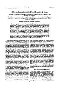
also examined the effects of HBV with or without TNF on human genes involved in metabolic pathways. Materials and methods Plasmids. The plasmid pCXN2 was kindly provided by Professor J. Miyazaki (17). The plasmid pCXN2-HBx was prepared as previously described (18). The plasmids pCMVHBV and pHBV were gratefully received from Professor A. McLachlan (19). The reporter plasmids pIGFBP1-luc and pNF-κ B-luc were purchased from Abcam (Tokyo, Japan) and Agilent Technologies (Tokyo, Japan), respectively. Cell culture. The human hepatoma cell lines HepG2, HepG2.2.15 and Huh7, which have been used in our laboratory (16,20), and the immortalized human hepatocyte (IHH) cell line (kindly provided by Professor R. Ray) (21) were cultured in Dulbecco's modified Eagle's medium (DMEM) (Sigma, St. Louis, MO, USA) supplemented with 10% fetal calf serum (FCS) at 37˚C and 5% CO2. PXB cells, fresh hepatocyte cells from chimeric mice with transplanted human hepatocytes, were purchased from PhoenixBio Co., Ltd. (Higashihiroshima, Japan) (22). PXB cells were infected with HBV genotype C strain as previously described (22). RNA extraction, cDNA synthesis and PCR array. HepG2 and HepG2.2.15. cells with or without 0.1 µg/ml TNF were seeded into 6-well plates. After 48 h, total cellular RNA was extracted using the RNeasy Mini kit (Qiagen, Hilden, Germany) according to the manufacturer's instructions. RNA samples were eluted in 60 µl elution buffer and quantified using a NanoDrop Lite spectrophotometer (Thermo Fisher Scientific, Madison, WI, USA), and were then stored at -80˚C until use. cDNA was synthesized from 25 ng total RNA using the SABiosciences RT2 First Strand kit (Qiagen) according to the manufacturer's instructions. Following a denaturation step at 42˚C for 5 min, RNA was reverse transcribed to single‑stranded cDNA using RT Enzyme Mix provided by the RT2 First Strand kit (Qiagen). The reverse transcription reaction was performed in a total volume of 20 µl at 42˚C for 15 min and 95˚C for 5 min. For the purpose of determining the expression of mRNAs associated with metabolic-related gene expression, quantitative PCR was performed using RT2 SYBR‑Green/ROX qPCR Master mix (PAHS-157Z, RT2 Profiler™ PCR Array Human Fatty Liver, Qiagen). The PCR array combines the quantitative performance of SYBR‑Green‑based real-time PCR with the multiple gene profiling capabilities of a microarray. Ninety-six-well plates containing gene-specific primer sets for 84 metabolic‑related genes, 5 housekeeping genes and 2 negative controls were used. After performing thermal cycling, amplification data were gathered using the ABI 7300 instrument for RT2 Profiler™ PCR arrays (Qiagen). Gene expression was normalized to internal controls (housekeeping genes) to determine the fold change in gene expression between the test sample (HepG2.2.15 with or without TNF) and the control sample (HepG2 with or without TNF) using the ∆∆CT (comparative cycle threshold) method (23). Transfection and reporter assay. Approximately 1.0x10⁵ cells were seeded into 6-well plates (AGC Techno Glass, Shizuoka,
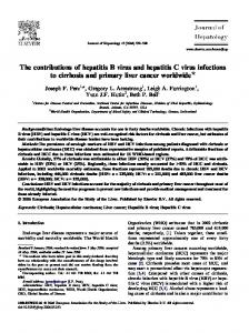
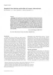
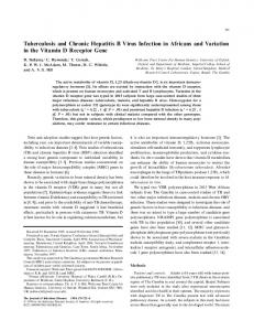
Japan) 24 h prior to transfection. For the purpose of detecting the relative activity of NF-κ B in Huh7 and IHH cells, the cells were co-transfected with 0.3 µg pHBV/pCXN2‑HBx and 0.1 µg pNF-κB-luc, respectively in Effectene transfection reagent (Qiagen). The cells were treated for 24 h with 10 ng/ml TNF-α and/or 10 ng/ml IL-1β, with or without 1 µM MG132 (Z-Leu‑Leu‑Leu-al; Sigma‑Aldrich) after transfection. For detecting the relative activity of IGFBP1‑promoter in HepG2 cells, the cells were cotransfected with 0.2 µg pCXN2, pCXN2HBX, pCMVHBV or pHBV and 0.1 µg pIGFBP1-luc in Effectene transfection reagent (Qiagen). At 30 h post-transfection, the cells were lysed with reporter lysis buffer (Promega, Madison, WI, USA) and the luciferase activities were determined using a luminometer (Luminescencer-JNR II AB-2300; ATTO Bio Instrument, Tokyo, Japan) as described previously (23). Antibodies and western blot analysis. IL-1R2 (ab97388) was purchased from Abcam. TNF-R1 (H-5) (sc-8436), TNF-R2 (D-2) (sc-8041), IL-1R1 (N-20) (sc-688), XBP1 (M-186) (sc-7160), IGFBP1 (H-120) (sc-13097), GAPDH (4G5) (sc-51906) were all purchased from Santa Cruz Biotechnology, Inc. (Santa Cruz, CA, USA). The cells were seeded into 6-well plates and cell lysates were prepared after 48 h using 50 µl sodium dodecyl sulfate sample buffer. After sonication, the lysed proteins were subjected to sodium dodecyl sulfate-polyacrylamide gel electrophoresis (SDS-PAGE) on 5-20% polyacrylamide gels and transferred onto polyvinylidene difluoride membranes (ATTO Bio Instrument) for western blot analysis. The membranes were incubated with primary specific antibodies. After washing, the membranes were incubated with secondary horse-radish peroxidase-conjugated antibodies. Signals were detected by means of enhanced chemiluminescence (GE Healthcare, Tokyo, Japan) and scanned using an image analyzer (LAS‑4000; FujiFilm, Tokyo, Japan). Statistical analysis. Results are expressed as the means ± SD. Comparisons were performed using the Student's t-test. All P-values were two-tailed, and a P-value