Snipported in part by an unirestricted grant from Researchi to Prevent ..... Several reportsat thisyear's ARVO meeting compared and defined differences between ...
CORNEAL PRESERVATION AT 40C WITH CHONDROITIN SULFATE CONTAINING MEDIUM* BY Richard L. Lindstrom, MD (BY INVITATION),
Donald J. Doughman, MD, Debra L. Skelnik, BS (BY INVITATION), AND (BY INVITATION) Elizabeth A. Mindrup,
BS
INTRODUCTION
THE FUNCTIONAL STATLTS OF THE ENDOTHELIUM AND SUSTAINED MAINTENANCE OF corneal deturgescence after corneal storage at 40C are of great clinical
importance. Limited short-term storage of corneas (4 days or less)",2 at 40C has been provided by McCarey-Kaufinan (M-K) medium and whole globe storage in moist chamber.4'5 Chondroitin sulfate, a negatively charged glycosaminoglycan, has been utilized in Japan to extend the storage and preservation of whole globes for up to 3 days.6 In addition, chondroitin sulfate containing preservation medium has significantly enhanced the quality of donor tissue and prolonged the length of storage at both 340C and 4°C in the Minnesota corneal preservation system.79 In a recent clinical study, 83 donor corneas were transplanted after 1 to 7 days storage at 4°C utilizing a commercially prepared corneal preservation medium formulation, CSM (Aurora Biologicals, Ltd), containing 1.35% chondroitin sulfate. Excellent endothelial cell preservation with up to 12 months postkeratoplasty follow-up was observed with no primary donor failures.") Clinical studies with K-Soll' (Cooper-Cilco, Inc), a 4°C corneal storage medium containing 2.5% chondroitin sulfate have also demonstrated successful transplantation after up to 14 days storage. 12 However, in a study by Bourne,13 significantly greater endothelial cell
*Fromn the Departmienit of Ophthalmology, University of Mlinn1esota. Miinnieapolis, Ninn1iesota. Snipported in part by an unirestricted grant from Researchi to Prevent Blindness, Inc.
TR. ANm. OPITH. Soc. vol. LXXXV, 1987
CQrneal Preservation
333
loss was found in patients whose corneas were stored in K-Sol at 4°C for more than 7 days with two primary donor failures reported. Corneal storage media are a composite of balanced salts, amino acids, energy sources, antioxidants, buffering agents, cell membrane stabilizers, deturgescents, and antibiotics. The lower temperature of the 4°C storage method reduces the metabolic rate of the cornea, but the storage medium must still support the basal requirements of the cornea. Temperature reduction changes membrane lipids, proteins, and water structures, each of which could alter barriers hindering the ease of passive diffusion, carrier-substrate interaction and energy coupling relationships of the active transport mechanism. 14 Thus, disturbances in membrane function as well as morphological and biochemical alterations may be of greater consequence at this temperature as a direct result of the lower metabolic rate. Therefore, critical evaluation of physiologic parameters such as ionic and amino acid composition, bicarbonate equilibrium, available energy sources, dissolved oxygen levels, osmolality and pH should be observed with respect to each preservation medium. Parameters for extended 4°C storage should be defined as to the reversibility of cell damage incurred during storage. We, therefore, designed a study to assess these two commercial 4°C storage media, CSM and K-Sol, analyzing length of storage time and ability to support corneal endothelial cell viability. MATERIALS AND METHODS
Sixteen pairs of human research corneas were obtained from the Lions Eye Bank of Washington and Northern Idaho for this comparative study of 4°C media. Paired human donor eyes, unsuitable for transplantation because of donor age or disease, were removed within 8 hours of death. Whole globes were decontaminated by a vigorous irrigation with sterile balanced salt solution. Excess conjunctival tissue was trimmed and the sclera was scored with a number 11 Bard-Parker blade 2 to 3 mm from the limbal area (360 degrees). The cornea was excised by making an initial incision onto the sclera to the suprachoroidal space, with the remainder of the corneal rim being excised with a corneal scissors. Corneoscleral rims were removed and placed endothelial side up in 20 ml of commercial CSM or K-Sol medium in a random fashion, and transported by automobile and commercial airlines surrounded by ice to the Minnesota Lions Eye Bank research laboratory. Four paired corneas were stored in each medium at 4°C for four different time intervals: 7, 10, 14, and 22 days. After 4°C storage, corneas were equilibrated for 2 hours at 34°C in closed
334
Linclstrom
bottles of test medium and then evaluated. The corneas were evaluated in a masked fashion following removal from unlabeled storage bottles. Corneal thickness measurements were made using a Leitz upright microscope fitted with a micrometer. The micrometer dial indicator was attached to the microscope stand above the stage with the set screw placement through the stage, directly under the foot of the dial indicator. The corneal thickness measurement involved focusing on the endothelium, setting the set screw to bring the dial micrometer to "zero", raising the stage to bring the epithelium into focus, and recording the dial indicator reading. Three thickness readings were made at two different central corneal sites to determine the mean corneal thickness. Endothelial cell viability was determined after staining the corneas with 0.1% trypan blue, whereas cell membrane integrity and areas of denuded endothelium were assessed after staining with alizarin red S. Areas of damaged or denuded endothelium were quantitated by computer assisted digitization and analysis. Central endothelial cell counts were determined from high power magnification (250 x) photographs. One way analysis of variance and the Newman-Keuls multiple range test were used to evaluate statistical significance (P < 0.05) between the two test groups. Evaluations of the medium components were conducted on sealed bottles of media as received from the manufacturers. Electrolyte analysis of Na+, K+, Cl-, Ca2 , glucose determinations, HCO3, pCO2, P02, and pH values were conducted by the Department of Laboratory Medicine and Pathology at the University of Minnesota under standard laboratory conditions. HCO3-, pCO2, P02, and pH values were determined at 34°C. The osmolality of the media was determined using a Precision Systems freezing point depression osmometer. Gentamycin concentrations were (quantitated by the Department of Toxicology at the University of MNinnesota, emploving a fluorescenice polarization immunoassav. RESULTS CORNEAL THICKNESS
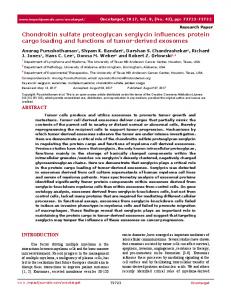
The mean (±+ SD) corneal thickness for corneas stored in CSM and K-Sol for 7, 10, and 14 days is presented in Fig 1. No statistical difference in corneal thickness (P < 0.05) was seen at 7, 10, or 14 days storage between CSM and K-Sol. K-Sol corneas at 7 days were thinner (463 + 27 ,um) than the CSM stored corneas (501 ± 42 ,um) at the same time period. K-Sol stored corneas increased in corneal thickness with time at 10 days (513 ±
Corneal Preservation
335
700 -/X
CsM
600 ::
L
K-So-
500
300 *0 400- 00
07
10
14
Days Storage at 40 C FIGURE 1
Mean corneal thickness of corneas stored in CSM or K-Sol at 4°C for 1, 10, and 14 days.
14 ,um) and 14 days (520 ± 26 ,um) storage. Corneas stored in CSM maintained similar corneal thickness throughout the 14 day storage time (10 days, 498 + 28 ,um; 14 days, 500 + 36 jim). ENDOTHELIAL CELL DAMAGE
Computerized digitization of the damaged endothelial areas allowed direct comparison of the two test media, where damaged areas were expressed as percent of total endothelial area. Endothelial cell damage was noted in the periphery of both the CSM and K-Sol corneas. Trypan blue and alizarin red S staining were present in lines radiating inwardly from the limbal edge, and are probably the result of folds or trauma to the cornea during preparation (Fig 2). The percent mean (+ SD) area of endothelial cell damage for corneas stored in CSM and K-Sol is presented in Fig 3. Although greater endothelial cell damage was consistently observed in the K-Sol stored corneas (7 days, 9.5% + 11.9%; 10 days, 9.8% + 7.4%; 14 days, 7.3% ± 6.6%) as compared to the CSM stored corneas (7 days, 2.9% ± 2.3%; 10 days, 4.9% ± 4. 1%; 14 days, 2.8% + 1. 9%) this difference was not statisticallv significant (P < 0.05) in this small series. The relative mean loss in both test groups was consistent, independent of storage time, up to 14 days.
~ ~ ~ ~ ~ ~ ~ ~ ~ ~ ~. Lindstrom
336
K-Sol
CSM
7 Days Storage
...
....
........~ ~~~~~~~~~~~~~~~~~~~~~~~~~~~~...
10 Days Storage
j_.
/_ 14 Days Storage
14 Days Storage
(epresentative Digitized Corneas) Mean Area of Damage: i.2 -
Mean Area of Damage
.FIGURE
6-30/
2
Trvpani blue staining of corneas stored in CSM
or
K-Sol at 40C for 7, 10, and 14 days.
Corneal Preservation
337 CSM
Eli K-Sol
E
j
]
as 20
0
0
0
10 Days Storage at 40 C
7
14
FIGURE 3
Computer digitization of area of endothelial cell damage of corneas stored in CSM or K-Sol at 4°C for 7, 10, and 14 days.
4000r-
I//A7
3500 E
300 _-
co
2500k
E E
K-Sol
2000C-
-a Z .5m0 1500CV0 c Lu
I-
TI
z 0
_
10001500 7
d
10 Days Storage at 40 C
TI /1
14
FIGURE 4
Mean central endothelial cell counts for corneas stored in CSM or K-Sol at 40C for 7, 10, and 14 days.
338
Lindstromn
FIGURE 5
Trvpan blue anid alizarini red S staininlg of endothelium of corneas stored in CSM or K-Sol at 4°C( for 7, 10, 14, and 21 days. Corneas stored in CSM at 4°C for 7, 10, 14, and 22 davs exhibit intact endothelial cell moinolayers anid mainitenianice of endothelial cell membrane integrity with distinctly stainied borders. Corneas stored in K-Sol at 4°C for 7 anid 10 days exhibit initact endothelial cell mioiolayers. At 14 days progressive reductioni in endothelial cell membrane integrity is demonistrated by alterniation in alizarin red staininlg and endothelial cell morphology. At 22 davs storage, D)escemet's membrane was devoid of cells.
Corneal Preservation
339
CENTRAL ENDOTHELIAL CELL COUNTS
Due to low temperature storage, and the lack of cell migration at this temperature, loss of endothelial cells in the periphery does not effect the central endothelial cell counts. Mean (+ SD) central endothelial cell counts are shown in Fig 4. CSM stored corneas had central endothelial cell counts of 2394 + 41, 2360 + 427, and 2390 + 227 cells/mm2 at 7, 10, and 14 days, respectively. Similarly K-Sol stored corneas had endothelial cell counts of 2177 ± 329, 2414 + 582, 2085 + 235 cells/mm2 for the same time periods. While the endothelial cell count at 14 days was 13% lower in K-Sol, no statistical differences (P < 0.05) in central endothelial cell counts were found at 7, 10, or 14 days storage in CSM and K-Sol. At 22 days storage the corneal endothelial monolayer was preserved in CSM (2140 + 296 cells/mm2), whereas all K-Sol corneas demonstrated virtually total cell death. EPITHELIAL AND ENDOTHELIAL CELL MORPHOLOGY
Epithelial cell layers appeared more edematous in corneas stored in CSM, and sloughing of epithelium was observed at 2 to 5 days. K-Sol stored corneas maintained epithelium more consistently during the first 2 to 3 days storage, but sloughing of epithelium was observed at 4 to 5 days. Microscopic evaluation of the epithelium at 7 days for both test media showed rounded, loosely adherent basal epithelium. In assessing cell morphology trypan blue staining allows a direct measurement of cell viability, while alizarin red S staining allows visualization of cell morphology and the determination of cell adhesion, tightness of intercellular spaces and gross membrane integrity. In the photographs shown in Fig 5, corneas stored in CSM at 4°C for 7, 10, 14, and 22 days exhibit intact endothelial cell monolayers and maintenance of endothelial cell mIembrane integrity with distinctly stained borders. Corneas stored in K-Sol at 4°C for 7 and 10 days exhibit intact endothelial cell monolayers. Cells in both media demonstrate slight alterations in the classic hexagonal pattern seen in vivo, appearing mildly dehydrated and exhibiting greater intercellular digitation. Wider and more irregular staining of intercellular spaces is seen more frequently with K-Sol stored corneas at 7 and 10 days storage as compared to CSM stored corneas for the same time periods. At 14 days storage in K-Sol, progressive reduction in endothelial cell membrane integrity is demonstrated by alteration in alizarin red S staining and endothelial cell morphology. At 22 days storage, K-Sol stored corneas exhibited large areas of centrally denuded Descemet's membrane with occasional clusters of intact cells.
340
Lindstromn
EVALUATION OF NIEDIUNI COMPONENTS
Components of CSM and K-Sol were compared as to their base medium and additional medium supplementation and are presented in Table I. CSM was formulated from experimental work supporting corneal preservation at 34°C for periods of up to 5 weeks.9 CSM utilizes Eagles Modified Minimal Essential Medium (MEM) with Earle's balanced salt solution and 25 mM HEPES supplemented with 1.35% chondroitin sulfate, 2 mM L-glutamine, 1 mM sodium pyruvate, 0.1 mM nonessential amino acids and an additional antioxidant. All concentrations of the MEM base medium are maintained as formulated. Sodium pyruvate is added as an alternative energy source, provided to enhance preservation of the cornea during 4°C storage. K-Sol is composed of TC-199 medium, with Hank's balanced salts and supplemented with 2.5% modified chondroitin sulfate and 50 mM HEPES. The base medium has been diluted approximately 26% with a balanced salt solution to maintain tonicity. The amino acid, vitamin, and other base medium component concentrations are similarly reduced by this dilution. This is demonstrated more clearly when we compare the chemical composition of human aqueous humor2' with CSM and K-Sol (Table II). The greatest differences in the concentration of components in K-Sol as compared to human aqueous humor are seen in reduced calcium, chloride, and bicarbonate concentrations. Most significant of these components is the low bicarbonate levels (< 4 mM/l) of K-Sol as compared to the physiologic concentration of 20.2 mM/l of bicarbonate found in aqueous humor. The concentration of components of CSM are similar to those found in human aqueous humor, with slightly higher concentrations of sodium, potassium, and glucose. Dissolved oxygen levels were similar for CSM (170 mm Hg) and K-Sol (168 mm Hg). The mean (±+ SD) gentamycin concentrations evaluated for CSM and K-Sol were 67.3 + 0.7 mg/l and 69.6 + 3.7 mg/l. The osmolality of CSM was 368 ± 3 mOsm as compared with 322 ± 4 mOsm for K-Sol. The pH of CSM and K-Sol was measured at 7.09 ± 0.1 and 7.31 ± 0.1, respectively. DISCUSSION
The ability of the cornea to maintain a relatively dehydrated state is essential to the maintenance of corneal transparency. Deturgescence is an energy dependent phenomenon performed primarily by the endothelial cells.22-24 The lower temperatures of 4°C storage methods reduce the rate of metabolic functions of the cornea, but still require the storage medium
Corneal Preservation
341
TABLE I: COMPARISON OF 4'C CORNEAL STORAGE MEDIA
MEDIUM CONIPONENTS
Base medium Chondroitin sulfate (%) L-Glutamine (mM) Sodium pyruvate (mM) Nonessential amino acids (mM) HEPES buffer (mM) Gentamycin (mg/i) Osmolality (mOsm) P02 (mm Hg)
CSM*
K-Sol
MEM 1.35 2.0 1.0
TC-199 2.5 (modified CS) 0 0
0.1
0
25.0 67.3 368
50.0 69.6 322
170 64
168 7
pCO2 (mm Hg)
*Additional antioxidant added. TABLE II: CHEMICAL COMPOSITION OF HUMAN AQUEOUS. CSM AND K-SOL HUMAN AQUEOUS
CONSTITUENT Sodium Potassium Chloride Glucose Bicarbonate Calcium pH
Osmolality
HUMOR (mM/I) 163 2.2-3.9 132 2.7-3.7 20.2 1.8 7.38 332
(mM/l) 206 5.3 132 5.2 19 1.8
CSM
7.09 368
(mM/l) 206 4.2 108 3.9