Coronary Artery Bypass Grafting Using the Radial Artery: Midterm Results in a Japanese Institute Atsushi Amano, MD, Hitoshi Hirose, MD, Akihito Takahashi, MD, and Naoko Nagano, MD Department of Cardiovascular Surgery, Shin-Tokyo Hospital, and Department of Cardiovascular Surgery, Kobari General Hospital, Chiba, Japan
Background. To avoid remote cardiac events associated with graft occlusions, arterial conduits are being increasingly utilized in coronary artery bypass grafting (CABG). The development of antispasmic agents has enabled the use of the radial artery as a graft conduit in CABG. Methods. Between December 1995 and December 1998, 920 consecutive isolated CABG operations were performed at Shin-Tokyo Hospital. The radial artery was used for graft conduits in 475 of these patients, and their data were analyzed in this study. The patients were followed to determine midterm graft patency, cardiac events, and survival. All data are given as mean ⴞ standard deviation. The end points were patient death or occurrence of cardiac events. Results. The radial artery was used in 475 patients (366 males and 109 females, with a mean age of 64.5 ⴞ 8.5 years). The left internal mammary artery was used in 94.9% of patients, the right internal mammary artery in 17.5%, the gastroepiploic artery in 50.9%, the inferior epigastric artery in 0.2%, and the saphenous vein in 39.2%. The in-hospital morbidity and mortality rates of
the studied group were 12.8% and 0.6%, respectively. A major complication related to radial artery harvesting, compartment syndrome of the arm due to postoperative bleeding, was observed in 1 patient. No postoperative myocardial infarction attributable to radial artery bypass was observed. During the late follow-up period of 3.5 ⴞ 0.9 years, cardiac events were observed in 63 patients, giving actuarial 2- and 3-year event-free rates of 92.8% and 89.6%, respectively. A total of 24 late deaths were noted, including seven cardiac deaths, giving actuarial 2- and 3-year survival rates of 98.1% and 97.2%, respectively. Postoperative angiography was performed in selected patients. The cumulative graft patency rate of the radial artery was 93.0% during the mean angiographical follow-up period of 1.5 ⴞ 1.1 years. Conclusions. No adverse effects were noted after CABG using a radial artery graft in this short- and midterm follow-up period.
T
dure. In this study, we carried out a retrospective analysis to confirm the safety of performing radial artery grafting and to determine midterm graft patency, cardiac events, and survival rates.
o avoid remote cardiac events associated with graft occlusions, which are known to occur more frequently in saphenous vein bypasses, arterial conduits are being increasingly utilized in coronary artery bypass grafting (CABG). The internal mammary arteries (IMAs) are the most frequently used, and their reported patency rates have been reported to be 90% or better even 10 years after CABG [1, 2]. Due to the anatomical location of IMAs and their excellent patency, they are normally used for bypass to the left anterior descending artery (LAD). The second choice for arterial conduits is controversial: the gastroepiploic artery (GEA) is often used as an in situ graft to the distal right coronary artery (RCA) [3], and since Acar and associates reported excellent outcomes in patients undergoing radial artery grafting in 1992 [4], the radial artery has also become more commonly utilized in CABG. At our institute, the technique of radial artery grafting was adopted in December 1995, and by the end of 1998, a total of 475 patients had undergone the proceAccepted for publication April 5, 2001. Address reprint requests to Dr Amano, Department of Cardiovascular Surgery, Shin-Tokyo Hospital, 473-1 Nemoto, Matsuda City, Chiba 2710077, Japan; e-mail:
[email protected].
© 2001 by The Society of Thoracic Surgeons Published by Elsevier Science Inc
(Ann Thorac Surg 2001;72:120 –5) © 2001 by The Society of Thoracic Surgeons
Patients and Methods Between December 1995 and December 1998, a total of 920 consecutive patients underwent isolated CABG at Shin-Tokyo Hospital. Those who underwent valvular surgery, left ventricular surgery, or additional general or vascular surgery at the time of CABG were excluded; thus, a total of 475 patients (51.6%) who underwent radial artery grafting were included in this study. The techniques of radial artery bypass are described elsewhere [5]. Briefly, preoperative exclusion criteria for radial artery harvesting were positive Allen’s test, serum creatinine greater than 1.8, and significant radial artery stenosis as revealed by Doppler ultrasound. Radial artery harvesting was performed using the standard technique described by Reyes and associates [6]. Prophylaxis of vasospasm was achieved by intravenous administration of calcium channel blockers such as diltiazem or nicorandil during the operation, as well as local spraying 0003-4975/01/$20.00 PII S0003-4975(01)02706-0
Ann Thorac Surg 2001;72:120 –5
with diluted papaverine (1:10). The harvested radial artery was preserved by soaking in warm papaverine solution. Shortly before anastomosis, the harvested pedicle was cannulated and systemic arterial pressure was applied in order to facilitate the release of spasm and to achieve further hemostasis. The radial artery was used as an aortocoronary bypass or as a composite Y-graft. When the radial artery was used as a composite graft, proximal anastomosis between the donor artery and the radial artery was first performed, followed by distal anastomosis onto the target coronary artery. The inflow conduit of the composite Y-graft was usually the left internal mammary artery (LIMA). Other graft conduits used with the radial artery were the LIMA, usually bypassed onto the LAD, the right internal mammary artery (RIMA) bypassed onto the proximal RCA or LAD, the GEA onto the distal RCA, and the saphenous vein onto the RCA or circumflex artery if arterial grafts were not available. Thus, the primary target of the radial artery was the circumflex artery (CX). CABG was performed under normothermia (36°C) with cardiopulmonary bypass. After September 1996, offpump CABG was adopted and selected cases were referred to off-pump CABG under beating-heart conditions [7]. Postoperative angiography was performed in selected patients. The first 50 patients systematically underwent coronary angiography within 3 months after surgery, and thereafter, early coronary angiography was performed either due to the poor quality of native coronary arteries or at the request of the referring cardiologists. Late coronary angiography was proposed for all patients followed at our institute; but was only performed with the patient’s permission. Follow-up angiography was strongly recommended for all patients with symptoms of angina. In addition, symptom-free patients underwent angiography at various times because the majority of patients (83%) were followed by referring physicians and were not under our direct observation. By retrospective chart review, the following parameters were collected: patients’ age, gender, results of preoperative angiography, cardiac profiles, preoperative risk factors, graft material, surgical data, postoperative complications, and mortality. Outpatient follow-up was completed by the referring cardiologists or hospital outpatient clinic. Any cardiac events after discharge from hospital, including myocardial infarction, angina, arrhythmia requiring hospitalization, congestive heart failure requiring hospitalization, native coronary artery or graft stenosis requiring any type of coronary intervention, and sudden death were counted as a cardiac events. These follow-up data were compiled by January 31, 2001. The end points were patient death or the occurrence of cardiac events. Results are expressed as mean ⫾ standard deviation. Postoperative patient survival, event-free rate, and longterm graft patency were calculated using the KaplanMeier method. All statistical analyses were performed using Statview version 5.0 (SAS Institute, Cary, NC).
AMANO ET AL CABG USING THE RADIAL ARTERY
121
Table 1. Preoperative Patient Demographics Number of patients using the n ⫽ 475 radial artery Clinical characteristics Age (years) 64.5 ⫾ 8.5 (34 – 86) Age over 75 49 10.3% Female 109 22.9% Cardiac profile Unstable angina 69 14.5% Acute myocardial infarction 21 4.4% Previous myocardial infarction 323 68.0% History of congestive heart failure 47 9.9% Poor ejection function (⬍ 40%) 31 6.5% Atrial fibrillation 13 2.7% Redo surgery 15 3.2% Emergency surgery 36 7.6% Preoperative IABP 25 5.3% Cardiogenic shock 5 1.1% Angiographic profile Left main disease 149 31.4% Number of diseased vessels 2.8 ⫾ 0.5 (1–3) Three-vessel disease 370 77.9% Coronary risk factors Hypertension 230 48.4% Diabetes 194 40.8% Insulin user 36 7.6% Hyperlipidemia 196 41.3% Smoking history 237 49.9% Obesity 17 3.6% Family history 59 12.4% Comorbidity Cerebral vascular accident 42 8.8% Calcified ascending aorta 40 8.4% Peripheral vascular disease 16 3.4% Chronic pulmonary obstructive disease 21 4.4%
Results Patients Demographics A total of 475 patients were included in this study (366 males and 109 females, with a mean age of 64.5 ⫾ 8.5 years). The demographic details of the patients are given in Table 1.
Operative Results Operative data are shown in Table 2. The mean number of distal anastomoses in this study group was 3.6 ⫾ 1.1. The LIMA, RIMA, GEA, inferior epigastric artery, and saphenous vein graft were additionally used in 94.9%, 17.5%, 50.9%, 0.2%, and 39.2%, respectively. Unexpected intraoperative radial artery atherosclerosis or calcification was found in nine cases (1.9%), and in these cases, other graft conduits were used instead. The distal anastomoses of the radial artery grafts were the CX (63.6%), the diagonal artery (19.1%), the RCA (15.7%), and the LAD (1.6%). Aortocoronary bypass using the radial artery was performed in 320 patients (67.4%), and composite Y-grafts were performed in 242 patients (50.9%). All composite Y-grafts were made using the LIMA. In 11
122
AMANO ET AL CABG USING THE RADIAL ARTERY
Ann Thorac Surg 2001;72:120 –5
patients, the radial artery was used for graft elongation purposes. Since the introduction of the radial artery bypass, total arterial bypass became the more common procedure at our institute: 23.3% before November 1995, rising to 57.3% after December 1995.
In-Hospital Results Postoperative courses are listed in Table 3. Morbidity and mortality rates were 12.8% and 0.6%, respectively. There was one major complication associated with radial artery harvesting, which was compartment syndrome of the forearm due to postoperative bleeding, requiring fasciotomy and the revision of the hemostasis. There were an additional four minor radial wound complications, including delayed healing and superficial wound infection treated by local wound care. There were no permanent sensory or motor deficits observed related to graft harvesting. No postoperative myocardial infarctions attributable to radial artery bypass were observed.
Remote Results Among the survivors, postoperative follow-up was completed in 459 patients (97.2%) with a mean follow-up period of 3.5 ⫾ 0.9 years. During this follow-up period cardiac events occurred in 63 patients (13.4%), which including angina recurrence in 33, congestive heart fail-
Table 2. Surgical Results of 475 Patients Undergoing Radial Artery Grafting n ⫽ 475 Number of distal anastomosis Aorta clamp time (minutes) Pump time (minutes) Off-pump CABG Average coronary clamp time (off-pump CABG) Operation time (minutes) Blood transfusion Packed red blood cells (units) Fresh-frozen plasma (units) Platelet (units) Summary of grafts used Left internal mammary artery Right internal mammary artery Radial artery AC bypass Composite Y-grafts Gastroepiploic artery Inferior epigastric artery Saphenous vein Total arterial bypass Implantation site of the radial artery graft Left anterior descending artery Diagonal artery Circumflex artery Right coronary artery CABG ⫽ coronary artery bypass graft.
3.6 ⫾ 1.1 67.8 ⫾ 21.4 102.2 ⫾ 29.5 14 16.2 ⫾ 9.1
(1– 8) (26 –158) (42–258) 2.9% (9 – 45)
319.0 ⫾ 76.9 85 4.3 ⫾ 1.8 18.5 ⫾ 4.9 15.6 ⫾ 5.7
(180 – 805) 17.9% (2–14) (10 –30) (10 –30)
451 83 475 320 145 242 1 186 289
94.9% 17.5% 100.0% 67.4% 30.5% 50.9% 0.2% 39.2% 60.8%
1.6% 19.1% 63.6% 15.7%
Table 3. Postoperative Data n ⫽ 475 Intubation (hours) ICU stay (days) Postop stay (days) Major complication (patients) Low-output syndrome Postoperative myocardial infarction Cerebral vascular accident Mediastinitis Reexploration for bleeding Pneumonia Respiratory failure Postop dialysis In hospital death
11.9 ⫾ 21.3 2.9 ⫾ 3.0 16.9 ⫾ 8.5 61 9 6 9 1 4 7 12 1 3
(1–264) (1–53) (6 – 83) 12.8% 1.9% 1.3% 1.9% 0.2% 0.8% 1.5% 2.5% 0.2% 0.6%
Some patients may have multiple complications.
ure in 10, percutaneous transluminal coronary angioplasty in 12, arrhythmia required hospitalization in 4, and sudden death in 4. Actuarial 1-, 2-, 3- and 4-year eventfree rates were 97.2%, 92.8%, 89.6%, and 85.8%, respectively. During the same follow-up period, there were 24 deaths (5.1%), including seven cardiac deaths. Actuarial 1-, 2-, 3-, and 4-year survival rates after surgery were 99.6%, 98.1%, 97.2%, and 93.7%, respectively.
Angiographic Study Early (within 3 months after surgery) postoperative angiography was performed in 98 patients (Table 4), which revealed two radial artery occlusions; one was a freeradial artery aortocoronary bypass to the CX, and the other was composite Y-grafting to the LCX. All patients with early graft failures were medically managed and no reoperations were performed. A total of 167 patients (36.4%) underwent late coronary angiography (more than 3 months after surgery), and revealed an additional 16 radial anastomotic occlusions. Nine occlusions were identified in composite radial artery grafts using the LIMA, and seven in aortocoronary bypasses using freeradial artery grafts. There were no significant differences in terms of composite or free radial graft occlusions. Among the patients who underwent late angiography, 26 patients had symptomatic angina and the other 141 patients were symptom free. Occlusion of the radial artery graft was related to the occurrence of angina in four cases (two composite grafts and two free grafts), and these patients were successfully managed by catheter interventions. The cumulative graft patency rate of the radial artery was 93.0% at the mean angiographical follow-up period of 1.5 ⫾ 1.1 years. The calculated patency rates of radial artery grafting at 1, 2, and 3 years were 98.2%, 91.0%, and 86.2%, respectively. String sign, which was defined as a severe and extensive narrowing of the whole body of the graft [4], was documented in a total of 13 radial artery grafts: five by early angiography and an additional eight by late angiography. No patients with string sign had symptomatic angina. Stenosis of the anastomosis was observed in a total of
Ann Thorac Surg 2001;72:120 –5
AMANO ET AL CABG USING THE RADIAL ARTERY
123
Table 4. Results of Postoperative Angiography Examined by Angiography
Angiography Performed Within 3 Months After Surgery
Angiography Performed Beyond 3 Months After Surgery
98 patients (20.8%) 354 anastomosis Within 3 month
167 patients (36.4%) 587 1.5 ⫾ 1.1 years
99/100 (99.0%) 27/27 (100.0%) 137/139 (98.6%) 96/97 (99.0%) 41/42 (97.6%) 48/50 (96.0%) 34/38 (89.5%) 5/98 (5.1%) 7/98 (7.1%)
168/171 (98.2%) 27/27 (100.0%) 213/229 (93.0%) 142/149 (95.3%) 71/80 (88.8%) 75/82 (91.4%) 71/79 (89.8%) 8/167 (4.8%) 6/167 (3.6%)
Number of patients Number of distal anastomosis Timing of angiography Distal anastomosis patency Left internal mammary artery Right internal mammary artery Radial artery AC bypass Composite graft Gastroepiploic artery Saphenous vein Radial artery string sign (patients) Radial artery stenosis at anastomosis (patients)
11 patients: 7 by early angiography and an additional 6 by late angiography.
Comment Coronary artery grafting using the radial artery was first described by Carpentier and associates in 1973. Since the initial results were poor, Carpentier concluded that the radial artery should have not been used due to the high frequency of early graft occlusion [8]. These unsuccessful results were mostly due to technical problems occurring while harvesting the radial artery and a lack of knowledge of the mechanism of vasospasm. Histologically, the radial artery is classified as a muscular artery, which is known to be liable to vasospasm [9]. To avoid graft injury, metal clips are utilized instead of electrocautery to maintain homeostasis. Recently, Harmonic scalpels (Ethicon Inc, Somerville, NJ) have been utilized at our institute to facilitate hemostasis [10]. For the prophylaxis of vasospasm, papaverine is used for local control of radial artery spasm while calcium channel blockers were administered systemically. In this study, calcium channel blockers, such as nicorandil or diltiazem, were continued postoperatively as well as intravenously and then by oral administration. We recommend their use for a minimum of 6 months after surgery to prevent remote vasospasm. Both chemical antivasospastic agents and mechanical antivasospastic care are essential to minimize the risk of radial artery vasospasm and to optimize the graft patency. In this study, the target of the radial artery graft was usually the CX, because the LAD was normally revascularized with the IMA, and the RCA with the GEA. The RCA can be revascularized with the radial artery if the GEA cannot be used or if the RCA system requires revascularization with more than two grafts. The use of saphenous vein grafts was reserved only if there was a lack of arterial grafts. One of the contraindications of radial artery harvesting is poor renal function and an increased risk of the need
for hemodialysis after surgery, because the radial artery is most commonly used for the inflow of the arteriovenous fistula for hemodialysis blood access. Another contraindication for radial artery use is poor collateral supply from the ulnar artery leading to a risk of postoperative hand ischemia. However, we avoided this problem using a combination of Allen’s test and preoperative Doppler examination. Emergency surgery is not a contraindication for radial artery grafting, because vasospasm occurring in the radial artery can also be successfully resolved under our harvesting protocol. The radial artery can be used safely in diabetic patients. Unlike wounds made to the leg, the wound resulting from radial artery harvesting usually heals rapidly and the occurrence of local infections is rare. Sensory deficit after radial artery harvesting usually due to superficial radial nerve injury, which rarely occurred, is considered to be the result of technical insufficiency during the harvesting. Redo patients may benefit from radial artery grafting, because the radial artery was not used before the 1980s. In our study, minimal radial artery graft-related complications were observed. No postoperative myocardial infarction attributable to the radial artery bypass was noted. One patient experienced unexpected postoperative renal failure requiring permanent dialysis access via a fistula created in the right arm. We experienced one case of compartment syndrome at the radial artery harvesting site. The wound was closed without any sign of the infection after evacuation of the hematoma and hemostasis of the bleeding vessel. The radial artery graft was examined by angiography in selected patients. Although the angiographical followup period (1.5 ⫾ 1.1 years) was relatively short, the graft patency was excellent. The calculated radial artery patency rates were 91.0% at 2 years and 86.2% at 3 years. These patency rates compared well with the patency of the other arterial graft conduits, such as the IMA or GEA. The longest angiographical follow-up of the radial artery was performed by Acar and associates, who reported radial artery grafts patency rates of 99% in the postoper-
124
AMANO ET AL CABG USING THE RADIAL ARTERY
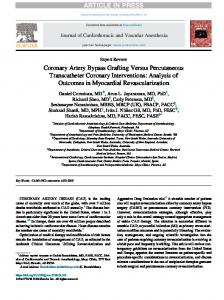
Fig 1. Flow competition with the native coronary artery. The right coronary artery (RCA) shows multiple but mild stenosis (left) and the radial artery (RA) has been grafted onto the posterior descending artery of the RCA (right). The distal flow is mainly provided by the native coronary artery. The grafted radial artery has poor graft flow and is showing a string sign.
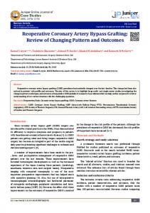
ative period, 92% at 1 year, and 83% at 5 years [11]. A recent review of radial artery grafting showed early patency rates of between 95.7% and 100%, and midterm patency rates of between 87.5% and 100% [12]. String sign occurs relatively frequently in muscular arterial conduits such as the radial artery, the GEA, and the inferior epigastric artery. Possati and associates reported that string sign of the radial artery was observed at a rate of 3.3% in his series of patients [13], which is in close agreement with our results. These string signs were observed under low-graft flow conditions, which resulted from flow competition between the graft and the native coronary artery [14]. Flow competition with the native coronary artery can also be observed in high-flow coronary artery with/without mild coronary stenosis (Fig 1). Another type of flow competition can develop among the composite grafts (Fig 2). If the vascular bed allotted to the radial artery graft is smaller than the territory of the composite IMA graft, and if the IMA requires higher runoff than the radial artery, the graft flow in the radial artery falls off. Furthermore, due to the greater vascular resistance in the radial artery than in the IMA, the low-flow state, angiographically called string sign, of the
Ann Thorac Surg 2001;72:120 –5
composite radial artery may lead to a no-flow condition or even total occlusion. Although low-flow volume is rarely related to late graft occlusion in IMA grafts [15], poor flow through the radial artery may result in graft failure. Thus, if poor runoff is expected, the composite radial artery graft should not be used and a free aortocoronary bypass should be considered. Free aortocoronary bypass using an adequately prepared radial artery can provide greater distal flow capacity than the IMA or the GEA [16]. Vein grafts also can provide high flow rates in the immediate postoperative period; however, vein graft disease may become evident 3 to 5 years after surgery. Intimal hyperplasia and atherosclerotic change of the graft has been reported to develop at the site of the valve and lead to venous graft failure [17]. The radial artery, which, unlike the saphenous vein, has no valves, will provide a better long-term patency rate than the saphenous vein graft. The best-case scenario for radial artery graft placement is bypassing of an occluded or tightly stenosed coronary artery with a large perfusion territory; however, the radial artery can be used in various locations in the manner of a composite or free graft. Good graft patency was reflected in the small number of patients experiencing remote cardiac events. Although the follow-up period was only 3.5 ⫾ 0.9 years, the calculated 3- and 4-year event-free rates were 89.6% and 85.8%, respectively. Remote death was observed in only 5.1% of our patients, and the calculated 3- and 4-year survival rates were 97.2% and 93.7%, respectively. These were similar to the results reported in a previous study by Acar and associates, who reported a 5-year event-free rate of 88.7% and a 5-year survival rate of 91.6% [11]. No adverse effects were noted after CABG using the radial artery in our midterm follow-up, and the patency of the radial artery graft was similar to other arterial grafts. The use of radial artery bypass contributed to increasing the chance of total arterial revascularization and to decreasing the occurrence of remote cardiac events or cardiac deaths.
References
Fig 2. Flow competition between the arms of the Y-graft. The distal perfusion territory of the radial artery (RA) is smaller than that of the left internal mammary artery (LIMA). The graft flow through the RA is smaller than the flow through the LIMA, and the RA is showing a string sign.
1. Reardon MJ, Conklin LD, Reardon PR, Baldwin JC. Coronary artery bypass conduits: review of current status. J Cardiovasc Surg (Torino) 1997;38:201–9. 2. Barner HB. Arterial grafting: techniques and conduits. Ann Thorac Surg 1998;66(Suppl):2–5. 3. Suma H, Amano A, Horii T, Kigawa I, Fukuda S, Wanibuchi Y. Gastroepiploic artery graft in 400 patients. Eur J Cardiothorac Surg 1996;10:6–11. 4. Acar C, Jebara VA, Portoghese M, et al. Revival of the radial artery for coronary artery bypass grafting. Ann Thorac Surg 1992;54:652– 60. 5. Yoshida S, Amano A, Egami J, Sunami H, Takahashi A. Early results of coronary artery bypass grafting using radial artery grafts. Nippon Kyobu Geka Gakkai Zasshi [in Japanese] 1998;45:166–7. 6. Reyes AT, Frame RN, Brodman RF. Technique for harvesting the radial artery as a coronary artery bypass graft. Ann Thorac Surg 1995;59:118–26. 7. Amano A, Hirose H, Takahashi A, Nagano N. Off-pump
Ann Thorac Surg 2001;72:120 –5
8.
9. 10. 11.
12.
coronary arterial bypass: midterm results. Jpn J Thorac Cardiovasc Surg 2001;49:67–78. Carpentier A, Guermonprez JL, Deloche A, Frechette C, Du Bost C. The aorta-to-coronary radial artery bypass graft. A technique avoiding pathological changes in grafts. Ann Thorac Surg 1973;16:111–21. Reardon MJ, Conklin LD, Reardon PR, Baldwin JC. Coronary artery bypass conduits: review of current status. J Cardiovasc Surg (Torino) 1997;38:201–9. Isomura T, Suma H, Sato T, Horii T. Use of the Harmonic Scalpel for harvesting arterial conduits in coronary artery bypass. Eur J Cardiothorac Surg 1998;14:101–3. Acar C, Ramsheyi A, Pagny JY, et al. The radial artery for coronary artery bypass grafting: clinical and angiographic results at five years. J Thorac Cardiovasc Surg 1998;116: 981–9. Parolari A, Rubini P, Alamanni F, et al. The radial artery:
AMANO ET AL CABG USING THE RADIAL ARTERY
13.
14.
15. 16. 17.
125
which place in coronary operation? Ann Thorac Surg 2000; 69:1288–94. Possati G, Gaudino M, Alessandrini F, et al. Midterm clinical and angiographic results of radial artery grafts used for myocardial revascularization. J Thorac Cardiovasc Surg 1998;116:1015–21. Calafiore AM, Di Giammarco G, Teodori G, et al. Radial artery and inferior epigastric artery in composite grafts: improved midterm angiographic results. Ann Thorac Surg 1995:60:517–24. Cooper GJ, Underwood MJ, Deverall PB. Arterial and venous conduits for coronary artery bypass. A current review. Eur J Cardiothorac Surg 1996;10:129– 40. Barner HB. Arterial grafting: techniques and conduits. Ann Thorac Surg 1998;66:S2–5. Mills NL, Everson CT. Vein graft failure. Curr Opin Cardiol 1995;10:562– 8.
REVIEW OF RECENT BOOKS Video-assisted Thoracic Surgery Edited by William S. Walker Oxford, United Kingdom, ISIS Medical Media, Ltd, 1999 250 pp, illustrated, £95 ($160) ISBN: 1 899066 09 8 Reviewed by R. Sudhir Sundaresan, MD This book on video-assisted thoracic surgery edited by William S. Walker is a welcome contribution to this field, given the recent expansion of minimal-access techniques in both general thoracic and cardiovascular surgery. This book has a fairly heavy emphasis on general thoracic surgery, with rather brief mention of video-assisted techniques in cardiovascular surgery toward the end. The book is enhanced by the early chapters that deal with the historic evolution of these techniques, along with a summary of the current status of instrumentation, imaging equipment, and strategies to equip the operating theatre for video-assisted thoracoscopic surgery.
© 2001 by The Society of Thoracic Surgeons Published by Elsevier Science Inc
In subsequent chapters, specific clinical problems and thoracoscopic approaches are outlined. In credit to this work, the background in each area is kept “to the point”, and exhaustive detail is omitted. Sufficient data from published series are provided to put the thoracoscopic approaches into appropriate context. Again, in credit to this work, a wide variety of thoracoscopic procedures are dealt with, including thoracoscopic approaches to lung volume reduction surgery, major pulmonary resections, esophageal resection, and treatment of benign esophageal disease. The illustrations and tables accompany the text very well. The operative photographs are of adequate quality, and in general are helpful. In summary, this book is a very readable and useful reference source for the practicing thoracic surgeon or for the thoracic surgical trainee. It is particularly appealing because of its ability to stay focused on the respective clinical problems, and to discuss the thoracic surgical approach succinctly. Chicago, Illinois
Ann Thorac Surg 2001;72:125 • 0003-4975/01/$20.00 PII S0003-4975(00)02381-X