NeuroImage: Clinical 3 (2013) 498–506
Contents lists available at ScienceDirect
NeuroImage: Clinical journal homepage: www.elsevier.com/locate/ynicl
Cortical motor activity and reorganization following upper-limb amputation and subsequent targeted reinnervation☆ Albert Chen a,b, Jun Yao b, Todd Kuiken c,d, Julius P.A. Dewald a,b,c,⁎ a
Department of Biomedical Engineering, Northwestern University, IL, USA Department of Physical Therapy and Human Movement Sciences, Northwestern University, IL, USA Department of Physical Medicine and Rehabilitation, Northwestern University, IL, USA d Sensory Motor Performance Program, Rehabilitation Institute of Chicago, IL, USA b c
a r t i c l e
i n f o
Article history: Received 4 August 2013 Received in revised form 23 September 2013 Accepted 1 October 2013 Available online 11 October 2013 Keywords: Cortical activity Brain reorganization Amputation Motor Targeted reinnervation Electroencephalography
a b s t r a c t Previous studies have postulated that the amount of brain reorganization following peripheral injuries may be correlated with negative symptoms or consequences. However, it is unknown whether restoring effective limb function may then be associated with further changes in the expression of this reorganization. Recently, targeted reinnervation (TR), a surgical technique that restores a direct neural connection from amputated sensorimotor nerves to new peripheral targets such as muscle, has been successfully applied to upper-limb amputees. It has been shown to be effective in restoring both peripheral motor and sensory functions via the reinnervated nerves as soon as a few months after the surgery. However, it was unclear whether TR could also restore normal cortical motor representations for control of the missing limb. To answer this question, we used high-density electroencephalography (EEG) to localize cortical activity related to cued motor tasks generated by the intact and missing limb. Using a case study of 3 upper-limb amputees, 2 of whom went through pre and post-TR experiments, we present unique quantitative evidence for the re-mapping of motor representations for the missing limb closer to their original locations following TR. This provides evidence that an effective restoration of peripheral function from TR can be linked to the return of more normal cortical expression for the missing limb. Therefore, cortical mapping may be used as a potential guide for monitoring rehabilitation following peripheral injuries. © 2013 The Authors. Published by Elsevier Inc. All rights reserved.
1. Introduction It has become well-established in the literature that changes in cortical organization often occur following injury to the peripheral nervous system. Several animal studies have demonstrated that motor and somatosensory representations of neighboring intact body parts expand into cortical areas previously devoted to an injured or missing limb (Donoghue and Sanes, 1987; Kaas, 2000; Kaas et al., 1983; Merzenich et al., 1978; Wall et al., 1986). Similar trends in cortical reorganization have been observed in humans that have sustained peripheral injuries such as upper-limb amputations (Elbert et al., 1994; Flor et al., 1995; Karl et al., 2001; Weiss et al., 2000). For example, face, lip, chin, and neck stimulations in upper-limb amputee subjects
☆ This is an open-access article distributed under the terms of the Creative Commons Attribution-NonCommercial-No Derivative Works License, which permits non-commercial use, distribution, and reproduction in any medium, provided the original author and source are credited. ⁎ Corresponding author at: Department of Physical Therapy and Human Movement Sciences, Northwestern University, IL, USA. Tel.: +1 312 908 6788; fax: +1 312 908 0741. E-mail address:
[email protected] (J.P.A. Dewald).
have been found to map to cortical areas corresponding to the missing extremity (Borsook et al., 1998; Elbert et al., 1997; Ramachandran et al., 1992). Despite numerous reports of cortical reorganization, other studies have shown that the motor representations of the missing limb persist following amputation, although not necessarily in their original areas. When voluntary executed movements of the missing limb are attempted, neuroimaging techniques such as functional magnetic resonance imaging (fMRI), positron emission tomography (PET) and magnetoencephalography (MEG) show that in general, the resulting movement representations of the missing limb may move out of the original cortical area and into neighboring areas (Giraux et al., 2001; Karl et al., 2004; Kew et al., 1994; Lotze et al., 2001). Similarly, the magnetic stimulation of motor cortical areas previously linked to the missing limb was also able to elicit contractions or movements from residual muscles adjacent to the site of amputation (Chen et al., 1998; Cohen et al., 1991; Irlbacher et al., 2002; Kew et al., 1994; Ridding and Rothwell, 1999; Roricht et al., 1999). These results beg the question of whether the persistent representation of the missing limb could return to its original area,
2213-1582/$ – see front matter © 2013 The Authors. Published by Elsevier Inc. All rights reserved. http://dx.doi.org/10.1016/j.nicl.2013.10.001
A. Chen et al. / NeuroImage: Clinical 3 (2013) 498–506
perhaps via surgical interventions that attempt to restore motor function. One such intervention is targeted reinnervation (TR), a technique that improves artificial limb function for amputees and successfully helped them to regain both motor and sensory functions related to the missing limb. In TR, the inactivated, residual sensorimotor nerves previously responsible for innervating the missing limb are surgically re-routed to alternative denervated muscle groups and skin areas over the chest or on the residual limb (Hijjawi et al., 2006; Kuiken, 2003; Kuiken et al., 2004). After a few months, new functional connections between the nerves, muscle and skin are created. The reinnervated muscles act as biological amplifiers for efferent motor command signals; surface electromyographic (EMG) signals at these new sites then provide control signals for an amputee to operate a motorized, myoelectric prosthesis (Kuiken et al., 2005). TR also returns sensations of touch, pressure, vibration and temperature for the missing limb to the skin overlying reinnervated muscles (Kuiken et al., 2007a,b). From a neural perspective, afferent traffic and efferent neural traffic are re-established for parts of the limb completely lost for months to years following amputation. The remarkable ability to completely rewire the peripheral neural connectivity of the arm and restore function of the lost limb makes TR a unique model for investigating a possible restoration of normal cortical expression in human amputees. In particular, we hypothesize that the return of new peripheral motor targets and voluntary muscle control can restore the original cortical representations for the missing limb. In this study, high-density electroencephalography (EEG) was used to investigate the locations of cortical activity related to cued motor tasks generated by the intact and missing limb. We present unique evidence for the restoration of cortical mapping that occur in the motor cortex from 2 individuals with upper-limb amputations who underwent TR. More specifically, we found that motor representations were re-mapped closer to presumed pre-amputation locations following TR. This suggests that an effective rehabilitation intervention such as TR is associated with the restoration of the cortical activity that normally controls them. Our results imply that cortical mapping can be used as a potential monitor and guide for future rehabilitation of peripheral injuries.
2. Methods 2.1. Subject selection Three subjects with upper-limb amputations were screened and selected to undergo TR at least 6 months following their injuries. All subjects suffered amputations due to trauma from automobile or electrocution accidents and had non-painful phantom limb sensations. In addition, all subjects were consistent users of upperarm prostheses — subject A1 used a cosmetic prosthesis while subjects A2 and A3 mainly used myoelectric prostheses. Information about the subjects, including amputation site and time of experimentation testing dates with respect to the date of amputation, is summarized in Table 1. Written consent was provided by each subject prior to participation in the study that was approved by the Institutional Review Board of Northwestern University and in compliance with the principles of the Declaration of Helsinki.
499
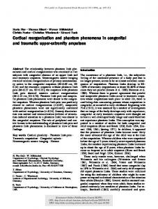
2.2. Targeted reinnervation procedures Graphical representations of each subject's TR surgical procedure are shown in Fig. 1 and are briefly described below. 2.2.1. Subject A1 — left transhumeral amputee Subject A1 was a 38 year old woman with a left transhumeral amputation due to injuries sustained in an automobile collision. During TR surgery performed 6 months after amputation (Fig. 1a), the median nerve was transferred to the denervated medial biceps brachii muscle, and the distal radial nerve was transferred to the denervated lateral triceps brachii muscle. The triceps long head and lateral biceps muscles remained intact for control of elbow extension and elbow flexion, respectively. Full muscle reinnervation took approximately 5 months. The intercostal brachial cutaneous nerve was also transected to denervate and numb the distal residual limb. Over the course of 7– 8 months, median and distal radial nerve afferents reinnervated this skin, producing sensations of her missing hand when touched at different points. For example, sensation referring to the index finger was found at the end of the residual limb near the reinnervated site for the median nerve. 2.2.2. Subject A2 — right shoulder disarticulation amputee Subject A2 is a 39 year old man who had his right arm amputated at the shoulder following an electrical accident. Four nerve transfers were made for the targeted reinnervation procedure (Fig. 1b). The musculocutaneous nerve was transferred to the clavicular head of the pectoralis major to control elbow flexion. The median nerve was transferred to the top half of the sternal head of the pectoralis major to control hand closing, while the distal radial nerve was transferred to the pectoralis minor to control hand opening/wrist extension. The ulnar nerve was also transferred to the lower half of the sternal head of the pectoralis major. Similarly to subject A1, muscle reinnervation and sensations for his missing limb returned over the course of 5– 7 months. 2.2.3. Subject A3 — bilateral shoulder disarticulation amputee Subject A3 is a 54-year old man with a bilateral arm amputation at the shoulder following electrical burn injuries. He underwent targeted reinnervation on the left side 9 months following the amputation. The remaining parts of 4 peripheral nerves were transferred to different segments of his left pectoral chest muscles (Fig. 1c). The musculocutaneous nerve was transferred to the clavicular head of the pectoralis major. The median nerve was transferred to the top half of the sternal head of the pectoralis major, while the radial nerve was transferred to the bottom half. The ulnar nerve was transferred to the pectoralis minor. Subcutaneous fat on the chest was removed to allow the muscle to come in close contact to the skin for producing stronger EMG signals. After 5 months, sensations for his missing limb returned to the skin overlying the reinnervated sites (Kuiken et al., 2007a), including feelings of pressure, temperature, and pain. 2.3. Experimental protocol In this study, cued motor tasks were used to determine motor cortical representations of the missing and intact limbs. In two subjects,
Table 1 Amputee subject information. Subject
Age
Sex
Dominant hand
Amputation site
Time from amputation to TR
Time from TR to post-TR testing
A1 A2 A3
38 39 54
F M M
R R R
Left transhumeral Right shoulder disarticulation Bilateral shoulder disarticulation
6 months 18 months 9 months*
10.5 months 18.5 months 4.5 years
Abbreviations: F = female, M = male, R = right, TR = targeted reinnervation, * = did not perform pre-TR experiments.
500
A. Chen et al. / NeuroImage: Clinical 3 (2013) 498–506
Fig. 1. Schematics of the targeted reinnervation (TR) procedures for all three subjects. (a) Subject A1 — left transhumeral amputee. (b) Subject A2 — right shoulder disarticulation amputee. (c) Subject A3 — bilateral shoulder disarticulation amputee.
these tasks were performed both pre-TR (1 week before surgery) and after TR. In subject A3 (bilateral amputee), the tasks were only performed after TR had already been performed on one side — however, he still had one side that did not undergo TR, which served to approximate a pre-TR representation for a missing limb. High-density (160 channels) EEG signals were recorded during performance of the tasks. The amputees all performed a subset of the following proximal and distal motor tasks with both sides of the body: shoulder abduction, elbow flexion or extension, and hand closing or opening. The specific motor tasks for each subject are listed in Table 2. For unilateral amputees A1 and A2, the motor tasks involved making different voluntary intended movements with both the missing limb and the intact arm. For the bilateral shoulder disarticulation amputee A3, movements were made on both sides, separately. For the missing limb, subjects were instructed to try to voluntarily execute the required movement, which differs from making a purely imaginary movement since it produces noticeable residual muscle activity. All movements were held for 5 s, and subjects were asked to make comfortably strong and consistent, but not maximal, movements. Most importantly, subjects were also instructed to try to activate the primary muscles used for the tasks as selectively as possible. Exact force levels of exertion were not required since they could not be measured at the amputated side. Pictures of the experimental setup are shown in Fig. 2. The amputee subjects were seated in a chair with their arms relaxed at their sides or resting on their lap before performing the task (Fig. 2a). A visual display in front of the subject was used to present timing information for each motor task (Fig. 2b). A yellow box with a countdown timer was shown on the screen; at the beginning of each trial, it would count down from 5 s to zero. When the timer reached zero, the box turned green, and the subject was supposed to initiate the movement. All the subjects went through a training session before data collection. During the training session, we monitored up to 15 separate muscles using electromyogram (EMG) electrodes, including forearm, upper arm, and Table 2 Experimental motor tasks for each amputee subject (both arms). Subject
Pre-TR
Post-TR
A1
Elbow extension Hand closing Shoulder abduction Hand closing Hand opening N/A
Elbow extension Hand closing Shoulder abduction Hand closing Hand opening Shoulder abduction Hand closing
A2
A3
Abbreviations: TR = targeted reinnervation, N/A = not available.
trunk muscles residing on the same side of the body performing movements and from the contralateral side, depending on availability. For experiments conducted after TR, sites over the reinnervated target muscles were also recorded. For training of the missing limb, we would first ask the subjects to bilaterally perform the movement and verify with the EMGs over the intact limb that they were performing the correct movement. Then we would ask them to only move the missing limb in the same way, and verify that the EMGs over the intact limb were then silent. This training was provided both pre-TR and postTR. EMG signals were filtered (high pass at 6 Hz and low pass at 500 Hz) and sampled at 1024 Hz. After some initial training of about 5–10 trials, sets of 30 trials were performed in succession, with about 90–120 trials performed in total for each task. Breaks of about 10 s were given between each trial, and longer breaks of 5–10 min separated each set to prevent fatigue. 2.4. High-density EEG collection and MRI Scalp voltage recordings were made during each motor task trial with a 160-channel EEG system using active silver/silver-chloride (Ag/AgCl) electrodes (Biosemi, Amsterdam, The Netherlands). This particular setup can be used concurrently with the EMG system and, advantageously, allows for overt and natural subject arm movements without needing to keep the head perfectly still, as is required with fMRI or MEG. The electrodes were evenly distributed on a stretchable fabric cap and fitted over the head of the subject. Electro-oculogram (EOG) electrodes were placed on the supra-orbital and infra-orbital margins for detection of eye movements. The skin under each electrode was lightly scratched and conductive gel was applied. The EEG data was sampled at 1024 Hz and low pass filtered for anti-aliasing (cutoff frequency = 400 Hz) for all 160 channels. EEG electrode positions and the anatomical landmarks (nasion and two preauricular points) were recorded using a 3D magnetic digitizer (Polhemus, Colchester, VT). The digitized electrode locations were used to co-register the EEG data with the subject's anatomical MRI. T1-weighted MR images were taken with a 3 Tesla (3 T) Siemens MAGNETOM Trio scanner (Siemens AG, Erlangen, Germany) at Northwestern Memorial Hospital. Approximately 176–192 contiguous images in the sagittal plane were taken, with voxel dimensions of 1.0 × 1.0 × 1.0 mm and voxel matrix of 256 × 256. 2.5. EEG signal analysis EEG signals were screened for the presence of eye and muscle movement artifacts in any of the channels, which eliminated that signal in an individual trial from further analysis. In general, about 5–10% of
A. Chen et al. / NeuroImage: Clinical 3 (2013) 498–506
501
Fig. 2. Experimental setup for the performance of motor tasks. (a) Experimental setup with EMG and EEG electrodes attached to amputee subject A1. (b) Visual display for indicating motor task timing information.
trials across channels were usually rejected. The remaining trials were aligned by EMG onset (determined by a statistical thresholding algorithm from the corresponding EMG signals), segmented, and averaged for each channel in the time window from −2000ms to +500 ms with respect to onset. The averaged EEG signals were imported into the CURRY software environment (Version 5.0, Compumedics Neuroscan, Charlotte, NC), for low pass filtering with a cutoff frequency of 50 Hz and baseline (−2000 to −1800 ms) correction. 2.6. EEG current density reconstruction and analysis Segmentations of the subject's MR images were performed in CURRY to reconstruct three-dimensional (3D) cortex, skin, and to build subject specific boundary element method (BEM) head models. The skin and cortex both had a 3.0 mm resolution. Specifically, the reconstructed skin was used to co-register the EEG electrodes by superimposing the locations of anatomical landmarks (nasion and two preauricular points). The BEM models consist of three compartments for the skin, skull, and brain with 10.0 mm, 9.0 mm, and 7.0 mm resolution, respectively. Coefficients of conductivity used for each compartment were 0.25 S/m for skin, 0.017 S/m for skull, and 1.79 S/m for brain (Yao and Dewald, 2005). The BEM model was used for inverse calculation to estimate the cortical activity related to motor tasks and sensory events. The Low Resolution Electromagnetic Tomography (LORETA) method was chosen as the inverse method to localize cortical generators from the scalp EEG potentials. In the CURRY program, a smooth current distribution was computed with LORETA, which assumes that neighboring neurons are activated simultaneously and synchronously, and thus have similar strengths (Pascual-Marqui et al., 1994, 2002). The LORETA Lp norm method with parameter p = 1 has been shown to provide better source localization ability than a variety of other inverse methods, including moving dipoles and minimum Lp norm (Bai et al., 2007; Grova et al., 2006; Yao and Dewald, 2005). The estimated spatial resolution is about 5 mm. Current density strengths were measured in units of μA/mm2, then normalized to the highest strength observed in the task to facilitate comparisons across subjects. Current density reconstructions exported from CURRY were loaded into MATLAB (The Mathworks, Natick, MA) for further processing and analysis. In CURRY, we manually chose the region of interest (ROI) as the primary motor cortex (M1) based on subject's anatomic MRI data. Subsequently, a MATLAB routine automatically extracted all sources from the current density reconstructions that resided in these regions. From the cortical activity sources, the center of gravity (COG), which finds the centroid of the locations of active sources weighted by their strengths, was calculated over a 100 ms window immediately prior to movement onset. This time window was chosen to encompass motor command release-related neural behavior before significant sensory feedback or muscle activity took place. An upper threshold for the
baseline was chosen as the value above which there was 95% confidence that the strength did not belong to the baseline activity in the time window. Active sources were then defined to be sources with strengths greater than 10 standard deviations above the mean of the baseline, corresponding to about the top 25% strongest sources (Naranjo et al., 2007). Sources were also required to be active for at least 10 consecutive ms, in order to eliminate sources most likely due to noise (Naranjo et al., 2007). This current-density reconstruction and analysis method has recently been tested on one amputee subject, who went through a series of longitudinal sensory tests, once before and three times after TR (Yao et al., 2011). During these experiments, a cutaneous stimulation experimental protocol was applied repetitively on the intact middle finger. Results from 4 different experiments showed highly repeatable source distributions over S1, providing preliminary evidence of the reliability of our source reconstruction methods (Yao et al., 2011). These results also implied that the COG calculation based on the reconstructed sources from amputee subjects is reliable, because the COG is directly calculated from the strengths and locations of the reconstructed sources. The same methods and analysis have been applied successfully in larger scale to hemiparetic stroke subjects as well (Yao et al., 2009). Independently, results of COG calculation based on EEG data were found to be consistent with results from other studies using fMRI (Grimm et al., 1998). 3. Results 3.1. Re-mapping of motor representations Motor representations for each movement task were reconstructed over the primary motor cortex (M1) from the EEG scalp potentials. The COG location with respect to the midline was then used as the basis of comparison between all the motor representations. In general, the motor representations for distal parts of the missing limb were displaced from their expected locations on the cortex after an amputation. However, following TR, the distal limb motor representations were mostly re-mapped closer to their expected locations. Below, the locations of motor representations and their COGs are described in more detail for each subject. 3.1.1. Motor representations for transhumeral amputee A1 For intact limb movements, active cortical sources for elbow extension were concentrated medially compared to sources for hand closing (see Fig. 3a), as was expected from somatotopic maps of movement representation in the motor cortex. Intact side COGs calculated for the elbow and hand distributions were clearly separated and located at distances of 18.4 mm and 31.3 mm away from the midline (interhemispheric fissure), respectively (Fig. 3b). In contrast, we observed a
502
A. Chen et al. / NeuroImage: Clinical 3 (2013) 498–506
Fig. 3. Motor representations for subject A1. (a) Active cortical sources within M1 for each task and side before and after TR surgery. Activity was measured in units of current density (μA/mm2), and then normalized to the maximum strength. (b) Centers of gravity (COG) for the elbow extension (triangle) and hand closing (square) motor representations with respect to the motor homunculus. The COGs for intact arm movements (orange) were mirrored across the midline for display purposes. (For interpretation of the references to color in this figure legend, the reader is referred to the web version of this article.)
stronger overlap of active sources between the two tasks on the amputated side, suggesting that control of the elbow and hand on this side may have involved overlapping cortical regions. Prior to surgery, the two COGs for the amputated side were located 35.2 mm and 37.8 mm away from the midline for the elbow and hand, respectively. These COGs then shifted medially after TR, with the elbow COG shifting 14.3 mm to reside 20.9 mm from the midline and the hand COG shifting 13.6 mm to reside 24.2 mm from the midline. If we assume that the missing limb representations were originally located where the mirrored intact limb representations are, then our results suggest that a large lateral shift occurred for the elbow representation after amputation. This was followed by a large medial shift after TR (represented by the large
arrow in Fig. 3b), returning it close to its expected pre-amputation position. However, a smaller lateral shift was observed for the hand COG after amputation, followed by a large medial shift past the mirrored intact hand COG by 8.1 mm after TR. This suggests that the elbow representation was able to re-map back closer to its expected preamputation location, while the hand representation re-mapped more medially than expected. 3.1.2. Motor representations for shoulder disarticulation amputee A2 For the intact limb, motor representations of the shoulder and hand were distributed in an orderly proximal-to-distal relationship according to normal somatotopic locations, as seen in Fig. 4. This somatotopic
Fig. 4. Motor cortical representations for subject A2. Centers of gravity (COG) for each motor representation before and after targeted reinnervation, with respect to the motor homunculus.
A. Chen et al. / NeuroImage: Clinical 3 (2013) 498–506
relationship was also preserved for the amputated limb before TR, but the shoulder abduction COG was located more medially compared to the mirrored intact shoulder COG (about 11.7 mm from the midline compared to 20.0 mm). Hand closing and opening before TR also activated cortical areas that were located much more medially than expected, even further medially than the mirrored intact shoulder representation. Distances between the midline and the hand COGs were 18.8 mm for hand closing and 20.6 mm for hand opening. Following TR, a lateral shift occurred for all the reinnervated limb's representations, bringing them closer to their expected locations. The shoulder representation moved laterally closer to the mirrored intact shoulder representation, with the new shoulder COG located 20.1 mm from the midline. The hand closing and opening representations also moved laterally to rest near the mirrored intact hand representations, about 35–40 mm from the midline. These results suggest that following TR, the shoulder and hand representations were able to move in a medial-to-lateral direction to return closer to their estimated original locations, while preserving their somatotopic organization.
3.1.3. Motor representations for bilateral shoulder disarticulation amputee A3 Motor representations for the bilateral shoulder disarticulation amputee A3 are displayed in Fig. 5. Representations for his nonreinnervated right amputated limb behaved similarly to those of subject A2's pre-TR amputated limb. The shoulder abduction representation was distributed more medially than the hand closing representation, with the shoulder COG about 24.4 mm from the midline and the hand COG about 31.9 mm away. For the reinnervated amputated limb, the shoulder abduction representation was located in approximately the same location as the mirrored non-reinnervated shoulder representation, with the COG also about 24 mm from the midline. The reinnervated hand closing representation was then found at a much more lateral site along the precentral gyrus than the non-reinnervated hand representation, with the COG about 41 mm from the midline. The COG location was close to the characteristic knob-like bend of the central gyrus where motor control of the hand is normally expected (Penfield and Rasmussen, 1950). Therefore, these results suggest that the shoulder representations for both amputated limbs remained about the same, even though TR was performed on one of the limbs. However, the reinnervated hand representation was able to re-map from a more medial location after injury to a more normal, lateral location after TR.
503
4. Discussion 4.1. Plasticity of motor representations after amputation Our results show that when voluntary movements of the missing limb are attempted after amputation, but before TR, movement representations of the missing limb shift out of the original cortical area into neighboring areas. Previous studies have also reported shifts in locations of movement representations following amputation (Giraux et al., 2001; Karl et al., 2004; Kew et al., 1994; Lotze et al., 2001). For transhumeral subject A1, a lateral shift of both the elbow and hand representations occurred following amputation. The elbow representation moved to encompass more of what used to be hand regions of the cortex, while the hand representation moved more laterally to that. In the shoulder disarticulation amputees (A2 and A3), hand representations appeared to shift medially closer to shoulder representations after injury. The variability of remapping of the motor cortex the afteramputation could be a result of the combined effects of various factors, such as the levels of amputation, the extent of usage of a prosthesis, and so on. Regardless, our results confirm the perseverance of longterm plasticity at the motor cortex after amputation, thus permitting remapping of motor cortices after TR. Another interesting result was that while missing limb representations did shift into neighboring areas, there was still considerable overlap with residual limb representations in the motor cortex after amputation. In previous studies, magnetic stimulation over these original cortical areas using TMS not only produced movements or muscle contractions in neighboring residual parts of the body (Cohen et al., 1991; Hall et al., 1990; Kew et al., 1994; Pascual-Leone et al., 1996; Roricht et al., 1999), but also produced a sense of phantom limb movement (Amassian et al., 1989; Cohen et al., 1991; Hess et al., 1986; Mercier et al., 2006). Thus, there may indeed be some cohabitation of a movement representation of the missing limb and a movement representation of a residual muscle in the same cortical area (Mercier et al., 2006; Reilly and Sirigu, 2008). However, our results show that the sole use of TMS does not provide a total picture of how the movement representations may shift to include other sources on the cortex, as seen through neuroimaging. 4.2. Plasticity of motor representations following targeted reinnervation Missing limb representations mostly shifted back closer to their original locations following TR. In the transhumeral amputee A1, the
Fig. 5. Motor cortical representations for subject A3. Centers of gravity (COG) are shown for each motor representation before and after targeted reinnervation, with respect to the motor homunculus.
504
A. Chen et al. / NeuroImage: Clinical 3 (2013) 498–506
elbow representation did shift back very close to its normal preamputation position, but the hand representation moved further medially past its normal location to reside near the elbow. Results from a previous study with a bilateral hand amputee who had undergone hand graft surgery suggest that post-amputation motor representations of the elbow and hand usually move medially closer to their normal locations following the surgery and are separated further apart (Giraux et al., 2001). There is one strong possibility for the difference between our result for A1 and those of the previous study. In the hand amputees, the majority of forearm muscles and nerves for controlling distal tasks remained intact prior to and following transplantation; only the end effector of the hand changed. A transhumeral amputee represents a very different case in which all of the normal innervations for the hand and wrist are gone, due to the much higher level of amputation. TR routes all the potential forelimb muscle innervations of a residual nerve to a single muscle instead of to many individual ones as in the intact arm. As a result, it becomes more difficult for the amputee to separate out activations for the hand and forelimb. In fact, the patient acknowledged that her wrist would flex unavoidably to assist in making her hand close. Therefore, this decrease in motor task selectivity likely recruited a broader area of cortical resources corresponding to the wrist that included more medial sources, lending a plausible explanation to why the hand closing task COG was centered more medially than one would expect. In shoulder disarticulation amputees A2 and A3, re-mapping of hand motor representations was found from medial locations back to their expected lateral locations in the characteristic hand region of M1 after TR. These results suggest that the subjects were able to reactivate cortical resources that previously controlled the missing hand, and hence agree more with previous results studying the reversibility of reorganization in hand or thumb amputees (Giraux et al., 2001; Ni et al., 2010; Vargas et al., 2009). One likely reason that the representations may have returned closer to their original locations is that the peripheral nerves may have had more time to regenerate into the muscle and for some individual fascicles to branch out and reinnervate separate segments. For example, in the bilateral shoulder amputee A3 who we tested 4.5 years after TR, median nerve activity for hand closing was mostly at one myoelectric site, but a portion of the median nerve corresponding to thumb abduction was found to innervate a separate portion of the pectoral muscle than the rest of the median nerve (Kuiken et al., 2004). This produced clearer activation of the reinnervated site and stronger sensation of performing the different hand movements, which may have translated into more normal expression of motor cortical activity for each movement. Similarly, clear activations of the reinnervated sites during hand closing and hand opening occurred for the other shoulder disarticulation amputee as well. This is in contrast to the transhumeral amputee A1, who after 10.5 months was still co-activating hand closing and wrist flexion together at the same site. It is possible that for subject A1, more time may be needed for motor activity to return closer to its normal location, which will then be reflected in the ability to more selectively close the hand. Therefore, our results suggest that the return of motor task execution following TR may correspond to the re-mapping of brain activity closer to their normal locations. This is in agreement with other nerve transfer studies, such as intercostal-to-musculocutaneous nerve transfers for restoring biceps control in root avulsion patients (Malessy et al., 1998, 2003). In those studies, intercostal nerves that normally help with respiration and posture control are transferred to the biceps. Initially, biceps control was brought about by means of a voluntary respiratory effort, but after several years, the subjects were able to contract the biceps while suppressing the respiratory effort and fMRI confirmed the presence of lateral brain activity. We believe that those results support our findings because brain activity is restored to the part of the cortex corresponding to the type of motor effort. Their subjects were instructed to make biceps contractions and could feel the biceps moving (there was no total deafferentation), and hence this could be mediated by a proposed
interneuronal connection between the upper extremity area (located laterally) and the normal intercostal area of the motor cortex (located medially). In our study, we take a less roundabout approach and directly connect the brain with an end effector (chest or arm muscle) via the original peripheral nerve pathway. Hence, when subjects perceive that they are contracting their amputated hand, we observe a return of brain activity closer to their original hand map in the cortex than before TR. Thus, reorganization in the motor cortex may correspond to the type of movement that is restored, and not necessarily the type of muscle used as an end effector. Overall, these examples of re-mapping indicate that peripheral interventions such as TR can further change the expression of cortical motor representations after amputation. The representations for the missing parts of the residual limb seem to move back to the expected motor cortical areas once the peripheral nerve is reconnected to new target muscles. This directly corresponds with the ability of the amputees to make voluntary muscle contractions with newly connected muscles. In addition, the re-mapping seemed to reestablish the somatotopic arrangement of the body parts in the motor cortex as a closely-linked proximal-to-distal representation following TR.
4.3. Possible limitations in the interpretation of our motor maps It is important to note here that we assume the original location of amputated limb representations to be the mirrored location of the intact limb representations following TR, since pre-amputation maps could not be obtained. It is possible that the intact limb representations could have been affected by the amputation or even by TR, via mechanisms such as changes in interhemispheric inhibition (Calford and Tweedale, 1990). Using averaged motor maps from healthy control subjects was considered but not deemed helpful for the following reasons: 1) no drastic inconsistencies between the motor maps from the intact limb and the normal homunculus map of the motor cortex (Penfield and Rasmussen, 1950), were observed as seen in Figs. 3–5 and 2) the superficial cortical anatomy of the brain is usually dissimilar between different subjects. Therefore, we have chosen to compare the cortical representation of the amputated side with the map from the intact side of the same subject to avoid increases in variability due to normalization to a standard brain. We argue that a withinsubject comparison represents a better control and requires fewer subjects than a cross-subject comparison between amputee and able-bodied subjects. Finally, given the very limited number of upper extremity amputee subjects available for participation in this study who were candidates for TR, the within-subject design was by far the best option. Another possible source of concern is the variance in the patients' amputations, which may result in different demands especially for shoulder tasks. In order to minimize this effect, the motor tasks themselves were kept relatively simple and all subjects acknowledged performing them in the manner that we requested. Furthermore, all the subjects were trained carefully before data collection. During training and data collection, EMG data were monitored to ensure that subjects performed the required motor tasks as selective as possible and at similar levels of magnitude. Given these precautions we argue that our within-subject comparisons are valid. Lastly, it is possible that the level of individual prosthetic use after amputation may have contributed to the expression of cortical reorganization. While subject A1 only used a cosmetic prosthetic, subjects A2 and A3 both used myoelectric prosthetics and it is not possible to quantify the extent of which this affected cortical representations without controlled longitudinal studies. However, it is expected that post-TR, the use of custom neuro-machine interfaces that allow the reinnervated muscles to drive a motorized arm may lead to even further beneficial re-mapping of representations than we have shown (Di Pino et al., 2009).
A. Chen et al. / NeuroImage: Clinical 3 (2013) 498–506
505
4.4. Central mechanisms underlying reorganization
5. Conclusions
To date, the mechanisms that allow for shifts in representations following peripheral injury and for re-mapping of representations following TR are not well understood. One of the most frequently accepted mechanisms postulated to be responsible for changes in the primary motor cortex (M1) following peripheral injury is the unmasking of latent synaptic connections (Jacobs and Donoghue, 1991; Kaas et al., 1983; Sanes and Donoghue, 2000; Sanes et al., 1988; Schieber and Deuel, 1997). These latent connections consist of horizontal connections spanning M1 that combine with M1 neurons to help construct a motor representation. This system of inhibitory and excitatory connections within M1 may provide the underlying connectivity responsible for the distributed nature of M1 subregion organization (Sanes and Donoghue, 2000). Particular neurons in M1 influence multiple muscles at the periphery instead of a single muscle. Thus, M1 regions controlling proximal and distal musculature are not strictly segregated and can overlap (He et al., 1993; Nudo et al., 2001; Sanes and Donoghue, 2000; Schieber, 1999; Schieber and Deuel, 1997). Recent studies have shown that these latent connections provide a possible substrate on which plasticity can occur. Motor output maps can rapidly change after procedures such as transient deafferentation (Brasil-Neto et al., 1993) or peripheral nerve transection (Donoghue et al., 1990). They can even rapidly change back soon after the transient deafferentation procedure is ended. Therefore, there must already be some underlying framework for rapid brain plasticity in place. The unmasking of existing connections may involve increasing the efficacy of synapses, such as by increasing the amount of excitatory neurotransmitter release, increasing the density of post-synaptic receptors, changing membrane conductances, decreasing inhibitory inputs, or reducing inhibition from excitatory inputs (Chen et al., 2002; Kaas, 1991; Wall, 1977). Of these, the main evidence has been that changes in motor maps can be produced by using agents that reduce GABA-mediated intracortical inhibition (Jacobs and Donoghue, 1991; Ridding and Rothwell, 1999; Ziemann et al., 1998a). What these studies have demonstrated pharmacologically may potentially take place spontaneously in the brain to bring about plastic changes and reorganization after peripheral injury. Further evidence to support the idea of a reduction in inhibition has been demonstrated in transient deafferentation and TMS studies following amputation in humans (Ridding and Rothwell, 1999; Ziemann et al., 1998b). Therefore, changes following upper-limb amputation may use these mechanisms to shift weights of latent connections, such that the weights for remaining intact muscles are strengthened at the expense of the missing parts of the upper limb (Giraux et al., 2001). In our study, we observed that representations for the missing parts of the limb were displaced following amputation for all subjects. After peripheral interventions to restore afferentation/ efferentation, such as a hand graft or targeted reinnervation, the weights of these connections may then be restored to their normal state over time. Longer-term changes following amputation may be mediated by additional mechanisms such as long-term potentiation (LTP) or long-term depression (LTD). LTP and LTD involving NMDA receptor activation or deactivation are longer-lasting mechanisms in which synapses stay strengthened or weakened. This may occur after peripheral injuries since long-lasting decreases (greater than an hour) in intracortical inhibition normally found after repetitive TMS (Ridding and Rothwell, 1999) were reversed after administering an NMDA receptor blocker (Ziemann et al., 1998a). Therefore, a combination of short and longer-term mechanisms may be responsible for the changes in motor representations following amputation. Peripheral interventions such as TR would then induce further changes through these same mechanisms.
In this case study, we were able to measure both the effects of a highlevel amputation and subsequent TR procedure on the expression of brain activity related to motor tasks of the upper limb. Primarily, we found that TR was able to re-map motor representations across a range of upper-limb amputees closer to their presumed preamputation states. These results have strong neural implications for rehabilitation, in particular highlighting the potential to assess the progress and effectiveness of surgical interventions and novel neuroprosthetics at restoring more natural control of motor functions for the missing limb. As we have shown, cortical mapping can reveal whether the amputee is ideally able to regain similar patterns of cortical behavior for the missing limb, such that the natural thought of moving the missing hand can be relayed via the reinnervated nerves to move a motorized hand in a neuro-prosthetic. Over time, the control of the prosthetic arm is done by the same regions that originally controlled the missing limb, which is more natural to amputees than other prosthetics that rely on indirect methods of control. Hence, the use of TR driven neuro-prosthetics that can take advantage of the brain's remarkable ability to reorganize is expected to generate enormous strides in restoring arm function to amputees in the future. Disclosure/conflict of interest None declared. Funding This study was supported by an American Heart Association Predoctoral Fellowship and National Institutes of Health grants N01-HD5-3402 and R01HD047569-05. Acknowledgments The authors would like to thank Carolina Carmona and Dan Krainak for assistance with experimental procedures and Paul Marasco for helpful comments about the manuscript. References Amassian, V.E., Cracco, R.Q., Maccabee, P.J., 1989. A sense of movement elicited in paralyzed distal arm by focal magnetic coil stimulation of human motor cortex. Brain Res. 479, 355–360. Bai, X., Towle, V.L., He, E.J., He, B., 2007. Evaluation of cortical current density imaging methods using intracranial electrocorticograms and functional MRI. Neuroimage 35, 598–608. http://dx.doi.org/10.1016/j.neuroimage.2006.12.026. Borsook, D., Becerra, L., Fishman, S., et al., 1998. Acute plasticity in the human somatosensory cortex following amputation. Neuroreport 9, 1013–1017. Brasil-Neto, J.P., Valls-Sole, J., Pascual-Leone, A., et al., 1993. Rapid modulation of human cortical motor outputs following ischaemic nerve block. Brain 116 (Pt 3), 511–525. Calford, M.B., Tweedale, R., 1990. Interhemispheric transfer of plasticity in the cerebral cortex. Science 249, 805–807. Chen, R., Corwell, B., Yaseen, Z., Hallett, M., Cohen, L.G., 1998. Mechanisms of cortical reorganization in lower-limb amputees. J. Neurosci. 18, 3443–3450. Chen, R., Cohen, L.G., Hallett, M., 2002. Nervous system reorganization following injury. Neuroscience 111, 761–773. Cohen, L.G., Bandinelli, S., Findley, T.W., Hallett, M., 1991. Motor reorganization after upper limb amputation in man. A study with focal magnetic stimulation. Brain 114 (Pt 1B), 615–627. Di Pino, G., Guglielmelli, E., Rossini, P.M., 2009. Neuroplasticity in amputees: main implications on bidirectional interfacing of cybernetic hand prostheses. Prog. Neurobiol. 88, 114–126. http://dx.doi.org/10.1016/j.pneurobio.2009.03.001 (S03010082(09)00035-5 [pii]). Donoghue, J.P., Sanes, J.N., 1987. Peripheral nerve injury in developing rats reorganizes representation pattern in motor cortex. Proc. Natl. Acad. Sci. U. S. A. 84, 1123–1126. Donoghue, J.P., Suner, S., Sanes, J.N., 1990. Dynamic organization of primary motor cortex output to target muscles in adult rats. II. Rapid reorganization following motor nerve lesions. Exp. Brain Res. 79, 492–503. Elbert, T., Flor, H., Birbaumer, N., Knecht, S., Hampson, S., Larbig, W., Taub, E., 1994. Extensive reorganization of the somatosensory cortex in adult humans after nervous system injury. Neuroreport 5, 2593–2597.
506
A. Chen et al. / NeuroImage: Clinical 3 (2013) 498–506
Elbert, T., Sterr, A., Flor, H., et al., 1997. Input-increase and input-decrease types of cortical reorganization after upper extremity amputation in humans. Exp. Brain Res. 117, 161–164. Flor, H., Elbert, T., Knecht, S., et al., 1995. Phantom-limb pain as a perceptual correlate of cortical reorganization following arm amputation. Nature 375, 482–484. Giraux, P., Sirigu, A., Schneider, F., Dubernard, J.M., 2001. Cortical reorganization in motor cortex after graft of both hands. Nat. Neurosci. 4, 691–692. http://dx.doi.org/10.1038/ 89472. Grimm, C., Schreiber, A., Kristeva-Feige, R., Mergner, T., Hennig, J., Lucking, C.H., 1998. A comparison between electric source localisation and fMRI during somatosensory stimulation. Electroencephalogr. Clin. Neurophysiol. 106, 22–29. Grova, C., Daunizeau, J., Lina, J.M., Benar, C.G., Benali, H., Gotman, J., 2006. Evaluation of EEG localization methods using realistic simulations of interictal spikes. Neuroimage 29, 734–753. http://dx.doi.org/10.1016/j.neuroimage.2005.08.053. Hall, E.J., Flament, D., Fraser, C., Lemon, R.N., 1990. Non-invasive brain stimulation reveals reorganized cortical outputs in amputees. Neurosci. Lett. 116, 379–386. He, S.Q., Dum, R.P., Strick, P.L., 1993. Topographic organization of corticospinal projections from the frontal lobe: motor areas on the lateral surface of the hemisphere. J. Neurosci. 13, 952–980. Hess, C.W., Mills, K.R., Murray, N.M., 1986. Magnetic stimulation of the human brain: facilitation of motor responses by voluntary contraction of ipsilateral and contralateral muscles with additional observations on an amputee. Neurosci. Lett. 71, 235–240 (doi: 0304-3940(86)90565-3 [pii]). Hijjawi, J.B., Kuiken, T.A., Lipschutz, R.D., Miller, L.A., Stubblefield, K.A., Dumanian, G.A., 2006. Improved myoelectric prosthesis control accomplished using multiple nerve transfers. Plast. Reconstr. Surg. 118, 1573–1578. http://dx.doi.org/10.1097/ 01.prs.0000242487.62487.fb. Irlbacher, K., Meyer, B.U., Voss, M., Brandt, S.A., Roricht, S., 2002. Spatial reorganization of cortical motor output maps of stump muscles in human upper-limb amputees. Neurosci. Lett. 321, 129–132 (doi: S0304394002000393 [pii]). Jacobs, K.M., Donoghue, J.P., 1991. Reshaping the cortical motor map by unmasking latent intracortical connections. Science 251, 944–947. Kaas, J.H., 1991. Plasticity of sensory and motor maps in adult mammals. Annu. Rev. Neurosci. 14, 137–167. http://dx.doi.org/10.1146/annurev.ne.14.030191.001033. Kaas, J.H., 2000. The reorganization of somatosensory and motor cortex after peripheral nerve or spinal cord injury in primates. Prog. Brain Res. 128, 173–179. http://dx.doi.org/10.1016/S0079-6123(00)28015-1. Kaas, J.H., Merzenich, M.M., Killackey, H.P., 1983. The reorganization of somatosensory cortex following peripheral nerve damage in adult and developing mammals. Annu. Rev. Neurosci. 6, 325–356. http://dx.doi.org/10.1146/annurev.ne.06.030183.001545. Karl, A., Birbaumer, N., Lutzenberger, W., Cohen, L.G., Flor, H., 2001. Reorganization of motor and somatosensory cortex in upper extremity amputees with phantom limb pain. J. Neurosci. 21, 3609–3618. Karl, A., Muhlnickel, W., Kurth, R., Flor, H., 2004. Neuroelectric source imaging of steadystate movement-related cortical potentials in human upper extremity amputees with and without phantom limb pain. Pain 110, 90–102. http://dx.doi.org/10.1016/ j.pain.2004.03.013. Kew, J.J., Ridding, M.C., Rothwell, J.C., et al., 1994. Reorganization of cortical blood flow and transcranial magnetic stimulation maps in human subjects after upper limb amputation. J. Neurophysiol. 72, 2517–2524. Kuiken, T.A., 2003. Consideration of nerve-muscle grafts to improve the control of artificial arms. Technol. Disabil. 15, 105–111. Kuiken, T.A., Dumanian, G.A., Lipschutz, R.D., Miller, L.A., Stubblefield, K.A., 2004. The use of targeted muscle reinnervation for improved myoelectric prosthesis control in a bilateral shoulder disarticulation amputee. Prosthet. Orthot. Int. 28, 245–253. Kuiken, T., Miller, L., Lipschutz, R., Stubblefield, K., Dumanian, G., 2005. Prosthetic command signals following targeted hyper-reinnervation nerve transfer surgery. Conf. Proc. IEEE Eng. Med. Biol. Soc. 7, 7652–7655. Kuiken, T.A., Marasco, P.D., Lock, B.A., Harden, R.N., Dewald, J.P., 2007a. Redirection of cutaneous sensation from the hand to the chest skin of human amputees with targeted reinnervation. Proc. Natl. Acad. Sci. U. S. A. 104, 20061–20066. http://dx.doi.org/10.1073/pnas.0706525104. Kuiken, T.A., Miller, L.A., Lipschutz, R.D., et al., 2007b. Targeted reinnervation for enhanced prosthetic arm function in a woman with a proximal amputation: a case study. Lancet 369, 371–380. http://dx.doi.org/10.1016/S0140-6736(07)60193-7. Lotze, M., Flor, H., Grodd, W., Larbig, W., Birbaumer, N., 2001. Phantom movements and pain. An fMRI study in upper limb amputees. Brain 124, 2268–2277. Malessy, M.J., van der Kamp, W., Thomeer, R.T., van Dijk, J.G., 1998. Cortical excitability of the biceps muscle after intercostal-to-musculocutaneous nerve transfer. Neurosurgery 42, 787–794 (discussion 794–785). Malessy, M.J., Bakker, D., Dekker, A.J., Van Duk, J.G., Thomeer, R.T., 2003. Functional magnetic resonance imaging and control over the biceps muscle
after intercostal-musculocutaneous nerve transfer. J. Neurosurg. 98, 261–268. http://dx.doi.org/10.3171/jns.2003.98.2.0261. Mercier, C., Reilly, K.T., Vargas, C.D., Aballea, A., Sirigu, A., 2006. Mapping phantom movement representations in the motor cortex of amputees. Brain 129, 2202–2210. http://dx.doi.org/10.1093/brain/awl180. Merzenich, M.M., Kaas, J.H., Sur, M., Lin, C.S., 1978. Double representation of the body surface within cytoarchitectonic areas 3b and 1 in “SI” in the owl monkey (Aotus trivirgatus). J. Comp. Neurol. 181, 41–73. http://dx.doi.org/10.1002/cne.901810104. Naranjo, J.R., Brovelli, A., Longo, R., Budai, R., Kristeva, R., Battaglini, P.P., 2007. EEG dynamics of the frontoparietal network during reaching preparation in humans. Neuroimage 34, 1673–1682. http://dx.doi.org/10.1016/j.neuroimage.2006.07.049. Ni, Z., Anastakis, D.J., Gunraj, C., Chen, R., 2010. Reversal of cortical reorganization in human primary motor cortex following thumb reconstruction. J. Neurophysiol. 103, 65–73. http://dx.doi.org/10.1152/jn.00732.2009. Nudo, R.J., Plautz, E.J., Frost, S.B., 2001. Role of adaptive plasticity in recovery of function after damage to motor cortex. Muscle Nerve 24, 1000–1019. Pascual-Leone, A., Peris, M., Tormos, J.M., Pascual, A.P., Catala, M.D., 1996. Reorganization of human cortical motor output maps following traumatic forearm amputation. Neuroreport 7, 2068–2070. Pascual-Marqui, R.D., Michel, C.M., Lehmann, D., 1994. Low resolution electromagnetic tomography: a new method for localizing electrical activity in the brain. Int. J. Psychophysiol. 18, 49–65. Pascual-Marqui, R.D., Esslen, M., Kochi, K., Lehmann, D., 2002. Functional imaging with low-resolution brain electromagnetic tomography (LORETA): a review. Methods Find. Exp. Clin. Pharmacol. 24 (Suppl. C), 91–95. Penfield, W., Rasmussen, T., 1950. The Cerebral Cortex of Man. The Macmillan Company, New York. Ramachandran, V.S., Stewart, M., Rogers-Ramachandran, D.C., 1992. Perceptual correlates of massive cortical reorganization. Neuroreport 3, 583–586. Reilly, K.T., Sirigu, A., 2008. The motor cortex and its role in phantom limb phenomena. Neuroscientist 14, 195–202. http://dx.doi.org/10.1177/1073858407309466. Ridding, M.C., Rothwell, J.C., 1999. Afferent input and cortical organisation: a study with magnetic stimulation. Exp. Brain Res. 126, 536–544. Roricht, S., Meyer, B.U., Niehaus, L., Brandt, S.A., 1999. Long-term reorganization of motor cortex outputs after arm amputation. Neurology 53, 106–111. Sanes, J.N., Donoghue, J.P., 2000. Plasticity and primary motor cortex. Annu. Rev. Neurosci. 23, 393–415. http://dx.doi.org/10.1146/annurev.neuro.23.1.393. Sanes, J.N., Suner, S., Lando, J.F., Donoghue, J.P., 1988. Rapid reorganization of adult rat motor cortex somatic representation patterns after motor nerve injury. Proc. Natl. Acad. Sci. U. S. A. 85, 2003–2007. Schieber, M.H., 1999. Somatotopic gradients in the distributed organization of the human primary motor cortex hand area: evidence from small infarcts. Exp. Brain Res. 128, 139–148. Schieber, M.H., Deuel, R.K., 1997. Primary motor cortex reorganization in a long-term monkey amputee. Somatosens. Mot. Res. 14, 157–167. Vargas, C.D., Aballea, A., Rodrigues, E.C., et al., 2009. Re-emergence of hand-muscle representations in human motor cortex after hand allograft. Proc. Natl. Acad. Sci. U. S. A. 106, 7197–7202. http://dx.doi.org/10.1073/pnas.0809614106. Wall, P.D., 1977. The presence of ineffective synapses and the circumstances which unmask them. Philos. Trans. R. Soc. Lond. B Biol. Sci. 278, 361–372. Wall, J.T., Kaas, J.H., Sur, M., Nelson, R.J., Felleman, D.J., Merzenich, M.M., 1986. Functional reorganization in somatosensory cortical areas 3b and 1 of adult monkeys after median nerve repair: possible relationships to sensory recovery in humans. J. Neurosci. 6, 218–233. Weiss, T., Miltner, W.H., Huonker, R., Friedel, R., Schmidt, I., Taub, E., 2000. Rapid functional plasticity of the somatosensory cortex after finger amputation. Exp. Brain Res. 134, 199–203. Yao, J., Dewald, J.P., 2005. Evaluation of different cortical source localization methods using simulated and experimental EEG data. Neuroimage 25, 369–382. http://dx.doi.org/10.1016/j.neuroimage.2004.11.036. Yao, J., Chen, A., Carmona, C., Dewald, J.P., 2009. Cortical overlap of joint representations contributes to the loss of independent joint control following stroke. Neuroimage 45, 490–499. http://dx.doi.org/10.1016/j.neuroimage.2008.12.002 (S1053-8119(08) 01263-9 [pii]). Yao, J., Carmona, C., Chen, A., Kuiken, T., Dewald, J., 2011. Sensory cortical re-mapping following upper-limb amputation and subsequent targeted reinnervation: a case report. Conf. Proc. IEEE Eng. Med. Biol. Soc. 2011, 1065–1068. http://dx.doi.org/ 10.1109/IEMBS.2011.6090248. Ziemann, U., Corwell, B., Cohen, L.G., 1998a. Modulation of plasticity in human motor cortex after forearm ischemic nerve block. J. Neurosci. 18, 1115–1123. Ziemann, U., Hallett, M., Cohen, L.G., 1998b. Mechanisms of deafferentation-induced plasticity in human motor cortex. J. Neurosci. 18, 7000–7007.