Acta Agronomica Hungarica, 61(1), pp. 23–35 (2013) DOI: 10.1556/AAgr.61.2013.1.3
COUMARIN AND SALICYLIC ACID ACTIVATE RESISTANCE TO Macrophomina phaseolina IN Helianthus annuus S. M. AL-WAKEEL, M. M. A. GABR, W. M. ABU-EL-SOUD and A. M. SALEH DEPARTMENT OF BOTANY, FACULTY OF SCIENCE, UNIVERSITY OF CAIRO, GIZA, EGYPT
Received: 13 October, 2012; accepted: 24 January, 2013
The induction of resistance to charcoal rot disease caused by Macrophomina phaseolina (Tassi) Goidanich in sunflower (Helianthus annuus L.) was studied after seed treatments with coumarin (COU) and salicylic acid (SA) at three different levels (0.3, 1.0 and 3.0 mM). The priming of sunflower seeds with 0.3 mM COU or 1.0 mM SA resulted in decreased disease severity and offered about 50% protection and more than 80% reduction in the length of stem lesions under greenhouse conditions. Both COU and SA treatments induced the accumulation of soluble sugars and phytoalexins, as well as stimulating the activity of β-1,3-glucanase and chitinase. Key words: coumarin, glucanase, Helianthus annuus, Macrophomina phaseolina, phytoalexins, salicylic acid Abbreviations: BTH, benzothiadiazole; COU, coumarin; DMAB, p-dimethylaminobenzaldehyde; IBA, indole butyric acid; GluNAc, N-acetylglucosamine; SA, salicylic acid
Introduction Sunflower (Helianthus annuus L.) is one of the most important oil seed crops in the world, characterized by its short growing period, high yield potential and high content of edible vegetable oil (Weiss, 2000). Unfortunately, sunflower is susceptible to a variety of fungal pathogens, which cause great losses in its yield and oil quality (Sangawan et al., 2005). Macrophomina phaseolina (Tassi) Goid., the causal agent of charcoal rot disease, is the most catastrophic fungal pathogen infecting sunflower (Bokor, 2007). In Egypt, the infection of sunflower by M. phaseolina greatly reduced the yield and caused negative effects on seed quality by reducing the oil, fat and protein contents (Aboshosha et al., 2007). Therefore, the Egyptian government is making great efforts to increase the cultivated area of sunflower and to increase its productivity via benefit programmes concerned with pathogen control. 0238–0161/$ 20.00©2013 Akadémiai Kiadó, Budapest
S. M. AL-WAKEEL et al.
24
Synthetic chemical fungicides do not provide adequate control of plant diseases, besides being toxic to the beneficial soil-microbial population and harmful to human and animal health (Chen et al., 2001; Bünemann et al., 2006). Natural plant products are important sources of new agrochemicals that control plant diseases (Walter et al., 2004; Reddy et al., 2009). These agrochemicals are non-phytotoxic, systemic and easily biodegradable (Kagale et al., 2004; Yamunarani et al., 2004). Moreover, the application of chemical inducers such as SA, acetylsalicylic acid, benzothiadiazole (BTH) and chitosan is an alternative and safe method for developing systemic resistance against various pathogens in sunflower (Roldán Serrano et al., 2007; Nandeeshkumar et al., 2008). Coumarin (COU; 1,2-benzopyrone) is the simplest member of the ohydroxycinnamic acid lactones and has been reported from many plant families (Brown and Zobel, 1990). The physiological effects of coumarins on plants were investigated at the organ, tissue and cellular levels (Macias et al., 1999; Abenavoli et al., 2003; Hossain et al., 2008). Coumarins are able either to promote or to inhibit plant growth, the response being concentration-dependent and species-specific (Murray et al., 1982; Brown and Zobel, 1990). The most obvious effects of coumarins were observed on germination, root growth and the ultrastructure of meristematic cells (Kapidlowska et al., 1994; Abenavoli et al., 2004). They also affect respiration and photosynthesis (Macias et al., 1999), nitrogen uptake and metabolism, as well as the activity of various enzymes (Abenavoli et al., 2003; Hossain et al., 2008). The present work investigates the effectiveness of COU in the induction of sunflower resistance to charcoal rot disease, comparing its effect with SA, a well-known inducer of systemic resistance. To the best of our knowledge this is the first investigation dealing with the potentiality of COU as a chemical inducer of systemic resistance. Materials and methods Plant material and seed pretreatment Seeds of a sunflower (Helianthus annuus L.) cultivar (Giza 53) susceptible to Macrophomina phaseolina (Tassi) Goidanich were surface sterilized using 0.1% (w/v) HgCl2, and then washed several times with distilled water. SA (o-hydroxybenzoic acid) and COU (1,2benzopyrone), obtained from the Sigma Chemical Co., were used as seed pretreatments at three different concentrations: 0.3, 1.0 and 3.0 mM. The sterilized seeds were soaked for 4 h either in the various concentrations of SA and COU or in water to provide a control. The primed seeds were air dried before sowing. Homogeneous healthy presoaked sunflower seeds were sown ten to a pot at a depth of 2 cm in plastic pots (25 cm diameter × 30 cm depth) containing a 2:1 mixture of clay : sand with ten pots per treatment. After emergence, the seedlings were thinned to 5 seedlings per pot. The pots were maintained in the greenhouse under natural light conditions with a 13 h photoperiod and an average day/night temperature of 28/19±2°C. The plants were irrigated with tap water as needed to maintain an optimal soil moisture regime throughout the experiment.
Acta Agronomica Hungarica, 61, 2013
CHARCOAL ROT DISEASE IN SUNFLOWER
25
Inoculum production and plant inoculation A pure strain of Macrophomina phaseolina (Tassi) Goidanich was grown on wooden toothpicks as described by Edmunds (1964). Round hard toothpicks (6.5 cm long with taper ends) were boiled for 15 min in distilled water to remove resin, gum and other toxic substances, then washed thoroughly and autoclaved at 121°C for 30 min. The sterilized toothpicks were placed in potato dextrose agar (PDA) plates inoculated with one disc (5 mm diameter) of M. phaseolina. The plates were incubated at 29°C, until extensive microsclerotial production was observed over the surface of the toothpicks. The toothpicks were then transferred aseptically to sterile Petri dishes, returned to the incubator for 24 h to remove excess moisture, then sealed with parafilm and stored at room temperature. Four weeks after sowing, the ten pots of each treatment were divided into two sets. The plants in the first set were inoculated by inserting a toothpick bearing the pathogen into the stem base, while the other set were inoculated with fungus-free toothpicks. The infected plants were observed daily for seven days to follow the progress of the disease symptoms, which appeared as a black stem lesion around the piercing position and then extended upwards and downwards. One week after infection the length of the stem lesions and the disease incidence were measured (for the latter, plants with visible stem lesions were considered as diseased). Plant leaves quickly frozen in liquid nitrogen, then stored at –70°C, were used for the assessment of systemic resistance parameters. Extraction and determination of carbohydrates Water-soluble carbohydrates were extracted by boiling a known weight of dry powdered leaf tissues in distilled water for 1 h in a water bath. The extract was cooled and centrifuged at 5000 g for 10 min, after which the supernatant was made up to a known volume. For the extraction of total carbohydrates, a known weight of dry tissue powder was boiled in 1 N HCl for 1.5 h in a water bath. The extract was cooled and centrifuged at 5000 g for 10 min. The supernatant was neutralized with 1 N NaOH and made up to a known volume with distilled water. The reducing sugar content was determined according to the method outlined by Clark and Switzer (1977). One ml of each sugar extract was mixed with 1 ml of freshly prepared Nelson’s alkaline copper reagent (Nelson's A:B; 25:1) and heated in a boiling water bath for 20 min, then rapidly cooled under running water. Nelson’s A consists of 12.5 g of anhydrous Na2CO3, 12.5 g potassium-sodium tartrate, 10 g NaHCO3 and 100 g anhydrous Na2SO4 in 500 ml distilled water, while Nelson’s B contains 7.5 g CuSO4 in 50 ml distilled water. Thereafter, 1 ml of arsenomolybdate reagent (25 g ammonium molybdate in 450 ml distilled water mixed with 21 ml concentrated sulphuric acid and 3 g sodium arsenate in 25 ml distilled water) was added and the mixture was shaken several times to dissolve Cu2O. When the effervescence stopped, the mixture was made up to 10 ml with distilled water and the colour intensity was measured at a wavelength of 540 nm against a water-reagent blank treated in the same manner as the samples. The reducing sugar content was determined from the glucose standard curve, and then calculated as mg sugar g–1 dry weight. Assay of β-1,3-glucanase and chitinase One gram of the liquid nitrogen-frozen leaves was homogenized at 4°C in prechilled 0.05 M sodium acetate buffer (pH 5.0) using a mortar and pestle. The slurry was centrifuged at 10,000 g at 4°C for 10 min and the clear supernatant was used for measuring β-1,3-glucanase activity. Instead of sodium acetate buffer, 0.1 M phosphate buffer (pH 7.0) was used for the extraction of chitinase. The method outlined by Zhu et al. (2003) was used to measure the β-1,3-glucanase activity, using laminarin as a substrate. The complete reaction mixture contained 450 µl crude enzyme extract, 50 µl laminarin stock solution (10 mg ml–1) and 500 µl 50 mM sodium acetate buffer (pH 5.0). After 60 min of incubation at 37°C, the reaction was terminated by boiling the reaction mixture for 5 min. The concentration of the reducing sugars produced was determined as described by Clark and Switzer (1977) and expressed as µg glucose min–1 g–1.
Acta Agronomica Hungarica, 61, 2013
26
S. M. AL-WAKEEL et al.
The chitinase (EC 3.2.1.14) activity in the crude extract was determined using the colorimetric assay described by Reissig et al. (1955) as the rate of hydrolysis of colloidal chitin into N-acetylglucosamine (GluNAc). Chitin powder (Sigma) was washed three times with 0.1 M sodium acetate buffer (pH 5.2) to remove coloured materials that would interfere with the enzyme assay. The reaction mixture contained 100 µl crude enzyme extract and 400 µl colloidal chitin (2 mg ml–1 0.1 M sodium acetate buffer, pH 5.2). The mixture was incubated in a water bath at 37°C for 1 h. Then the reaction was terminated by the addition of 100 µl 0.8 M boric acid (pH 9.2) and boiling in a water bath for 3 min. After cooling and centrifugation at 10,000 g for 30 min to remove the chitin substrate, 300 µl of the supernatant was mixed with 3 ml pdimethylaminobenzaldhyde (DMAB). The absorbance of the mixture was measured at a wavelength of 585 nm after an incubation period of 20 min at 37°C. The activity of chitinase was expressed as µM GluNAc min–1 g–1 fresh weight using a standard GluNAc curve. HPLC analysis Phenolic compounds were extracted for high performance liquid chromatography (HPLC) analysis as described by Prats et al. (2006) with some modifications. One gram oven-dried tissue was ground in a mortar with a mixture of acetone/methanol (1:1, v/v) at room temperature. After centrifugation at 3,000 g for 10 min, the clear extract was evaporated under a vacuum (40°C) and the pellet was resuspended in 1 ml of HPLC spectral grade methanol. The procedure outlined by Weisz et al. (2009) was used with some modifications for the separation of the individual phenolic compounds in each sample. The analysis was carried out using a Perkin-Elmer HPLC system (USA) equipped with a binary LC-250 gradient pump and a LC-290 UV/Vis detector. The samples were separated on a C18 Hypersil ODS column (100 × 4.6 mm) with 5 μm particle size. The mobile phase consisted of eluent A: 2% acetic acid in water (v/v) and eluent B: 0.5 % acetic acid in water : acetonitrile (50:50, v/v). The elution gradient was started using 90% solvent A : 10% solvent B and the ratio was changed linearly over 30 min to reach 100% solvent B, at a constant flow rate of 1 ml min–1. The phenolic compounds were monitored by absorption at 254 nm. All the measurements were performed in triplicate. The individual phenolic compounds in each sample were identified by comparing their relative retention times with those of the standard mixture chromatogram. The concentration of each identified compound was calculated by comparing its peak area with that of the comparable standard and then converted to μg phenolics g–1 dry weight. All the standards and solvents were HPLC spectral grade.
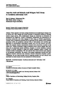
Results Effect of SA and COU treatments on the severity of charcoal rot disease A significant reduction in disease severity was observed in all the SA and COU treatments as compared with the infected but untreated plants, particularly in the case of those treated with 1.0 mM SA or 0.3 mM COU (Fig. 1). Seed pretreatment with 1.0 mM SA resulted in the lowest disease incidence and the maximum reduction in the length of necrotic stem lesions (48 and 83%, respectively). This was followed by the 0.3 mM COU treatment, which gave values of 52% disease incidence and 82% reduction in lesion length (Table 1).
Acta Agronomica Hungarica, 61, 2013
CHARCOAL ROT DISEASE IN SUNFLOWER (a)
(b)
27
(c)
Fig. 1. Development of necrotic lesions on the stem of (a) untreated, (b) 1.0 mM SA-treated and (c) 0.3 mM COU-treated 5-week-old sunflower plants inoculated with M. phaseolina using microsclerotia-bearing toothpicks Table 1 Effects of SA and COU seed treatments on the development of charcoal rot disease symptoms on 5-week-old sunflower plants Seed treatment (mM) Control SA COU
Disease incidence (%) g
0.3 1.0 3.0 0.3 1.0 3.0
100±0.00 64±1.15d 48±0.88a 60±1.15c 52±1.76b 68±0.66e 72±0.88f
Length of stem lesion (cm) 3.60±0.21d 0.76±0.07a 0.60±0.12a 0.70±0.09a 0.64±0.11a 0.88±0.05b 1.06±0.07c
All values are mean ± standard error of five independent replicates. Data within a column followed by different letters are significantly different at the 0.05 level according to Duncan’s test.
Effect of M. phaseolina infection, SA and COU treatments on carbohydrate content The results revealed that infection with M. phaseolina increased the level of soluble sugars in both control and treated plants; however, the enhancement was more obvious in SA- and COU-treated plants (Table 2). This was accompanied by a great reduction in the level of the insoluble and total sugar fractions in the infected but untreated plants, amounting to about 50 and 33%, respectively, as compared with the uninfected control plants (Table 2). Both SA and COU treatments caused a substantial increase in the level of soluble sugars in both uninfected and infected plants, but the increment compared with untreated plants was greater in COU-treated plants (Table 2). Seed pretreatment with the various SA concentrations increased the level of insoluble sugars both in uninfected plants and to a higher extent in infected plants as compared with their respective untreated controls. An obvious reduction in the level of insoluble sugars was produced in healthy plants by the higher doses of COU (1.0 and 3.0 mM), but not by the lowest dose (0.3 mM). On the other hand, all the COU treatments resulted in the accumulation of insoluble sugars in infected plants as compared with the untreated hosts.
Acta Agronomica Hungarica, 61, 2013
S. M. AL-WAKEEL et al.
28
Effect of M. phaseolina infection, SA and COU treatments on the activity of β-1,3-glucanase and chitinase Chitinase activity was induced in response to fungal infection and seed treatment with either SA or COU (Table 3). A similar pattern of induction was observed in both infected and uninfected sunflower plants, while a more obvious increase was caused by the combined effect of seed treatment and infection. The most effective treatments for the induction of chitinase activity were the higher doses of SA (1.0 and 3.0 mM) and the lower dose of COU (0.3 mM). β-1,3glucanase exhibited a response similar to that of chitinase, with a much higher level of induction in the COU treatments, especially at the higher concentrations (Table 3). Table 2 Effects of SA and COU seed treatments on the carbohydrate composition (mg glucose g–1 dry wt) in the leaves of 5-week-old healthy and M. phaseolina-infected sunflower plants Soluble sugars
Seed treatment (mM) Control SA COU
Healthy a
0.3 1.0 3.0 0.3 1.0 3.0
Insoluble sugars
Infected
Healthy a
Total sugars
Infected b
Healthy a
Infected ab
3.08±0.11 4.01±0.10 11.96±0.32 6.01±0.20 15.04±0.40 10.02±0.38a 3.50±0.10b 4.88±0.11b 12.60±0.31bc 8.97±0.25c 16.10±0.42bc 13.85±0.29c 4.13±0.05c 5.96±0.13d 13.73±0.37d 10.35±0.31e 17.86±0.44d 16.31±0.42e 3.82±0.11b 5.36±0.12c 13.16±0.35c 9.46±0.29d 16.98±0.51cd 14.82±0.46d 4.51±0.13cd 6.29±0.16de 11.93±0.29b 8.74±0.27cd 16.44±0.45cd 15.03±0.42d 4.87±0.15d 6.51±0.15e 10.14±0.31a 6.91±0.19b 15.01±0.47ab 13.42±0.35bc 5.13±0.16e 6.82±0.17f 9.69±0.24a 6.23±0.16ab 14.82±0.41a 13.05±0.33b
All values are mean ± standard error of three independent replicates. Data within a column followed by different letters are significantly different at the 0.05 level according to Duncan’s test. Table 3 Effects of SA and COU seed treatments on the chitinase and β-1,3-glucanase activities in the leaves of 5-week-old healthy and M. phaseolina-infected sunflower plants Seed treatment (mM)
Chitinase activity (µM GluNac min-1g-1 fresh wt) Healthy
Control SA COU
Infected a
0.3 1.0 3.0 0.3 1.0 3.0
5.86±0.13 7.34±0.08c 8.80±0.26d 7.93±0.17c 8.09±0.20c 6.94±0.58b 6.17±0.31ab
β-1,3-Glucanase activity (µg glucose min–1g–1 fresh wt) Healthy
a
8.73±0.72 11.64±0.80c 14.52±0.40e 12.79±0.47cd 13.21±0.41cd 10.78±0.44b 9.45±0.10ab
Infected a
7.62±0.36 8.43±0.44ab 9.74±0.27b 9.01±0.45ab 10.32±0.49c 11.09±0.95d 12.15±0.74d
9.84±0.79a 12.07±0.29b 14.12±0.12c 13.23±0.60bc 15.24±0.16d 16.16±0.66e 17.01±0.47f
All values are mean ± standard error of three independent replicates. Data within a column followed by different letters are significantly different at the 0.05 level according to Duncan’s test
Acta Agronomica Hungarica, 61, 2013
CHARCOAL ROT DISEASE IN SUNFLOWER
29
HPLC analysis HPLC analysis on the phenolic compounds extracted from the leaves of control, 1.0 mM SA- and 0.3 mM COU-treated healthy or M. phaseolinainfected sunflower plants revealed qualitative and quantitative variations in their phenolic profiles (Table 4). The HPLC analysis of the leaf extract of healthy control plants revealed the presence of five phenolic compounds, including coumarin and salicylic acid as the major components, while chlorogenic, caffeic and ferulic acids were less abundant. Treatment with 1.0 mM SA or 0.3 mM COU increased the levels of these phenolic compounds, with the exception of caffeic acid, the level of which was reduced, while leading to the formation of scopolin, ayapin and scopoletin. The levels of salicylic, chlorogenic and caffeic acids were higher in the SA treatment, while COU-treated plants contained higher amounts of coumarin, scopolin, ayapin, scopoletin and ferulic acid. Infection of sunflower with M. phaseolina increased the levels of salicylic, chlorogenic, ferulic and caffeic acids as compared with their levels in the healthy control, while that of coumarin decreased in the infected plants. This reduction in the coumarin content was accompanied by the production of scopoletin and ayapin. In infected plants treated with SA or COU the levels of ayapin and coumarin, and those of salicylic, chlorogenic and ferulic acids were higher than in untreated but infected plants. Moreover, the reduction in the scopoletin level caused by infection with M. phaseolina in the leaves of SA- and COU-treated plants was accompanied by the production of its glycoside form (scopolin), a higher level of which was induced by COU than by SA. Table 4 Qualitative and quantitative analysis of phenolic compounds in the leaves of control, 1.0 mM SA- and 0.3 mM COU-treated 5-week-old healthy and M. phaseolina-infected sunflower plants using HPLC Standards Control 1.0 mM SA 0.3 mM COU Retention Retention Concentration Retention Concentration Retention Concentration –1 –1 time (min) time (min) (µg g dry wt) time (min) (µg g dry wt) time (min) (µg g–1dry wt) Healthy plants Chlorogenic acid 03.09 03.10 37.0±0.77 03.08 103.6±0.61 03.11 70.6±0.47 Scopolin 03.63 – – 03.61 71.5±0.25 03.52 101.2±0.72 Ayapin 18.53 – – 18.29 62.6±0.31 18.64 81.6±0.54 Caffeic acid 19.45 19.57 63.1±1.09 19.30 43.0±0.28 19.19 35.8±0.17 Scopoletin 22.86 – – 23.01 262.1±0.98 22.93 304.0±0.93 Coumarin 23.80 23.56 204.3±1.51 23.89 234.9±2.03 23.65 362.5±2.14 Salicylic acid 26.11 26.32 138.6±1.12 26.09 305.5±1.48 25.74 223.9±1.30 Ferulic acid 27.23 27.20 97.4±0.87 27.58 161.4±0.83 27.19 183.6±0.91 Infected plants Chlorogenic acid 03.09 02.94 87.2±0.92 02.96 123.0±0.83 3.08 97.8±0.67 Scopolin 03.63 – – 03.68 98.4±0.50 3.78 148.2±1.03 Ayapin 18.53 18.56 41.5±0.63 18.58 84.1±0.39 18.86 117.9±0.82 Caffeic acid 19.45 19.44 96.3±0.85 19.48 90.4±0.32 19.37 70.3±0.25 Scopoletin 22.86 23.11 283.4±1.25 23.12 237.6±0.51 22.60 209.6±0.47 Coumarin 23.80 24.25 117.5±1.33 23.73 203.5±1.91 23.58 314.5±1.13 Salicylic acid 26.11 26.13 183.1±0.81 25.95 381.0±1.42 26.15 268.3±1.07 Ferulic acid 27.23 26.98 135.8±1.07 27.06 192.3±0.92 27.10 164.5±0.82 All values are mean ± standard error of three independent replicates Compound
Acta Agronomica Hungarica, 61, 2013
30
S. M. AL-WAKEEL et al.
Discussion The plant carbohydrate metabolism is greatly affected by compatible interactions between plants and pathogenic fungi. One common feature is a reduction in the rate of photosynthesis (Tang et al., 1996; Chou et al., 2000). An increase in extracellular invertase activity results in the accumulation of soluble sugars and the inverse regulation of photosynthesis (Roitsch et al., 2003). Furthermore, carbohydrates are withdrawn from the plant by the pathogen, which acts as an additional sink competing with the natural sink organs of the host plant, resulting in a considerable modification of the production, partitioning and utilization of photoassimilates within the host tissue (Fotopoulos et al., 2003). This depletion in the carbohydrates affects plant health, as the greatest part of the sugars generated during photosynthesis is stored in the plant for later use in energy-requiring processes. Furthermore, carbohydrates are the basic building blocks for the synthesis of various defence chemicals such as phenolics, phytoalexins and lignin. Hence, the quality and quantity of sugars play an important role in disease resistance. Specific soluble sugars, such as sucrose, glucose, trehalose, fructose and galactose, are correlated with disease resistance in some plant–pathogen interactions. According to the present results, the infection of sunflower plants with M. phaseolina resulted in an increase in the level of soluble reducing sugars (Table 2). This increase was accompanied by a reduction in the levels of other sugar fractions and may well be correlated with the induction of the host invertases and amylases as a result of fungal infection (Voegele et al., 2006; Singh et al., 2009). In this respect, Lobato et al. (2009) reported an increase in the total soluble carbohydrate content in common bean plants after inoculation with the necrotrophic fungus Colletotrichum lindemuthianum. Furthermore, Singh et al. (2009) reported that the leaves of barley plants infected with leaf blight showed lower contents of endogenous total and non-reducing sugars, but a higher level of reducing sugars as compared to healthy leaves. A similar result was reported for sunflower plants infected with sunflower chlorotic mottle virus (Arias et al., 2003). The priming of sunflower seeds with various concentrations of SA and COU resulted in the accumulation of soluble and insoluble sugars in the infected plants. The induction of soluble sugars may supply energy for defence reactions and/or may act as signals inducing defence gene expression (Sinha et al., 2002). As expected, the treatments resulted in considerable protection against charcoal rot disease as compared with the untreated plants (Fig. 1). In this regard, El-Fiki et al. (2004) revealed that presoaking sesame seeds in indole butyric acid (IBA) and SA were very effective treatments for reducing charcoal rot disease symptoms. They also recorded a significant induction of soluble reducing and total sugars in the leaves of mature sesame plants as a result of seed treatment with SA. Furthermore, El-Tayeb et al. (2006) reported the induction of soluble, insoluble and total carbohydrate in the leaves and stems of sunflower treated with 0.5 mM SA. Acta Agronomica Hungarica, 61, 2013
CHARCOAL ROT DISEASE IN SUNFLOWER
31
Chitinases and β-1,3-glucanases seem to play an important role in plant defence against phytopathogenic microorganisms (Boller, 1993). Reports have shown that the regulation of chitinases and β-1,3-glucanases is co-ordinated and that they act synergistically in enhancing plant resistance against fungal infection (Jongedijk et al., 1995). These two enzymes target different structures in the cell walls of the fungal pathogen and greater protection was observed in transgenic plants over-expressing both chitinase and glucanase genes (Zhu et al., 1994; Chen et al., 2006). The results revealed that the activity of both chitinase and β-1,3-glucanase increased in response to infection with M. phaseolina, and to a much higher extent as the result of seed treatment with either SA or COU as compared with untreated plants (Table 3). These results are in accordance with the results obtained by Cachinero et al. (1996) and Nandeeshkumar et al. (2008), who reported that the inoculation of both resistant and susceptible sunflower cultivars with downy mildew (Plasmopara halstedii) resulted in a marked increase in the activity of chitinase and β-1,3-glucanase. Furthermore, the induction of chitinase and β-1,3-glucanase was demonstrated in several plant species after the exogenous application of chemical inducers such as SA (Jayaraj et al., 2004), BTH (Roldán Serrano et al., 2007) and chitosan (Nandeeshkumar et al., 2008). The HPLC analysis revealed the presence of five constitutive phenolic compounds, namely: salicylic, chlorogenic, caffeic and ferulic acids and coumarin, in the leaves of healthy control sunflower plants. Similarly, many phenolic compounds, such as chlorogenic, caffeic, cinnamic, ferulic, syringic and vanillic acids, have been identified in extracts from different plant organs of sunflower (Pedrosa et al., 2000; Ghafar et al., 2001; Weisz et al., 2009). The increase in the majority of constitutive phenolics, particularly salicylic acid and coumarin, along with the production of scopoletin, scopolin and ayapin in the leaves of sunflower plants treated with 1.0 mM SA or 0.3 mM COU, indicates their potential role in activating defence responses in sunflower. In this respect, the exogenous application of methyl salicylate, methyl jasmonate and other abiotic elicitors was found to increase the levels of scopoletin and ayapin and of coumaric, caffeic and ferulic acids in sunflower and poplar leaves (Gutierrez et al., 1995; An et al., 2006). In sunflower, coumarins such as scopolin, scopoletin and ayapin have been described as phytoalexins, since their synthesis is only induced in response to abiotic elicitors and fungal infection (Prats et al., 2003; Shimizu et al., 2005). Moreover, Saftić-Panković et al. (2006) reported the accumulation of chlorogenic, ferulic and caffeic acids, along with scopoletin, after the infection of sunflower with downy mildew. The defensive role of coumarin phytoalexins in sunflower is well documented and their contents were higher in resistant than in susceptible varieties (Prats et al., 2007). Similarly, the present results clearly confirmed that the increase in preformed phenolics, as well as the production of scopoletin and ayapin in the leaves of sunflower plants have a role in defence against M. phaseolina. Acta Agronomica Hungarica, 61, 2013
32
S. M. AL-WAKEEL et al.
Moreover, the reduction in the level of scopoletin in infected plants treated with SA or COU was related to the increase in the production of its glycoside form, scopolin. Since scopolin is known to be less phytotoxic than scopoletin or ayapin (Gutierrez et al., 1996; Prats et al., 2006), its accumulation in sunflower leaves indicates the ability of SA and COU treatments to confer charcoal rot disease resistance without direct damage to the host plants. In this regard, Sauerborn et al. (2002) and Dmitriev et al. (2003) demonstrated the accumulation of scopoletin and ayapin in sunflower treated with SA, INA and BTH, which enhanced resistance against pathogenic fungi and parasitic weeds. To conclude, the significant reduction in the severity of charcoal rot after SA and COU treatments suggests their efficiency in activating systemic resistance to M. phaseolina in sunflower. Therefore, priming seeds with SA and COU could be used commercially as an alternative approach to improve the resistance of sunflower to charcoal rot disease. References Abenavoli, M. R., Sorgonà, A., Albano, S., Cacco, G. (2004): Coumarin differentially affects the morphology of different root types of maize seedlings. J. Chem. Ecol., 30, 1871–1880. Abenavoli, M. R., Sorgonà, A., Sidari, M., Badiani, M., Fuggi, A. (2003): Coumarin inhibits the growth of carrot (Daucus carota L. cv. Saint Valery) cells in suspension culture. J. Plant Physiol., 160, 227–237. Aboshosha, S. S., Atta-Alla, S. I., El-Korany, A. E., El-Argawy, E. (2007): Characterization of Macrophomina phaseolina isolates affecting sunflower growth in El-Behera governorate, Egypt. Inter. J. Agric. Biol., 9, 807–815. An, Y., Shen, Y., Wum, L., Zhang, Z. (2006): A change of phenolic acids content in poplar leaves induced by methyl salicylate and methyl jasmonate. J. Forest Res., 17, 107–110. Arias, M. C., Lenardon, S., Taleisnik, E. (2003): Carbon metabolism alterations in sunflower plants infected with the sunflower chlorotic mottle virus. J. Phytopathol., 151, 267–273. Bokor, P. (2007): Macrophomina phaseolina causing a charcoal rot of sunflower through Slovakia. Biologia Bratislava, 62, 136–138. Boller, T. (1993): Antimicrobial functions of the plant hydrolases chitinase and β-1,3-glucanase. pp. 391–400. In: Fritig, B., Legrand, M. (eds.), Mechanisms of Plant Defense Responses. Kluwer Acad. Publ., Dordrecht. Brown, S. A., Zobel, A. M. (1990): Biosynthesis and distribution of coumarins in the plant. pp. 15–37. In: Proc. Conf. Coumarins: Research and Applications. Padua, Italy, 20–22 September 1990, Bünemann, E. K., Schwenke, G. D., Van Zwieten, L. (2006): Impact of agricultural inputs on soil organisms – A review. Aust. J. Soil Res., 44, 379–406. Cachinero, J. M., Cabello, F., Jorrin, J., Tena, M. (1996): Induction of different chitinase and βl,3-glucanase isozymes in sunflower (Helianthus annuus L.) seedlings in response to infection by Plasmopara halstedii. Eur. J. Plant Pathol., 102, 401–405. Chen, S. C., Liu, A. R., Zou, Z. R. (2006): Over-expression of glucanase gene and defensin gene in transgenic tomato enhances resistance to Ralstonia solanacearum. Russ. J. Plant Physiol., 53, 671–677. Chen, S. K., Edwards, C. A., Subler, S. (2001): Effects of the fungicides benomyl captan and chlorothalonil on soil microbial activity and nitrogen dynamics in laboratory incubations. Soil Biol. Biochem., 33, 1971–1980.
Acta Agronomica Hungarica, 61, 2013
CHARCOAL ROT DISEASE IN SUNFLOWER
33
Chou, H. M., Bundock, N., Rolfe, S. A., Scholes, J. D. (2000): Infection of Arabidopsis thaliana with Albugo candida (white blister rust) causes a reprogramming of host metabolism. Mol. Plant Pathol., 1, 99–113. Clark, J. M., Switzer, R. L. (1977): Experimental Biochemistry. 2nd edn. W. H. Freeman and Company, San Francisco. pp. 265–273. Dmitriev, A., Tena, M., Jorrin, J. (2003): Systemic acquired resistance in sunflower (Helianthus annuus L.). Tsitol. Genet., 37, 9–15. Edmunds, L. K. (1964): Combined relation of plant maturity texture and soil moisture to charcoal stalk rot development in grain sorghum. Phytopathology, 54, 514–517. El-Fiki, A. I. I., Mohamed, F. G., El-Deeb, A. A., Khalifa, M. M. A. (2004): Some applicable methods for controlling sesame charcoal rot disease (Macrophomina phaseolina) under greenhouse conditions. Egypt. J. Phytopathol., 32, 87–101. El-Tayeb, M. A., El-Enany, A. E., Ahmed, N. L. (2006): Salicylic acid-induced adaptive response to copper stress in sunflower (Helianthus annuus L.). Plant Growth Regul., 50, 191–199. Fotopoulos, V., Gilbert, M. J., Pittman, J. K., Marvier, A. C., Buchanan, A. J., Sauer, N., Hall, J. L., Williams, L. E. (2003): The monosaccharide transporter gene AtSTP4 and the cell-wall invertase Atbetafruct1 are induced in Arabidopsis during infection with the fungal biotroph Erysiphe cichoracearum. Plant Physiol., 132, 821–829. Ghafar, A., Saleem, B., Haq, A. U., Qureshi, M. J. (2001): Isolation and identification of allelochemicals of sunflower (Helianthus annuus L.). Inter. J. Agric. Biol., 3, 21–22. Gutierrez, M. C., Edwards, R., Tena, M., Cabello, F., Serghini, K., Jorrin, J. (1996): The production of coumarin phytoalexins in different plant organs of sunflower (Helianthus annuus L.). J. Plant Physiol., 149, 261–266. Gutierrez, M. C., Parry, A., Tena, M., Jorrin, J., Edwards, R. (1995): Abiotic elicitation of coumarin phytoalexins in sunflower. Phytochemistry, 38, 1185–1191. Hossain, M. D., Jing, L., Shirong, G., Fujita, M. (2008): Suppressive effects of coumarins on pumpkin seedling growth and glutathione-S-transferase activity. J. Crop Sci. Biotech., 11, 187–192. Jayaraj, J., Muthukrishnan, S., Liang, G. H., Velazhahan, R. (2004): Jasmonic acid and salicylic acid induce accumulation of β-1,3-glucanase and thaumatin-like proteins in wheat and enhance resistance against Stagonospora nodorum. Biol. Plant., 48, 425–430. Jongedijk, E., Tigelaar, H., van Roekell, J. S. C., Bres-Vloemans, S. A., Dekker, I., van den Elzen, P. J. M., Cornelissen, B. J. C., Melchers, L. S. (1995): Synergistic activity of chitinases and β-1,3-glucanases enhances fungal resistance in transgenic tomato plants. Euphytica, 85, 173–180. Kagale, S., Marimuthu, T., Thayumanavan, B., Nandakumar, R., Samiyappan, R. (2004): Antimicrobial activity and induction of systemic resistance in rice by leaf extract of Datura metel against Rhizoctonia solani and Xanthomonas oryzae pv oryzae. Physiol. Mol. Plant. Pathol., 65, 91–100. Kapidlowska, E., Kowalec, M., Sulkowski, G., Zobel, A. M. (1994): The effect of coumarin on root elongation and ultrastructure of meristematic cell protoplast. Ann. Bot., 73, 525–530. Lobato, A. K. S., Gonçalves-Vidigal, M. C., Vidigal Filho, P. S., Costa, R. C. L., Cruz, F. J. R., Santos, D. G. C., Silva, C. R., Silva, L. I., Sousa, L. L. (2009): Changes in photosynthetic pigment and carbohydrate content in common bean cultivars infected by Colletotrichum lindemuthianum. Plant Soil Environ., 55, 58–61. Macias, M., Irma, L., Rojas, S., Mata, R., Lotina-Hennsen, B. (1999): Effect of selected coumarins on spinach chloroplast photosynthesis. J. Agric. Food Chem., 47, 2137–2140. Murray, R. D. H., Mendez, H. J., Brown, S. A. (1982): The Natural Coumarins: Occurrence, Chemistry and Biochemistry. John Wiley and Sons, New York, USA. Nandeeshkumar, P., Sudisha, J., Ramachandra, K. K., Prakash, H. S., Niranjana, S. R., Shekar, S. H. (2008): Chitosan induced resistance to downy mildew in sunflower caused by Plasmopara halstedii. Physiol. Mol. Plant Pathol., 72, 188–194.
Acta Agronomica Hungarica, 61, 2013
34
S. M. AL-WAKEEL et al.
Pedrosa, M. M., Muzquiz, M., Garcia-Vallejo, C., Burbano, C., Cuadrado, C., Ayet, G., Robredo, L. M. (2000): Determination of caffeic and chlorogenic acids and their derivatives in different sunflower seeds. J. Sci. Food Agric., 80, 459–464. Prats, E., Bazzalo, M. E., Léon, A., Jorrin, J. V. (2003): Accumulation of soluble phenolic compounds in sunflower capitula correlates with resistance to Sclerotinia sclerotiorum. Euphytica, 132, 321–329. Prats, E., Bazzalo, M. E., Leon, A., Jorrin, J. V. (2006): Fungitoxic effect of scopolin and related coumarins on Sclerotinia sclerotiorum. A way to overcome sunflower head rot. Euphytica, 147, 451–460. Prats, E., Llamas, M. J., Jorrin, J., Rubiales, D. (2007): Constitutive coumarin accumulation on sunflower leaf surface prevents rust germ tube growth and appressorium differentiation. Crop Sci., 47, 1119–1124. Reddy, K. R. N., Reddy, C. S., Muralidharan, K. (2009): Efficacy of certain agrochemicals on Aspergillus spp. and subsequent aflatoxin production in rice. Pest. Biochem. Physiol., 93, 53–57. Reissig, J., Strominger, J., Leloir, L. (1955): A modified colorimetric method for the estimation of n-acetyl-glucosamine sugars. J. Biol. Chem., 217, 959–967. Roitsch, T., Balibrea, M. E., Hofmann, M., Proels, R., Sinha, A. K. (2003): Extracellular invertase: Key metabolic enzyme and PR protein. J. Exp. Bot., 54, 513–524. Roldán Serrano, A., Luna Del Castillo, J., Jorrín Novo, J., Fernández Ocana, A., Gómez Rodríguez, M. V. (2007): Chitinase and peroxidase activities in sunflower hypocotyls: Effects of BTH and inoculation with Plasmopara halstedii. Biol. Plant., 51, 149–152. Saftić-Panković, D., Veljović-Jovanović, S., Pucarević, M., Radovanović, N., Mijić, A. (2006): Phenolic compounds and peroxidases in sunflower near-isogenic lines after downy mildew infection. HELIA, 29, 33–42. Sangawan, M., Metha, N., Saharan, G. (2005): Diseases of Oil Seed Crops. Indus Publ. Co., India. Sauerborn, J., Buschmann, H., Ghiasi, K. G., Kogel, K. H. (2002): Benzothiadiazole activates resistance in sunflower (Helianthus annuus) to the root-parasitic weed Orobanche cumana. Phytopathology, 92, 59–64. Shimizu, B. I., Miyagawa, H., Ueno, T., Sakata, K., Watanabe, K., Ogawa, K. (2005): Morning glory systemically accumulates scopoletin and scopolin after interaction with Fusarium oxysporum. Z. Naturforsch., 60, 83–90. Singh, S., Asthir, B., Bains, N. S., Mann, S. K. (2009): Induction of carbohydrate metabolism in relation to leaf blight in barley (Hordeum vulgare). Adv. Biol. Res., 3, 61–66. Sinha, A. K., Hofmann, M. G., Römer, U., Köckenberger, ِ W., Elling, L., Roitsch, T. (2002): Metabolizable and non-metabolizable sugars activate different signal transduction pathways in tomato. Plant Physiol., 128, 1480–1489. Tang, X., Rolfe, S. A., Scholes, J. D. (1996): The effect of Albugo candida (White blister rust) on the photosynthetic and carbohydrate metabolism of leaves of Arabidopsis thaliana. Plant Cell Environ., 19, 967–975. Voegele, R. T., Wirsel, S., Möll, U., Lechner, M., Mendgen, K. (2006): Cloning and characterization of a novel invertase from the obligate biotroph Uromyces fabae and analysis of expression patterns of host and pathogen invertases in the course of infection. Mol. Plant-Microbe Interact., 19, 625–634. Walter, M., Harris-Virgin, P., Thomas, W., Tate, G., Waipara, N. W., Langford, G. (2004): Agrochemicals suitable for downy mildew control in New Zealand boysenberry production. Crop Protec., 23, 327–333. Weiss, E. A. (2000): Oil Seed Crops. 2nd Edn. Blackwell Sci. Publ., UK. Weisz, G. W., Kammerer, R. D., Carle, R. (2009): Identification and quantification of phenolic compounds from sunflower (Helianthus annuus L.) kernels and shells by HPLCDAD/ESI-MSn. Food Chem., 115, 758–765.
Acta Agronomica Hungarica, 61, 2013
CHARCOAL ROT DISEASE IN SUNFLOWER
35
Yamunarani, K., Jaganathan, R., Bhaskaran, R., Govindaraju, P., Velazhahan, R. (2004): Induction of early blight resistance in tomato by Quercus infectoria gall extract in association with accumulation of phenolics and defense-related enzymes. Acta Physiol. Plant., 26, 281–290. Zhu, Q., Maher, E. A., Masoud, S., Dixon, R. A., Lamb, C. J. (1994): Enhanced protection against fungal attack by constitutive co-expression of chitinase and glucanase genes in transgenic tobacco. Biotechnol., 12, 807–812. Zhu, Y. J., Qiu, X., Moor, P. H., Borth, W., Hu, J., Ferreira, S., Albert, H. H. (2003): Systemic acquired resistance induced by BTH in papaya. Physiol. Mol. Plant Pathol., 63, 237–248. Corresponding author: A. M. Saleh Phone: 00201003676683; 00966558424680 E-mail:
[email protected]
Acta Agronomica Hungarica, 61, 2013