Phytopathol. Mediterr. (2006) 45, S101–S109
Cryptovalsa ampelina on grapevines in N.E. Spain: identification and pathogenicity JORDI LUQUE1, DOLORES SIERRA2, ESTER TORRES3 and FRANCESC GARCIA3 1
Departament de Protecció Vegetal, Institut de Recerca i Tecnologia Agroalimentàries (IRTA), Centre de Cabrils, Ctra. de Cabrils s/n, E-08348 Cabrils, Spain 2 Departament de Biologia Vegetal (Botànica), Facultat de Biologia, Universitat de Barcelona, Avda. Diagonal 645, E-08028 Barcelona, Spain 3 Laboratori de Sanitat Vegetal, DARP, Via Circulació Nord, Tram VI, Carrer 3, Zona Franca, E-08040 Barcelona, Spain
Summary. Surveys conducted in diseased vineyards in Catalonia (N.E. Spain) showed that Cryptovalsa ampelina was very abundant on pruned canes, although it was isolated occasionally from necrotic wood of living plants. Identification of C. ampelina from the pruned canes was based on the morphology of the teleomorph. Its polysporous asci and pigmented allantoid ascospores distinguish it from Eutypa lata, the causal agent of eutypiose. However, cultures of C. ampelina are practically indistinguishable from cultures of other diatrypaceous species, therefore a PCR-based test was developed to identify cultures isolated from cankered wood. The designed species-specific primer pair (Camp1/Camp-2R) allowed for the unambiguous identification of C. ampelina in all tested cases involving cultures of diatrypaceous fungi. Additionally, the specificity of the primer pair to C. ampelina was confirmed by testing it on the host and on several other fungi known to occur on grapevine, namely species in the genera Botryosphaeria, Fomitiporia, Phaeoacremonium, Phaeomoniella and Phomopsis. The pathogenicity of C. ampelina on grapevine was confirmed through the observation of significant vascular lesions in artificial inoculations of grapevine plants, but the low frequencies of both mycelium reisolation and wound canker extension would suggest a low virulence for this fungus. Although C. ampelina does not appear to be a major pathogen of grapevine, its implication as a contributing factor to the decline of grapevines should deserve further investigations.
Key words: Diatrypaceae, grapevine decline, molecular identification, species-specific primers, Vitis vinifera.
Introduction Several fungi in the Diatrypaceae are known to occur on grapevines worldwide, with species in the genera Cryptovalsa, Diatrype, Diatrypella, Eutypa, and Eutypella (Farr et al., 1989; Trouillas et al., 2001). Eutypa lata, the causal agent of eutypiose,
Corresponding author: J. Luque Fax: +34 93 7533954 E-mail:
[email protected]
is the major grapevine pathogen in this group (Carter, 1988; Dubos, 1996; Larignon and Dubos, 1997). This fungus is well known as a cosmopolitan, plurivorous species on many fruit trees of economic interest, such as almond, apple, apricot, cherry, olive, and walnut (Glawe et al., 1983; Rumbos, 1988, 1993, 1997; Farr et al., 1989; Munkvold and Marois, 1991). While identification, pathogenicity, epidemiology and control of E. lata have been studied thoroughly, little is known about the other diatrypaceous fungi that occur on grapevines. Recent reports on the identification and patho-
S101
J. Luque et al.
genicity of Eutypa leptoplaca (Trouillas and Gubler, 2004; Safodien et al., 2005), Eutypella vitis (Catal et al., 2005), and Cryptovalsa ampelina (Mostert et al., 2004) are contributing to a better knowledge about these fungi. Thus, for example, while the genus Cryptovalsa has been considered for some time as incertae sedis in the Ascomycota (Hawksworth et al., 1995), Mostert et al. (2004) confirmed, by phylogenetic analysis of the 5.8S nuclear rRNA gene and its flanking regions ITS1 and ITS2, that C. ampelina could be well accommodated in the Diatrypaceae. In recent years C. ampelina has been found repeatedly on grapevine canes in Australia and South Africa (Mostert et al., 2004). Recent surveys conducted in Catalonia (N.E. Spain) have shown this fungus to be very abundant on old pruned canes, although it was also isolated from diseased wood of living plants (Luque, unpublished data). Since co-occurrence of diatrypaceous fungi in diseased wood of grapevines (Trouillas et al., 2001) could lead to misidentification of the species, the purpose of the work reported here was focused on the identification of C. ampelina and the study of its pathogenicity on grapevine. The first objective was achieved through a morphological characterization of the fungus and subsequent design of a speciesspecific primer pair to be used in a molecular PCRbased diagnostic test. Pathogenicity was assessed from artificial inoculations conducted in a greenhouse experiment.
Materials and methods Geographic survey and isolations
Surveys were conducted in Catalonia (N.E. Spain) during the spring and summer seasons of 2003–2004 to study the fungi associated with grapevine trunk diseases. Sampled plants included individuals showing symptoms known to be associated with grapevine trunk diseases (reduced growth and foliar chlorosis, foliar necroses characteristic of esca, and both branch and trunk cankers with longitudinal splits). Additionally, old pruned canes were collected when possible at each sampling point to determine the presence of pathogenic fungal species in the samples. Isolations of C. ampelina were made either from single ascospores or by directly plating out pieces of diseased tissue after surface sterilization (70%
S102
Phytopathologia Mediterranea
ethanol, 4 min). Isolates were cultured on potato dextrose agar (PDA; Difco Laboratories, Detroit, IL, USA) at 25°C with 12/12 hours light/dark photoperiod. Isolates were maintained on PDA plugs in tubes of sterile distilled water at 4°C until further use. Morphological and molecular characterization
Microscope examinations were carried out on a Zeiss Axiophot microscope fitted with DIC optics. The following parameters were used to describe dimensions of conidia and ascospores (n=50 unless specified): mean, standard deviation, 95% confidence intervals and minimum and maximum sizes. Dimensions of other fungal structures are given as the range of at least 20 measurements. Identification of C. ampelina was based on the descriptions of Nitschke (1867) [basion. Valsa ampelina Nitschke], Fuckel (1870), Saccardo (1882), and the one recently published by Mostert et al. (2004). The following representative herbarium samples of C. ampelina were used to aid in the identification: BR-Myc 093345,30 (Fuckel, Fungi Rhenani No. 1824; Ad Vitis viniferae sarmentos aridos), and BR-Myc 093344,31 (Rabenhorst, Herbarium Mycologicum Ed. II No. 147; on Vitis sarmenta). For molecular characterization of the isolates, fungal DNA was extracted following the modified protocol of Martin et al. (1998), using CTAB as extraction buffer and later precipitating DNA with isopropanol. Amplification of the ITS1 and ITS2 regions flanking the 5.8S ribosomal RNA gene was carried out by using the universal primers ITS1 and ITS4 (White et al., 1990). PCR reactions were performed on a GeneAmp® PCR System 9700 thermal cycler (PE Applied Biosystems, Foster City, CA, USA). Each reaction contained about 100 ng genomic DNA template, 20 µl Eppendorf Mastermix 2.5⫻ (Eppendorf AG, Hamburg, Germany), 0.4 µm of each primer, and made up to a final volume of 50 µl with sterile HPLC-grade water. The following temperature profile was used: one cycle of initial denaturation at 95°C for 3 min, followed by 35 cycles of denaturation (20 sec at 95°C), annealing (30 sec at 55°C) and extension (40 sec at 72°C), and a final extension at 72°C for 5 min. PCR products were purified using the High Pure PCR Product Purification Kit (Roche Diagnostics
Cryptovalsa ampelina on grapevines in N.E. Spain
GmbH, Mannheim, Germany). The purified amplicons were sequenced in both directions using the ITS primers and the BigDye™ Terminator v1.1 Cycle Sequencing Kit (Applied Biosystems, Foster City, CA, USA). The resulting fragments were analyzed on an ABI Prism 377 automated DNA sequencer (Perkin Elmer, Norwalk, CT, USA). Specific primers design
ITS sequences of the representative isolates of C. ampelina CBS 117484 and CBS 117485, both obtained from single ascospore isolations, were used to design the species-specific primers targeting the rRNA gene. Additional ITS sequences of C. ampelina STE-U 5621 and STE-U 5622 were provided by L. Mostert (CBS, Utrecht, Netherlands), while those from other diatrypaceous fungi were obtained from GenBank: E. lata (AF099911, AF455427, AJ302450, AJ302451, AJ302452, AJ302459), Eutypa leptoplaca (AJ302453, AY684237), Eutypella vitis (AJ302466, AY462566, AY462574, AY462576, AY462578), Diatrypella spp. (AJ302440, AJ302441, AJ302442, AJ302443, AJ302444), and Diatrype spp. (AJ302422, AJ302437, AJ302439, AY684241). Nucleotide sequences of all taxa included in this study were aligned using the BioEdit Sequence Alignment Editor (Hall, 1999). Alignments were
checked visually and manual adjustments were made where necessary. The specific primer pair was designed from the aligned sequences with the aid of the Primer3 software (Rozen and Skaletsky, 2000). The designed primer pair Camp-1/Camp-2R was tested for amplification in 25 µl PCR reactions with reactives in the same concentrations as described above. Primer sequences and the optimal temperature conditions for the PCR reaction are given below. The specificity of the primer pair Camp-1/Camp2R was tested on all isolates provisionally assigned to Diatrypaceae after morphological examination. Species-specific primers for the identification of E. lata and the corresponding PCR conditions as designed by Lecomte et al. (2000) were used on the same isolates to identify this latter species. All specific PCR reactions included a negative control (sterile water) and two positive controls (C. ampelina CBS 117484 and E. lata JL355). Isolates showing negative amplifications for both specific primer pairs were later processed for ITS amplification and direct sequencing following the methods described above. Additionally, PCR reactions using the Camp-1/Camp-2R pair were tested for specificity on DNA extracts from other fungi occurring on Vitis and the host plant (Table 1). All PCR reactions were performed twice for each DNA sample.
Table 1. Specificity of the primer pair Camp-1/Camp-2R for the identification of Cryptovalsa ampelina. Legend of symbols: +, band of expected size; -, no bands; n.t., not tested. Species Cryptovalsa ampelina Eutypa lata Other diatrypaceous fungi Botryosphaeria dothidea Botryosphaeria lutea Botryosphaeria obtusa Botryosphaeria parva Botryosphaeria stevensii Fomitiporia mediterranea Phaeoacremonium aleophilum Phaeomoniella chlamydospora Phomopsis cf. viticola Vitis vinifera ‘Macabeu’
Specific primer pair C. ampelina
E. lata
+ -
+ n.t. n.t. n.t. n.t. n.t. n.t. n.t. n.t. n.t. n.t.
Isolates tested 11 69 4 2 1 12 15 2 8 10 9 1 1
Vol. 45, Supplement, 2006
S103
J. Luque et al.
Pathogenicity test
Results
Artificial inoculations were conducted on oneyear old grapevine plants (cv. Macabeu grafted onto Richter 110 rootstocks). Plants were maintained in a greenhouse in 3-liter pots filled with a sand:peat mixture (6:1, v:v; peat Floratorf, from Floragard, Oldenburg, Germany) with sufficient water supply. The pathogenicity test was performed in a completely randomized experimental design, with 18 inoculated plants per treatment. Two isolates of C. ampelina were used, namely CBS 117484 and CBS 117486. A superficial wound (15⫻5 mm, 1 mm depth) was made on the bark of each plant with a sterilized scalpel, 10 cm above the graft union. A mycelial plug (5 mm diameter) obtained from the margin of a fungal colony was placed in the wound with the mycelium facing the stem, and the wound was wrapped with Parafilm® (American National Can, Greenwich, CT, USA). Control plants were inoculated with sterile PDA plugs instead of the fungal inoculum. Additionally, plants inoculated with the grapevine pathogens E. lata and Botryosphaeria obtusa (JL411 and JL398, respectively) were included for comparison purposes. Results were recorded over two different periods, with one half of the inoculated plants processed at each time. Nine plants were chosen randomly for the first reading, six months after inoculations (late autumn). The second reading was three months later (late winter). External symptoms such as foliar chlorosis, plant wilting and canker expansion were noted at each reading period. The length of the internal vascular lesions was recorded by removing the bark from the stem and measuring the necrotic lesions upwards and downwards from the site of inoculation. Reisolations were attempted by plating on PDA several surface sterilized wood pieces (70% ethanol, 4 min) taken from the necrotic tissues. Cultures were incubated at 25°C for further identification of the inoculated fungi. Data from the assays were analysed using the SPSS v.10 statistical package. Data were checked for normality and equal variance distributions, transformed if necessary, and analyzed using multifactorial analysis of variance (ANOVA) procedures. After ANOVA, mean treatment values were compared against their respective controls with the Dunnett two-tailed test.
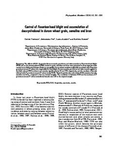
Cryptovalsa ampelina was found on pruned canes in 84% of the 25 localities sampled for pruning fragments. The fungus was identified from its teleomorph, which was present in all samples. It was also isolated from eight plants (about 4% of the total sampled plants), from diseased wood showing occasional wedge-shaped necroses (three plants). Cryptovalsa ampelina was the only fungus isolated in one plant, while in the remaining cases this species coincided with two or more of the following fungi: Botryosphaeria obtusa, B. parva, Fomitiporia mediterranea, Phaeoacremonium aleophilum and Phaeomoniella chlamydospora. However, no fruiting bodies of C. ampelina were detected in the field samples, therefore identification was later accomplished with the examination of the anamorph morphology in culture and the use of the Camp-1/Camp-2R primers in specific PCR reactions. The ascostromata of C. ampelina is poorly developed, embedded in the substrate, with a textura intrincata showing two kinds of constitutive hyphae: hyaline hyphae 2–3 µm wide, and dematiaceous hyphae 7–8 µm wide. Perithecia arranged in a single layer, singly arising, in rows or in small groups (Fig. 1a), 200–500 µm diam., subglobose, with cylindrical necks 50–100 µm long, and ostiole circular and periphysate. Perithecial wall comprised of two layers: the external of textura globulosa with dematiaceous, thick-walled cells, 8–20 µm diam., and the internal, textura angularis, with hyaline compressed cells. Paraphyses abundant, 1.5–3 µm wide, cylindrical, wider at the base, hyaline and rarely septate. Asci (90–)110–122(–155) ⫻ (10–)13–15(–18) µm (n=75), cylindrical to spindle-shape, long stipitate (70–100 µm), polysporous (Fig. 1b). Ascospores (9–)11–12(–13) ⫻ (2–)2.6– 2.7(–3) µm (n=150), unicellular, allantoid to reniform, wall smooth, with shades of pale brown (Fig. 1c). The fungus can be readily distinguished from other members in the family by its polysporous asci and the coloured ascospores. The anamorph of C. ampelina, a species in the genus Libertella (Mostert et al., 2004), was not seen on the field samples. Cultures in PDA were white to cream-white, cottony, with diffuse margins. Reverse of colonies first pale yellow, later (>20 days) developing irregular, mostly central, dark areas. Colony growth was rapid, covering the plate sur-
S104
Phytopathologia Mediterranea
Cryptovalsa ampelina on grapevines in N.E. Spain
face in 3–4 days at 25°C. Conidiomata sporodochium-like, consisting of conidiophores aggregated on blackened mycelial crusts, and producing cream coloured conidial masses in 4 weeks. Conidiophores hyaline and branched. Conidiogenous cells hyaline, subcylindrical, slightly tapered to the apex, proliferating sympodially, 5–16⫻1–2 µm (n=20). Conidia (17)–20–23–(30)⫻1–1.5 µm, unicellular, hyaline, filiform, slightly curved to hamate, with a truncate, flattened base (Fig. 1d). PCR amplifications of the DNA extracts of C. ampelina using the ITS1/ITS4 primers gave a prod-
uct of approximately 600 bp (GenBank accession numbers AY920390 and AY920391 for CBS 117484 and CBS 117485, respectively). Both nucleotide sequences were identical and matched exactly the sequences from the isolates STE-U 5621 and STEU 5622. After the alignment of all nucleotide sequences included in this study (data not shown), a primer pair was designed to amplify part of the ITS1-5.8S-ITS2 rDNA region (~300 bp) with the following sequences: Camp-1 (forward, ITS1 region): 5’-CCT ACC CTG TAG CTA CCC TA-3’, Camp-2R (reverse, ITS2 region): 5’-CAG CGT CTA
Fig. 1. Cryptovalsa ampelina. a) Erumpent perithecia on grapevine canes. b) Asci and ascus tips (insert). c) Ascospores. d) Conidia. Scale bars in µm, except in 1a (mm).
Vol. 45, Supplement, 2006
S105
J. Luque et al.
TAG CTA GGC GA-3’. Optimal temperature conditions for the specific PCR amplification in 25 µl reactions were determined as follows: initial denaturation at 95°C for 3 min, followed by 35 cycles of denaturation (20 sec at 95°C), annealing (30 sec at 58°C) and extension (40 sec at 72°C), and a final extension at 72°C for 5 min. A total of 84 isolates obtained from diseased wood of grapevines and identified from their morphology as potentially belonging to the Diatrypaceae were processed for molecular identification using the specific primers Lata 1/Lata 2-2 (E. lata) and Camp-1/Camp-2R (C. ampelina) in separate PCR reactions. According to the results of the specific PCR reactions, 69 isolates were identified as E. lata and 11 as C. ampelina (Fig. 2 and Table 1). None of these 80 isolates gave a double positive PCR reaction, thus confirming the discrimination between E. lata and C. ampelina in the tests. Only four isolates showed negative results with both primer pairs (from two independent repeated tests). These isolates were later processed for direct sequencing of the ITS region. Sequence homologies for these isolates obtained from GenBank ranged from 92 to 98% and were related to other diatrypaceous taxa (Eutypa leptoplaca, Eutypella vitis and Eutypella sp.). The test for specificity of the Camp1/Camp-2R primer pair on other fungi occurring on Vitis and the host plant DNA showed no DNA amplification in any case (Table 1). No banding patterns other than those expected for each specific primer pair, either for identification of C. ampelina or E. lata, were seen. All plants in the pathogenicity test, regardless of the inoculation treatment, grew adequately and showed no external symptoms during the trial. Inoculation wounds in all control plants healed
Fig. 2. PCR amplifications using the species-specific primers Lata 1/Lata 2-2 and Camp-1/Camp-2R on DNA extracts from different sources. M, molecular weight marker, 100 bp DNA ladder (New England BioLabs, Beverly, MA, USA).
within the experimental period. Wound cankers, as defined by the death of the bark and the underlying tissues surrounding the inoculation wound were less abundant in plants inoculated with C. ampelina (11–16% of inoculated plants), but more frequent in E. lata (33%) and B. obtusa (56%) (Table 2). Internal lesions, as characterized by dark brown discolourations were found in all plants, although those from plants of the control group were slightly paler. Original measurements of lesion lengths were log-transformed before ANOVA to homogenize group variances. A preliminary ANOVA showed non-significant effects of the period af-
Table 2. Pathogenicity of Cryptovalsa ampelina on one-year old grapevine plants cv. Macabeu (n=18). Data on Eutypa lata and Botryosphaeria obtusa included for comparison purposes. Treatment
Wound canker (n)
Mycelium recovery (n)
2 3 6 10 0
9 5 17 18 0
Cryptovalsa ampelina CBS 117484 Cryptovalsa ampelina CBS 117486 Eutypa lata JL411 Botryosphaeria obtusa JL398 Control a
Necrosis lengtha (mm) 14.83 16.61 18.61 23.88 9.44
Mean values different from control are shown in bold according to Dunnett’s test (P