JOURNAL OF BACTERIOLOGY, Aug. 2006, p. 5532–5540 0021-9193/06/$08.00⫹0 doi:10.1128/JB.00469-06 Copyright © 2006, American Society for Microbiology. All Rights Reserved.
Vol. 188, No. 15
Crystal Structure of a Type III Pantothenate Kinase: Insight into the Mechanism of an Essential Coenzyme A Biosynthetic Enzyme Universally Distributed in Bacteria† Kun Yang,1 Yvonne Eyobo,1 Leisl A. Brand,2 Dariusz Martynowski,1 Diana Tomchick,1 Erick Strauss,2* and Hong Zhang1* Department of Biochemistry, University of Texas Southwestern Medical Center, Dallas, Texas 75390-8816,1 and Department of Chemistry, Stellenbosch University, Matieland 7602, South Africa2 Received 4 April 2006/Accepted 22 May 2006
Pantothenate kinase (PanK) catalyzes the first step in the five-step universal pathway of coenzyme A (CoA) biosynthesis, a key transformation that generally also regulates the intracellular concentration of CoA through feedback inhibition. A novel PanK protein encoded by the gene coaX was recently identified that is distinct from the previously characterized type I PanK (exemplified by the Escherichia coli coaA-encoded PanK protein) and type II eukaryotic PanKs and is not inhibited by CoA or its thioesters. This type III PanK, or PanK-III, is widely distributed in the bacterial kingdom and accounts for the only known PanK in many pathogenic species, such as Helicobacter pylori, Bordetella pertussis, and Pseudomonas aeruginosa. Here we report the first crystal structure of a type III PanK, the enzyme from Thermotoga maritima (PanKTm), solved at 2.0-Å resolution. The structure of PanKTm reveals that type III PanKs belong to the acetate and sugar kinase/heat shock protein 70/actin (ASKHA) protein superfamily and that they retain the highly conserved active site motifs common to all members of this superfamily. Comparative structural analysis of the PanKTm active site configuration and mutagenesis of three highly conserved active site aspartates identify these residues as critical for PanK-III catalysis. Furthermore, the analysis also provides an explanation for the lack of CoA feedback inhibition by the enzyme. Since PanK-III adopts a different structural fold from that of the E. coli PanK—which is a member of the “P-loop kinase”superfamily—this finding represents yet another example of convergent evolution of the same biological function from a different protein ancestor. type of PanK (PanK-III) was identified which represents the only known pantothenate kinase activity in many pathogenic bacteria, including Helicobacter pylori, Pseudomonas aeruginosa, and Bordetella pertussis, as well as the category A biodefense pathogen Francisella tularensis (9). Moreover, some bacteria, such as Bacillus subtilis and Mycobacterium tuberculosis, have genes that code for both PanK-I and PanK-III. To distinguish these from one another, the gene that codes for PanKIII was dubbed coaX, in contrast to coaA genes that produce PanK-I. Substantial biochemical data have been accumulated for both PanK-I and PanK-II. These data show that although evolutionarily unrelated, both types of PanK are feedback inhibited by the end product of the pathway, CoA, as well as its thioesters, although the extent of inhibition depends on the system and the specific inhibitor (12, 41–43, 47, 52, 58). This feedback inhibition of PanK activity by CoA and its derivatives represents a key regulatory mechanism that controls intracellular CoA levels in response to a cell’s metabolic status (32). One exception to this observation is the PanK enzyme from S. aureus, which is not inhibited either by CoA or its thioesters, most probably due to this organism’s unique redox biology that depends on high concentrations of CoA and an NADPHdependent CoA disulfide reductase to maintain its intracellular redox balance (17, 31, 34). In contrast to the large body of data gathered on type I and II PanKs, relatively little is known about the mechanism and regulation of PanK-III. Current knowledge does, however, clearly indicate a unique position for these enzymes among
Pantothenate kinase (PanK; EC 2.7.1.33) catalyzes the ATPdependent phosphorylation of pantothenate (vitamin B5) to give 4⬘-phosphopantothenate. This reaction represents the first and committed step in the universal biosynthetic pathway of coenzyme A (CoA) (6, 27, 32). Because CoA is a ubiquitous and essential cofactor in all organisms, genes coding for the five enzymes that make up this pathway—including PanK—are essential for their survival and growth (6, 27). Three distinct types of PanK, as differentiated by primary sequence analysis and kinetic properties, have been characterized so far. Type I PanKs (PanK-I) are found exclusively in eubacterial species and are exemplified by the Escherichia coli enzyme encoded by the coaA gene (46, 47). The second type of PanK (PanK-II) is found mainly in eukaryotes, including yeast and various fungi, plants, and mammals (12, 41–43). Interestingly, PanKs from a few gram-positive bacteria, such as Staphylococcus aureus (PanKSa) and several bacilli, are also included in this group based on sequence homology, although the bacterial enzymes exhibit certain catalytic characteristics different from their eukaryotic counterparts (15, 31). Recently, a third * Corresponding author. Mailing address for H. Zhang: Department of Biochemistry, University of Texas Southwestern Medical Center, Dallas, TX 75390-8816. Phone: (214) 645-6372. Fax: (214) 645-5948. E-mail:
[email protected]. Mailing address for E. Strauss: Department of Chemistry, Stellenbosch University, Matieland 7602, South Africa. Phone: 27-21-808-3355. Fax: 27-21-808-3360. E-mail:
[email protected]. † Supplemental material for this article may be found at http://jb .asm.org/. 5532
VOL. 188, 2006
PanKs: while their kcat and Km values for pantothenate are comparable to those of PanK-I and -II, they exhibit an unusually high (in the mM range) Km for ATP, a 30- to 100-fold increase over the other types (9). Furthermore, unlike other PanKs the type III enzymes are not inhibited by CoA or any of its thioesters, a characteristic that might be singularly significant to organisms that predominantly harbor this type of PanK. Numerous studies have identified the potential of CoA biosynthetic enzymes as targets for drug development. Most recently, a comprehensive in vivo analysis of Salmonella enterica has highlighted the five enzymes of CoA biosynthesis to be among those previously known but as yet unexploited antimicrobial targets of important human pathogens (5). This analysis is based on the essential requirement of these enzymes for survival and/or virulence and on the lack of homology between bacterial PanK-I enzymes and their mammalian PanK-II counterparts. Development of inhibitors targeting these enzymes is being actively pursued (15, 20, 49, 54, 59, 60). Among these, the N-substituted alkylpantothenamides have shown the greatest promise as growth inhibitors of both E. coli and S. aureus (15, 31, 49, 59). These compounds act as alternative substrates of PanK and two other CoA biosynthetic enzymes, allowing their conversion to CoA analogs that subsequently inhibit CoA- and acetyl-CoA-utilizing enzymes and inactivate proteins with CoA-derived prosthetic groups, such as the acyl carrier protein (ACP) (31, 49, 59). Importantly, PanK-III enzymes are not affected by the N-alkylpantothenamide family of inhibitors; neither do they accept these compounds as alternate substrates (9). A structural characterization of PanK-III active site configuration will, therefore, greatly facilitate the development of inhibitors targeting this type of PanK. Such structure-based drug development strategies targeting PanK-I are already possible due to the availability of the threedimensional structure of E. coli PanK-I protein (PanKEc) (26, 58) and have allowed a structure-activity relationship (SAR) analysis to be performed on the pantothenamide-type inhibitors of this enzyme (54). While the sequence and structure of PanK-I indicate that it belongs to the “P-loop kinase” superfamily (13, 14), no structures of any PanK-II or PanK-III were previously known. Using state-of-the-art fold prediction methods, we have predicted that both these PanK types adopt an RNase H-like fold (9, 13) and are distantly related to the acetate and sugar kinase/heat shock protein 70 (hsp70)/actin (ASKHA) superfamily (8, 23). Because ASKHA and P-loop superfamilies belong to two different protein folds, presumably PanK-II and PanK-III will have completely different active site architectures from that of PanK-I. To verify our fold predictions and to address the general lack of knowledge of type III PanKs, we have determined the crystal structure of the PanK-III enzyme from Thermotoga maritima (PanKTm) at a 2.0-Å resolution. The structure confirms that PanK-III belongs to the ASKHA superfamily, which allowed us to identify its active site and to model the interactions between the substrates and the active site residues by comparison with other members of this superfamily. Based on this model, mutagenesis and kinetic analysis of highly conserved aspartate residues were carried out to investigate their roles in catalysis. Finally, we provide a comprehensive survey of the phylogenetic distribution of all three types of PanK and show that PanK-III has a much wider distribution in the bacterial
STRUCTURE OF TYPE III PANTOTHENATE KINASE
5533
kingdom than originally anticipated. Taken together, these results add significantly to our current knowledge of this key metabolic enzyme.
MATERIALS AND METHODS Cloning, expression, and purification of PanKHp and PanKTm. The cloning, expression and purification of Helicobacter pylori PanK (PanKHp; previously named CoaXHp) are described elsewhere (9). The Thermotoga maritima coaX gene (accession no. NC_000853, region 905791 . . . 906531) was amplified from T. maritima genomic DNA (ATCC 43589D) by PCR and was cloned into the BamHI and XhoI restriction sites of the pProEX-HTa expression vector (Invitrogen, Carlsbad, CA) containing a trc promoter, N-terminal His6 tag, and a tobacco etch virus (TEV) protease cleavage site. The resulting plasmid was transformed into E. coli BL21(DE3) for protein expression. Cells were grown in liquid Luria-Bertani (LB) medium containing 100 g/ml ampicillin at 37°C until the optical density at 600 nm reached 0.6 and were subsequently induced with 0.8 mM isopropyl-1-thio--D-galactopyranoside (IPTG). Growth was continued overnight at 20°C. The cells were harvested by centrifugation and frozen at ⫺80°C. The thawed cells were resuspended in the lysis buffer (20 mM imidazole, 0.1 M NaCl, 20 mM HEPES, pH 8.0, 0.03% Brij-35, 5 mM -mercaptoethanol, 2 mM phenylmethylsulfonyl fluoride, and protease inhibitor cocktail (SigmaAldrich, St. Louis, MO) and were passed twice through a high-pressure homogenizer (AVESTIN Inc., Ottawa, Canada). The clarified cell extract was then loaded onto a nickel-nitrilotriacetic acid-agarose column (QIAGEN, Valencia, CA) and eluted with a gradient of imidazole (0 to 250 mM). The N-terminal His6 tag was removed by treatment with TEV protease produced in house using a vector kindly provided by Dave Waugh (NCI, Frederick, MD) (30), followed by purification of the cleaved protein by ion-exchange chromatography on a Resource Q column (GE Healthcare Life Sciences, Piscataway, NJ). The selenomethionine-substituted PanKTm protein was expressed in minimal medium supplemented with selenomethionine and other nutrients following standard protocols (18) and was purified as described above. Crystallization and data collection. We have attempted the crystallization of both PanKHp and PanKTm proteins. However, only crystals of PanKTm gave diffraction data of sufficient quality to allow its structural analysis. Crystals of PanKTm were grown at 4°C using a sitting drop vapor diffusion method. Drops containing 1.5 l of PanKTm protein (concentration of 18 mg/ml in 20 mM HEPES, pH 8.0, and 200 mM NaCl) mixed with an equal volume of the reservoir solution containing 15% polyethylene glycol 3350 were equilibrated against the reservoir over a period of several days. PanKTm crystals of around 0.2 to 0.4 mm appeared typically within 1 week. Prior to data collection, crystals were transferred sequentially to the cryoprotectant solutions containing 20 mM HEPES, pH 8.0, 100 mM NaCl, and additional polyethylene glycol 3350 at final concentrations of 15%, 25%, and 35% before flash-freezing in liquid propane. Diffraction data were collected at beamline 19-BM at the Advanced Photon Source, Argonne National Laboratory (Argonne, IL). The diffraction data were indexed, integrated, and scaled using the HKL2000 program package (39). The crystals belong to the primitive monoclinic space group P21 with cell dimensions a ⫽ 75.11 Å, b ⫽ 137.79 Å, c ⫽ 75.22 Å, and  ⫽ 109.22°. Structure determination and refinement. The initial phases were obtained by the multiwavelength anomalous dispersion (MAD) phasing method from a crystal of the selenomethionyl variant of PanKTm. An X-ray fluorescence scan of a selenomethionyl PanKTm crystal was conducted near the absorption edge (Kedge) of selenium. The MAD data were collected at two wavelengths that correspond to the peak and inflection points of the K-edge of selenium. Twentyeight selenium sites were located by the program SHELXD (45). Refinement of the heavy atom parameters and phase calculation were performed using the program MLPHARE in the CCP4 package (16, 38). Initially a fivefold noncrystallographic symmetry was identified by program RESOLVE (50, 51) from the 28 Se sites. Density modification including a fivefold molecular averaging was subsequently carried out using RESOLVE. The resulting map was of excellent quality and revealed that there are actually a total of six PanKTm monomers in the asymmetric unit. The majority of the model was automatically built by RESOLVE and was completed manually using the O program (28). The crystallographic refinement was carried out with Refmac5 (36) of the CCP4 package. The current model contains six PanKTm monomers, each from residue 1 to residue 245 (of total 246 residues) plus 3 additional residues, Met⫺2-Asp⫺1-Pro0, which are introduced upstream of the first methionine during cloning. There are thus a total of 1,488 protein residues and 1,366 water molecules in the current model. The crystal data and refinement statistics are listed in Table 1.
5534
YANG ET AL.
J. BACTERIOL.
TABLE 1. Data collection, phasing, and refinement statistics Result for SeMet: Parameter Peak
a
Inflection
General Wavelength (Å) 0.97872 0.97886 Resolution (Å) 50-2.00 Å 50-2.00 Å Total no. of observations 377,376 367,288 No. of unique reflections 95,595 95,673 % Completeness 97.7 (93.3) 96.3 (88.7) (% in outer shell) b Rsym (outer shell) 0.077 (0.226) 0.097 (0.881) I/ (outer shell) 28.78 (5.59) 21.46 (2.10) Figure of merit 0.78 Refinement Resolution range (Å) Rworkc Rfreed Protein atoms (avg B factor) Solvent atoms (avg B factor) rmsd bond length (Å) rmsd bond angle (°) Ramachandran plot % in most favored region % in additional allowed region % in disallowed region
30-2.00 Å 17.7(%) 24.8(%) 11,574 (22.78) 1,345 (39.41) 0.012 1.338 92.8 7.1 0.1
a
Bijvoet pairs were treated as equivalent reflections during data processing. Rsym ⫽ ⌺hkl{(⌺j 兩 Ij ⫺ ⬍I⬎兩 )⌺j 兩 Ij 兩} Rwork ⫽ ⌺hkl 兩 Fo ⫺ Fc 兩/⌺hkl 兩 Fo 兩 , where Fo and Fc are the observed and calculated structure factors, respectively. d Five percent of the reflections were used in the calculation of Rfree. b c
Active site mutagenesis. Mutagenesis studies were performed on H. pylori PanK-III enzyme, since the enzymatic properties of PanKHp are the best characterized so far and the structural analysis of this protein is also under way. The coaX gene in plasmid pET28a (encoding the native H. pylori PanK-III protein) was mutated by mutagenesis PCR to introduce changes from aspartate to asparagine and glutamate at amino acid residues 17, 87, and 102. The pET28aHpCoaX plasmid (9) was used as template for the PCRs except for the PCRs creating the mutants D17E and D87E, where the plasmids pET28aHpCoaX(D17N) and pET28a-HpCoaX(D87N) were used, respectively, as templates. The resulting expression vectors were named pET28a-HpCoaX(D17N), pET28aHpCoaX(D17E), pET28a-HpCoaX(D87N), pET28a-HpCoaX(D87E), pET28aHpCoaX(D102N), and pET28a-HpCoaX(D102E). The pET28a-HpCoaX mutants were transformed into E. coli BL21 Star(DE3) (Invitrogen), and overnight cultures (500 l) of these transformants were used to inoculate LB media (500 ml) supplemented with 30 g/ml kanamycin sulfate. Cultures were grown at 37°C to an A600 of ⬃0.6 and subsequently induced by the addition of 100 M IPTG. After growing overnight at 37°C and shaking at 200 rpm, the cells were harvested by centrifugation, suspended in sonication buffer (5 mM imidazole, 0.5 M NaCl, and 20 mM Tris-HCl, pH 7.9, using 10 ml/g cell paste), disrupted by sonication, and centrifuged at 15,000 ⫻ g for 30 min to clarify the cell extract. The extract was applied to a 1-ml HiTrap chelating HP column charged with Ni2⫹ as described by the supplier (GE Healthcare Life Sciences). Weakly bound proteins were removed by washing with sonication buffer, followed by sonication buffer containing 60 mM imidazole. Protein was eluted by using sonication buffer containing 500 mM imidazole. The purified protein solutions were exchanged to gel filtration buffer (5 mM MgCl2, 25 mM Tris, pH 8.0, and 5% glycerol) using HiTrap desalting columns (GE Healthcare Life Sciences) preequilibrated in the same buffer. The protein concentration was determined by the Bradford method using bovine serum albumin as a standard. Aliquots of the purified proteins were stored at ⫺80°C until needed. Enzyme activity assay. The enzyme activity assay and steady-state kinetics were carried out as described before (9). Briefly, a continuous spectrophotometric assay was used which couples the production of ADP to the reactions
catalyzed by pyruvate kinase and lactate dehydrogenase. The consumption of NADH was monitored by changes in absorption at 340 nm. An extinction coefficient of 6,220 M⫺1 cm⫺1 for NADH was used. Each 600-l reaction mixture contained 100 mM HEPES, pH 7.6, 20 mM KCl, 10 mM MgCl2, 2 mM phosphoenolpyruvate, 0.3 mM NADH, 5 U of lactate dehydrogenase, 2.5 U of pyruvate kinase, and 0.27 M of the PanK-III protein. ATP concentrations were varied from 0.5 to 15 mM, while the pantothenate concentrations ranged between 5 and 500 M. Reactions were initiated by the addition of pantothenate. Reactions with PanKTm were conducted at 50°C. The PanKHp mutant proteins were assayed in the same manner, and the initial rate of reaction was compared to the initial rate determined for the native PanKHp protein under identical conditions. The reaction mixtures for the mutant protein assays contained 100 mM HEPES (pH 7.6), 20 mM KCl, 10 mM MgCl2, 2 mM phosphoenolpyruvate, 0.3 mM NADH, 5 U of lactate dehydrogenase, 2.5 U of pyruvate kinase, 15 mM ATP, and 4.5 g of mutant pantothenate kinase. Reactions were initiated by the addition of 500 M pantothenate and were conducted in a total volume of 300 l at 25°C. Modeling of the substrates in the PanKTm active site. The structural superposition of PanKTm with the following ASKHA proteins was performed manually using the O program (28): 2-hydroxyglutaryl-CoA dehydratase component A (Protein Data Bank [PDB] code 1hux) (33), acetate kinase (1g99) (11), the C-terminal half of human hexokinase (1dgk) (2), hsp70 (1ba1) (56), and glycerol kinase (1glc) (24). Each of the two domains of these template structures was superimposed separately onto the corresponding domain of PanKTm guided by the multiple sequence alignment. In general, when the second or the C-terminal domains are superimposed, the bound ATP or ADP in the templates falls into the cleft of the PanKTm active site without any serious steric clash. An ATP molecule was then placed in the PanKTm active site at the consensus position for ATP that is commonly shared among all members of the superfamily. The pantothenate molecule taken from the complex structure of E. coli PanK (PanKEc) with ADP and pantothenate (PDB code 1sq5) (26) was placed in the general location of the second substrate of the ASHKA superfamily. The following assumptions were made during the modeling. Assumption 1 was that the Asp6 side chain directly coordinates an Mg2⫹ ion which would interact with the - and ␥-phosphates of ATP. Assumption 2 was that Asp105 acts as the catalytic base and is within hydrogen bond distance from the 4-hydroxyl group of pantothenate. Assumption 3 was that the two C3 methyl groups of pantothenate would fit into a small hydrophobic pocket formed by residues Ala129, Ile145, and Leu163⬘ from the second monomer of the dimer. This manually docked model was then energy minimized using the CNS program (10). Protein structure accession number. Coordinates of PanKTm have been deposited in the Research Collaboratory for Structural Bioinformatics (RCSB) Protein Data Bank under accession code 2GTD.
RESULTS AND DISCUSSION Description of PanKTm monomer structure. The crystal structure of PanKTm was solved using the MAD phasing method and refined to a 2.0-Å resolution. Six PanKTm monomers are found in the crystallographic asymmetric unit of the PanKTm crystal. The enzyme’s gel filtration profile indicates that PanKTm exists in solution as a dimer, which is likely the functional unit of PanKTm (data not shown). The hexameric appearance of PanKTm in the crystal could be a consequence of the given crystallization conditions. The structure of the PanKTm monomer confirmed our previous prediction that it adopts the “RNase H-like fold” as classified in the SCOP database (3, 37). More specifically it belongs to the “actin-like ATPase domain” superfamily of proteins, often referred to as the ASKHA (acetate and sugar kinase/hsp70/actin) superfamily (8, 23). Proteins in this superfamily include the ATPase domain of actin (22, 29) and hsp70 (19), acetate kinase (11), several sugar kinases, such as hexokinase (44, 48), glycerol kinase (24), ATP- and ADP-dependent glucokinases (25, 35), as well as 2-hydroxyglutaryl-CoA dehydratase component A (2-HG-CoA dehydratase Comp A) (33). Similar to all other members of the ASKHA superfamily, the PanKTm monomer contains two domains that have the
VOL. 188, 2006
STRUCTURE OF TYPE III PANTOTHENATE KINASE
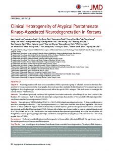
FIG. 1. Ribbon diagram of PanKTm monomer. The corresponding secondary structure elements in the two duplicate domains (N- and C-terminal domains) are labeled 1 to 5 and 1⬘ to 5⬘ for -strands and ␣1 to ␣3 and ␣1⬘ for ␣3⬘ for ␣-helices, respectively. The two helices ␣i1 and ␣i2 between 3⬘ and ␣1⬘ are considered to be insertions to the core of the fold. Three highly conserved aspartate residues, Asp6, Asp105, and Asp125, are shown in the ball-and-stick representation. The loop regions corresponding to the three conserved motifs PHOSPHATE 1 (between strands 1 and 2), PHOSPHATE 2 (between strands 1⬘ and 2⬘), and ADENOSINE (between strands 4⬘ and ␣2⬘) are colored orange.
same fold and are considered to be a result of gene duplication (Fig. 1). The core of each domain consists of a five-stranded mixed -sheet with strand order 3-2-1-4-5, where strand 2 is antiparallel to the rest of the sheet. The topology of the core of this fold is 321␣14␣25␣3, with the first helix ␣1 following strand 3. Two of the helices (␣1 and ␣2) are located on one side of the -sheet, while the third helix (␣3) is on the other side. Notably, the last helix ␣3 interacts intimately with the -sheet of the C-terminal domain and should be considered as part of the C-terminal domain, while the corresponding helix near the C terminus (␣3⬘) actually contributes to the core of
5535
the N-terminal domain (Fig. 1). This structural arrangement is characteristic of all members of the ASKHA superfamily. The search for similar structures in the protein data bank using program DALI (21) returns as its top hit 2-HG-CoA dehydratase Comp A (PDB code 1hux) with a Z-score of 14.5 and overall root mean square deviation (rmsd) of 202 superimposed C␣s of 3.7 Å (Table 2). The structural similarities of PanKTm to other members of the ASKHA superfamily are also high as shown in Table 2. Clearly, PanK-III proteins share the same fold as the ASKHA proteins and are likely to have evolved from the same ancestral protein as the rest of the superfamily. Notably, the sequence identities between PanKIII and the rest of the family are low (17% and below) and the rmsd’s between the superimposed C␣ atoms are quite large, ranging from 3.6 Å to 5.0 Å. To some extent, this large structural deviation is due to the difference in relative orientations between the two domains of the structures, but it also reflects the large evolutionary distance between PanK-III and the rest of the ASKHA superfamily. In PanKTm, the only deviation from the minimum core of the RNase H-like fold is a small insertion between strands 3⬘ and ␣1⬘ of the C-terminal domain, which consists of a pair of antiparallel helices and a long connecting loop (Fig. 1). Insertion at this particular site has been universally observed in all members of the ASKHA superfamily characterized so far. While many members in the superfamily are extensively decorated by long insertions at various sites of the RNase H-like fold core, PanKTm and 2HG-CoA dehydratase Comp A (1hux) appear to contain minimally inserted elements aside from the core. Structure of PanKTm dimer. PanKTm forms a tight dimer in the crystal as well as in solution, as demonstrated by the size exclusion chromatography profile (data not shown). The helical insertion (␣i1 and ␣i2) between 3⬘ and ␣1⬘ along with helix ␣1⬘ form an extensive dimer interface with the corresponding region of the second monomer (Fig. 2). This dimer interface buries about a 2,034-Å2 surface area and is largely hydrophobic in nature. The conformations of each monomer in the dimer are very similar, with an average rmsd in C␣ positions of 0.2 to 0.3Å even though no noncrystallographic symmetric restraints are imposed during refinement. Several other members of the ASKHA superfamily also form dimers through insertions located at the same site (between 3⬘ and ␣1⬘) and through a helix corresponding to the ␣1⬘ helix of the second domain, such as those observed in 2-HG-CoA dehydratase Comp A (33) and acetate kinase (11). However, the details of this interface differ, and the relative orientations of the two monomers are also different between
TABLE 2. Proteins structurally similar to PanKTm and structural alignment statistics from DALIa Protein (PDB code chain identifier)
Z-score
Rmsd (Å)
No. of aligned C␣s
Sequence identity (%)
Total no. of residues in protein
HG-CoA DH Comp A (1hux-A) FtsA (1e4f-T) Glycerol kinase (1glc-G) Acetate kinase (1g99-A) Hexokinase (1qha-A)
14.5 14.0 13.2 13.1 11.8
3.7 3.9 3.7 3.6 4.2
202 216 222 220 226
16 10 13 11 11
259 378 489 398 903
a
For details, see reference 21.
5536
YANG ET AL.
J. BACTERIOL.
FIG. 2. Ribbon diagram of PanKTm dimer. The two monomers are colored cyan and magenta, respectively. The active site of each monomer is marked by the ball-and-stick representation of the conserved aspartate residues. Modeled pantothenate (in green; see “Materials and Methods” for details) is also shown to indicate its location near the dimer interface.
PanK-III and the other members of the superfamily. As will be discussed later, the proposed pantothenate binding site in PanKTm contains residues from both monomers of the dimer, strongly suggesting that the dimer is the functional unit for PanKTm and probably for all other PanK-III enzymes as well. The active site of PanK-III. Several structures in the ASKHA superfamily have been solved in the presence of the bound substrates, those being either the ATP (or ADP) nucleotide or the phosphoryl acceptor substrate. In glycerol kinase and hexokinase, the ternary complexes with ADP and the phosphoryl acceptor substrate (or product in glycerol kinase) were also obtained (2, 24). In all these structures, the nucleotide binds at the same general location in a cleft formed between the two domains (Fig. 3). The structure-based multiple sequence alignment of representative PanK-III enzymes and a diverse set of the structures in the ASKHA superfamily (Fig. 4) shows that several conserved motifs that interact with the bound substrate, in particular ATP, are also conserved in PanK-III (8). These include the so-called PHOSPHATE 1 motif that encompasses the loop connecting strands 1 and 2 of the N-terminal domain and contains an invariant aspartate residue (Asp6 in PanKTm). This Asp residue has been shown to coordinate the Mg2⫹ ion that interacts with the - and ␥-phosphates of ATP (24, 53). The second highly conserved aspartate residue (Asp105 of PanKTm) is located at the beginning of helix ␣3. This Asp residue is close to the phosphoryl acceptor group and has been proposed to act as a catalytic base (23, 24). The PHOSPHATE 2 motif, also present in PanK-III, is located in the C-terminal domain between 1⬘ and 2⬘ and is somewhat structurally symmetrical to the PHOSPHATE 1 motif in the N-terminal domain. The third conserved motif, ADENOSINE, located in a loop after strand 4⬘ in the C-terminal domain, forms part of the pocket that binds the adenosine moiety of the nucleotide. The conservation of these sequence and structural motifs in the PanK-III proteins indicates that these motifs are likely to play similar roles in substrate binding and catalysis as in other members of the superfamily. Since the complex structure of PanK-III with substrate is at present not yet available, super-
position of PanKTm with several ASKHA proteins enabled us to model both the ATP and pantothenate substrates in the PanKTm active site (Fig. 5). This model provided a general placement of the substrates based on which further mutagenesis and kinetic analysis may be carried out to investigate the precise roles of the active site residues. Proposed ATP and pantothenate binding sites. In the current model of the PanKTm-substrate complex (Fig. 5), the ATP molecule interacts directly with the PHOSPHATE 1, PHOSPHATE 2, and ADENOSINE motifs. Asp6 of the PHOSPHATE 1 motif is in position to coordinate the divalent metal ion that interacts with the ATP phosphates, while Asp125 in the PHOSPHATE 2 motif may coordinate Mg2⫹ indirectly through a water molecule. The ADENOSINE motif in PanKTm adopts a conformation very similar to that in other members of the family and is predicted to play a similar role in binding the adenosine moiety of ATP. The modeling of pantothenate binding in PanKTm revealed that the second monomer of the dimer likely contributes to the pantothenate binding site of the first monomer and vice versa (Fig. 5). The loop following helix ␣i1 in the C-terminal domain insertion of the second monomer (hereafter termed “Pan cap,” for “pantothenate binding site cap”) is in close contact with the PHOSPHATE 1 loop of the first monomer. A hydrogen bond is formed between the side chain of Asn9 of the first monomer to the backbone amide group of residue Ala161⬘ of the second monomer. Additionally, Leu163⬘ of the second monomer, together with Ala129 and Ile145 of the first monomer, forms a small hydrophobic site that would accommodate the two methyl groups of the pantothenate substrate. As a result, this “Pan cap” loop forms a cap that would close in on the bound pantothenate and sequester the substrate from the surrounding solvent. The three hydrophobic residues (Ala129, Ile145, and Leu163) are highly conserved among all PanK-III proteins, suggesting their importance in either dimerization or the formation of the pantothenate binding site. The shared active site between two monomers of the PanK-III homodimer appears to be a novel feature unique among members of ASKHA superfamily. The phosphoryl transfer reactions catalyzed by ASKHA enzymes are believed to proceed via nucleophilic attack by
VOL. 188, 2006
STRUCTURE OF TYPE III PANTOTHENATE KINASE
5537
FIG. 3. Fold comparison of PanK-III with representative members of the ASKHA superfamily. The corresponding secondary structure elements in the core of each structure are colored accordingly, with -strands in magenta and ␣-helices in cyan. The regions that are considered insertions to the RNase H-like fold core are gray. Bound substrates in each structure are shown in a stick representation. The modeled substrates of PanKTm are also shown.
the phosphoryl acceptor group on the ␥-phosphoryl moiety of ATP, followed by the direct transfer of the terminal phosphate to the acceptor molecule (7, 23). Residues corresponding to Asp105 of PanKTm have been proposed to act
as a catalytic base activating the hydroxyl group of the phosphoryl acceptor for the nucleophilic attack. In the PanKTmsubstrate complex model, Asp105 is in good position for playing such a role (Fig. 5).
FIG. 4. Multiple sequence alignment of representative sequences of PanK-III (group I) and actin/hsp70/sugar kinase superfamily with known structures (group II). Sequences are labeled according to the gi number or PDB code and species name. The first and last residue numbers are indicated before and after each sequence, with the lengths of insertions specified in square brackets and the total sequence lengths of proteins following in parentheses. Residue conservation is denoted by the following scheme: uncharged, highlighted in yellow; charged/polar, in gray; small, in red; identical, bold and highlighted in black. The PHOSPHATE 1, PHOSPHATE 2, and ADENOSINE motifs are indicated at the bottom of the alignment. The secondary structure elements (E, -strand; H, ␣-helix) for PanKTm (gi 15611833) and (PDB 1hux_A) are marked above each sequence block, respectively. Abbreviations of species names are as follows: Hp, Helicobacter pylori; Ps, Pseudomonas syringae, Dv, Desulfovibrio vulgaris; Ch, Cytophaga hutchinsonii; Bc, Bacillus cereus; En, Emericella nidulans; Mm, Mus musculus; Ce, Caenorhabditis elegans; Hs, Homo sapiens; Sa, Staphylococcus aureus; Af, Acidaminococcus fermentans; Tm, Thermotoga maritima; and Ec, Escherichia coli.
5538
YANG ET AL.
FIG. 5. Model of MgATP and pantothenate binding in PanKTm active site. The color scheme is the same as that in Fig. 3 for the first monomer of the dimer, while the region corresponding to the second monomer is in yellow. The substrates ATP and pantothenate are shown as thick bonds, while the side chains of several active site residues are shown in the ball-and-stick representation. The Mg2⫹ ion is shown as a purple ball.
Roles of conserved aspartate residues. To investigate the proposed roles for the highly conserved Asp residues, we have carried out mutagenesis and kinetic analysis on PanKHp, a PanK-III closely related to PanKTm with which it shares about 32% sequence identity (Fig. 4). To confirm their kinetic similarity, we determined the apparent steady-state kinetic parameters for PanKTm in comparison to those of PanKHp, which have previously been reported (9). Since T. maritima is a thermophilic organism, kinetic data for PanKTm were obtained at 50°C. Our results show that the kinetic properties of PanKTm are very similar to those of PanKHp, with an apparent kcat of 0.29 ⫾ 0.02 s⫺1, a Km-Pantothenate of 40.3 ⫾ 3.3 M, and a high Km-ATP of 6.04 ⫾ 0.71 mM (compared to PanKHp values of 2.09 ⫾ 0.26 s⫺1, 101 ⫾ 26 M, and 9.59 ⫾ 2.14 mM, respectively (9)). The lower turnover number obtained for PanKTm probably reflects its requirement for a higher optimum temperature, which has not yet been determined. Based on sequence and structural analysis, it is almost certain that the conserved residues in these two proteins have the same function. PanKHp residues Asp17, Asp87, and Asp102 (corresponding to Asp6, Asp105, and Asp125, respectively, in PanKTm) were each mutated to either Asn or Glu. With the exception of the Asp17Glu mutant, all the mutant proteins expressed well and were purified by immobilized metal ion affinity chromatography. The enzymatic activities of the mutants were subsequently measured and compared to that of the native enzyme (Table 3). These results show that even a conservative substitution of any of the three proposed active site aspartate residues reduced enzyme activity drastically to less
J. BACTERIOL.
than 6% of that of the wild type. Two of these residues, Asp17 and Asp102, are proposed to be the metal ligands, while Asp87 is proposed to be the catalytic base. The mutagenesis data underscore the critical roles these residues play in the PanKIII-catalyzed reaction and are consistent with the mutant data for hexokinases and other members of the ASKHA superfamily (4, 55). Structural comparison of type I and type III PanKs. Comparison of the crystal structure of PanKTm and its active site configuration with those of PanKEc may provide a structural explanation for its lack of feedback inhibition by CoA and its inability to phosphorylate the N-alkylpantothenamide antimetabolites. As illustrated in a series of structures of PanKEc complexed with CoA, MgATP, as well as with both ADP and pantothenate (26, 58), CoA binds to PanKEc tightly in a site that partially overlaps with the ATP site, with 5⬘-phosphate of CoA overlapping with the ␥-phosphate of ATP. Surprisingly, the 3⬘-phosphoadenosine moiety of CoA binds at a completely different site from that of the adenosine group of ATP (58). In contrast, the pantetheine tail of CoA largely overlaps with the pantothenate binding site. The additional thiol group of CoA is accommodated in a hydrophobic pocket lined with mostly aromatic residues (58). It is hypothesized that this hydrophobic pocket would also be able to accommodate the longer hydrophobic tail of the N-substituted alkylpantothenamides (26), which can thus serve as alternative substrates of the enzyme and are converted to CoA antimetabolites (49). Inspection of a potential pantothenate binding site in PanKTm reveals no such hydrophobic pocket that could accommodate the longer tail of either CoA or N-alkylpantothenamides, which may explain, at least partially, why these molecules are not inhibitors or substrates of PanK-III and why PanK-III is not feedback inhibited by CoA or its thioesters. At present, it is difficult to speculate why PanK-III has such a high Km for ATP. It should be noted that PanK-III is not the only enzyme in the ASKHA superfamily that possesses such a high Km towards its substrate. The Kms of Methanosarcina thermophila acetate kinase for its substrate are also quite high: 2.8 mM for ATP and 22 mM for acetate (1). The consequences and implications of such steady-state kinetic properties on the metabolic fluxes in the organism remain to be illustrated. Clearly, further kinetic and structural studies are required to fully understand the underlying mechanisms of the PanK-IIIcatalyzed reaction.
TABLE 3. Effect of mutation of the active site aspartate residues on the activity of PanKHpa PanKHp protein
Relative rate of activity (%)
Native ........................................................................... 100 ⫾ 2.1 Asp17Asn..................................................................... 4.7 ⫾ 1.4 Asp87Asn..................................................................... 2.8 ⫾ 1.1 Asp87Glu ..................................................................... 5.0 ⫾ 1.7 Asp102Asn................................................................... 2.7 ⫾ 2.7 Asp102Glu ................................................................... 2.7 ⫾ 1.3 a The native enzyme and mutants were assayed under identical conditions in reaction mixtures containing 15 mM ATP plus 500 M pantothenate in 100 mM HEPES, pH 7.6, in three separate experiments for each protein. The initial rates of reaction were determined and are reported relative to the rate of the native enzyme, which was set at 100. Reported errors are the standard deviation of the three experiments. For experimental details, see Materials and Methods.
VOL. 188, 2006
STRUCTURE OF TYPE III PANTOTHENATE KINASE
TABLE 4. Phylogenetic distributions of different types of PanK in bacteriaa Presence of PanK Bacterial group Type I
Actinobacteria Aquificae Bacteroidetes/Chlorobi Chlamydiaeb Chloroflexi Cyanobacteria Deinococcus/Thermus Firmicutes Fusobacteria Planctomycetes Proteobacteria ␣  ␦/ε ␥ Spirochetes Thermotogae
Type II
Type III
Yes
Yes Yes Yes
Yes
Yes Yes Yes Yes Yes Yes
Yes
Yes Yes
Yes
Yes Yes Yes Yes Yes Yes
a The presence of each type of PanK is indicated. For a more detailed distribution of different types of PanK in individual species, see the supplemental material. b No candidate for any type of PanK can as yet be identified in the Chlamydiae (41).
5539
present in most of the major bacterial groups. The crystal structure of PanKTm revealed that type III PanK belongs to the ASKHA superfamily and adopts an entirely different fold from that of type I PanK. Mutagenesis and comparative structure analysis of PanK-III uncovered features of the enzyme and provided a structural explanation for the lack of product feedback inhibition of PanK-III. Since the currently established inhibitors of type I and type II PanKs are ineffective against PanK-III, the biochemical and structural elucidation of PanKIII not only is important for understanding the fundamental metabolic pathways in many PanK-III-harboring organisms but also provides a structural basis for the computer-aided design of specific inhibitors targeting PanK-III which may lead to novel antibacterial therapeutics. ACKNOWLEDGMENTS We thank Andrei Osterman for help with use of the SEED database and many enlightening discussions and Nick Grishin for critical reading of the manuscript. This work is supported by NIH grant GM63689 (to H.Z.), grants from the National Research Foundation of South Africa (GUN 2054218 and FA2005040600033 to E.S.), and in part, by a grant (I1505) from The Welch Foundation. Use of the Argonne National Laboratory Structure Biology beamline at Advanced Photon Source was supported by the U.S. Department of Energy, Office of Biological and Environmental Research, under contract no. W-31-109-ENG-38. REFERENCES
Type III PanK occurs in a wide range of bacterial species. It has been noted previously that PanK-III appears to be more common in the bacterial world than the “classical” PanK-I (40, 57). To examine this assertion, we conducted a comprehensive survey of the phylogenetic distribution of type I, II and III PanKs in over 300 complete or nearly complete genomes from the Archaea, Eukarya, and 13 major groups of Bacteria using the expert annotated SEED genomic integration platform (http://theseed.uchicago.edu/FIG/index.cgi). The same database has previously been used to establish the existence of the five-step universal CoA biosynthetic pathway in the majority of these organisms (40, 57). The results of this survey clearly show that PanK-III exists in 12 of the 13 major bacterial groups, the exception being the Chlamydiae, for which no candidate PanK has yet been identified (Table 4; for more details, see Table S1 in the supplemental material). PanK-I, on the other hand, is present in only four groups of Bacteria: the Actinobacteria, Chloroflexi (green non-sulfur bacteria), Firmicutes (gram-positive bacteria), and Proteobacteria (purple bacteria and relatives). This surprisingly widespread distribution of PanK-III further underscores the importance of understanding the mechanism of this important enzyme and the regulation of CoA biosynthesis in organisms harboring PanK-III. Interestingly, a number of bacteria have more than one type of PanK: in mycobacteria and several bacilli both type I and type III PanKs are present, while Bacillus anthracis and B. cereus contain both PanK-II and PanK-III. The physiological significance of this functional redundancy is currently unclear. Moreover, genes coding for the PanK activity have not been identified in the Archaea kingdom, suggesting the existence of another, as yet uncharacterized, type of PanK (40). In summary, we have demonstrated that type III PanK encoded by coaX has a more widespread phylogenetic distribution than the long-known PanK-I and is nearly universally
1. Aceti, D. J., and J. G. Ferry. 1988. Purification and characterization of acetate kinase from acetate-grown Methanosarcina thermophila. Evidence for regulation of synthesis. J. Biol. Chem. 263:15444–15448. 2. Aleshin, A. E., C. Kirby, X. Liu, G. P. Bourenkov, H. D. Bartunik, H. J. Fromm, and R. B. Honzatko. 2000. Crystal structures of mutant monomeric hexokinase I reveal multiple ADP binding sites and conformational changes relevant to allosteric regulation. J. Mol. Biol. 296:1001–1015. 3. Andreeva, A., D. Howorth, S. E. Brenner, T. J. Hubbard, C. Chothia, and A. G. Murzin. 2004. SCOP database in 2004: refinements integrate structure and sequence family data. Nucleic Acids Res. 32:D226–D229. 4. Arora, K. K., C. R. Filburn, and P. L. Pedersen. 1991. Glucose phosphorylation. Site-directed mutations which impair the catalytic function of hexokinase. J. Biol. Chem. 266:5359–5362. 5. Becker, D., M. Selbach, C. Rollenhagen, M. Ballmaier, T. F. Meyer, M. Mann, and D. Bumann. 2006. Robust Salmonella metabolism limits possibilities for new antimicrobials. Nature 440:303–307. 6. Begley, T. P., C. Kinsland, and E. Strauss. 2001. The biosynthesis of coenzyme A in bacteria. Vitam. Horm. 61:157–171. 7. Blattler, W. A., and J. R. Knowles. 1979. Stereochemical course of phosphokinases. The use of adenosine [␥-(S)-16O,17O,18O]triphosphate and the mechanistic consequences for the reactions catalyzed by glycerol kinase, hexokinase, pyruvate kinase, and acetate kinase. Biochemistry 18:3927–3933. 8. Bork, P., C. Sander, and A. Valencia. 1992. An ATPase domain common to prokaryotic cell cycle proteins, sugar kinases, actin, and hsp70 heat shock proteins. Proc. Natl. Acad. Sci. USA 89:7290–7294. 9. Brand, L. A., and E. Strauss. 2005. Characterization of a new pantothenate kinase isoform from Helicobacter pylori. J. Biol. Chem. 280:20185–20188. 10. Bru ¨nger, A. T., P. D. Adams, G. M. Clore, W. L. DeLano, P. Gros, R. W. Grosse-Kunstleve, J. S. Jiang, J. Kuszewski, M. Nilges, N. S. Pannu, R. J. Read, L. M. Rice, T. Simonson, and G. L. Warren. 1998. Crystallography & NMR system: a new software suite for macromolecular structure determination. Acta Crystallogr. Sect. D Biol. Crystallogr. 54:905–921. 11. Buss, K. A., D. R. Cooper, C. Ingram-Smith, J. G. Ferry, D. A. Sanders, and M. S. Hasson. 2001. Urkinase: structure of acetate kinase, a member of the ASKHA superfamily of phosphotransferases. J. Bacteriol. 183:680–686. 12. Calder, R. B., R. S. Williams, G. Ramaswamy, C. O. Rock, E. Campbell, S. E. Unkles, J. R. Kinghorn, and S. Jackowski. 1999. Cloning and characterization of a eukaryotic pantothenate kinase gene (panK) from Aspergillus nidulans. J. Biol. Chem. 274:2014–2020. 13. Cheek, S., K. Ginalski, H. Zhang, and N. V. Grishin. 2005. A comprehensive update of the sequence and structure classification of kinases. BMC Struct. Biol. 5:6. [Online.] doi:10.1186/1472-6807-5-6. 14. Cheek, S., H. Zhang, and N. V. Grishin. 2002. Sequence and structure classification of kinases. J. Mol. Biol. 320:855–881. 15. Choudhry, A. E., T. L. Mandichak, J. P. Broskey, R. W. Egolf, C. Kinsland, T. P. Begley, M. A. Seefeld, T. W. Ku, J. R. Brown, M. Zalacain, and K.
5540
16. 17.
18. 19. 20.
21. 22. 23. 24. 25.
26.
27.
28. 29. 30. 31.
32. 33. 34. 35. 36. 37.
YANG ET AL.
Ratnam. 2003. Inhibitors of pantothenate kinase: novel antibiotics for staphylococcal infections. Antimicrob. Agents Chemother. 47:2051–2055. Collaborative Computational Project Number 4. 1994. The CCP4 Suite: programs for protein crystallography. Acta Crystallogr. Sect. D Biol. Crystallogr. 50:760–763. delCardayre, S. B., K. P. Stock, G. L. Newton, R. C. Fahey, and J. E. Davies. 1998. Coenzyme A disulfide reductase, the primary low molecular weight disulfide reductase from Staphylococcus aureus. Purification and characterization of the native enzyme. J. Biol. Chem. 273:5744–5751. Doublie, S. 1997. Preparation of selenomethionyl proteins for phase determination. Methods Enzymol. 276:523–530. Flaherty, K. M., C. DeLuca-Flaherty, and D. B. McKay. 1990. Three-dimensional structure of the ATPase fragment of a 70K heat-shock cognate protein. Nature 346:623–628. Gerdes, S. Y., M. D. Scholle, M. D’Souza, A. Bernal, M. V. Baev, M. Farrell, O. V. Kurnasov, M. D. Daugherty, F. Mseeh, B. M. Polanuyer, J. W. Campbell, S. Anantha, K. Y. Shatalin, S. A. K. Chowdhury, M. Y. Fonstein, and A. L. Osterman. 2002. From genetic footprinting to antimicrobial drug targets: examples in cofactor biosynthetic pathways. J. Bacteriol. 184:4555– 4572. Holm, L., and C. Sander. 1995. Dali: a network tool for protein structure comparison. Trends Biochem. Sci. 20:478–480. Holmes, K. C., D. Popp, W. Gebhard, and W. Kabsch. 1990. Atomic model of the actin filament. Nature 347:44–49. Hurley, J. H. 1996. The sugar kinase/heat shock protein 70/actin superfamily: implications of conserved structure for mechanism. Annu. Rev. Biophys. Biomol. Struct. 25:137–162. Hurley, J. H., H. R. Faber, D. Worthylake, N. D. Meadow, S. Roseman, D. W. Pettigrew, and S. J. Remington. 1993. Structure of the regulatory complex of Escherichia coli IIIGlc with glycerol kinase. Science 259:673–677. Ito, S., S. Fushinobu, J. J. Jeong, I. Yoshioka, S. Koga, H. Shoun, and T. Wakagi. 2003. Crystal structure of an ADP-dependent glucokinase from Pyrococcus furiosus: implications for a sugar-induced conformational change in ADP-dependent kinase. J. Mol. Biol. 331:871–883. Ivey, R. A., Y.-M. Zhang, K. G. Virga, K. Hevener, R. E. Lee, C. O. Rock, S. Jackowski, and H.-W. Park. 2004. The structure of the pantothenate kinase. ADP.pantothenate ternary complex reveals the relationship between the binding sites for substrate, allosteric regulator, and antimetabolites. J. Biol. Chem. 279:35622–35629. Jackowski, S. 1996. Biosynthesis of pantothenic acid and coenzyme A, p. 687–694. In F. C. Neidhardt, R. Curtiss III, J. L. Ingraham, E. C. C. Lin, K. B. Low, B. Magasanik, W. S. Reznikoff, M. Riley, M. Schaechter, and H. E. Umbarger (ed.), Escherichia coli and Salmonella: cellular and molecular biology, 2nd ed., vol. 1. ASM Press, Washington, D.C. Jones, T. A., J.-Y. Zou, S. W. Cowan, and M. Kjeldgaard. 1991. Improved methods for building protein models in electron density maps and the location of errors in these models. Acta Crystallogr. Sect. A 47:110–119. Kabsch, W., H. G. Mannherz, D. Suck, E. F. Pai, and K. C. Holmes. 1990. Atomic structure of the actin:DNase I complex. Nature 347:37–44. Kapust, R. B., and D. S. Waugh. 1999. Escherichia coli maltose-binding protein is uncommonly effective at promoting the solubility of polypeptides to which it is fused. Protein Sci. 8:1668–1674. Leonardi, R., S. Chohnan, Y.-M. Zhang, K. G. Virga, R. E. Lee, C. O. Rock, and S. Jackowski. 2005. A pantothenate kinase from Staphylococcus aureus refractory to feedback regulation by coenzyme A. J. Biol. Chem. 280:3313– 3322. Leonardi, R., Y. M. Zhang, C. O. Rock, and S. Jackowski. 2005. Coenzyme A: back in action. Prog. Lipid Res. 44:125–153. Locher, K. P., M. Hans, A. P. Yeh, B. Schmid, W. Buckel, and D. C. Rees. 2001. Crystal structure of the Acidaminococcus fermentans 2-hydroxyglutaryl-CoA dehydratase component A. J. Mol. Biol. 307:297–308. Luba, J., V. Charrier, and A. Claiborne. 1999. Coenzyme A-disulfide reductase from Staphylococcus aureus: evidence for asymmetric behaviour on interaction with pyridine nucleotides. Biochemistry 38:2725–2737. Lunin, V. V., Y. Li, J. D. Schrag, P. Iannuzzi, M. Cygler, and A. Matte. 2004. Crystal structures of Escherichia coli ATP-dependent glucokinase and its complex with glucose. J. Bacteriol. 186:6915–6927. Murshudov, G. N., A. A. Vagin, and E. J. Dodson. 1997. Refinement of macromolecular structures by the maximum-likelihood method. Acta Crystallogr. Sect. D Biol. Crystallogr. 53:240–255. Murzin, A. G., S. E. Brenner, T. Hubbard, and C. Chothia. 1995. SCOP: a structural classification of proteins database for the investigation of sequences and structures. J. Mol. Biol. 247:536–540.
J. BACTERIOL. 38. Otwinowski, Z. 1991. Maximum likelihood refinement of heavy atom parameters, p. 80–86. In W. Wolf, P. R. Evans, and A. G. W. Leslie (ed.), Isomorphous replacement and anomalous scattering. Daresbury Laboratory, Warrington, United Kingdom. 39. Otwinowski, Z., and W. Minor. 1997. Processing of X-ray diffraction data collected in oscillation mode. Methods Enzymol. 276:307–326. 40. Overbeek, R., T. Begley, R. M. Butler, J. V. Choudhuri, H. Y. Chuang, M. Cohoon, V. de Crecy-Lagard, N. Diaz, T. Disz, R. Edwards, M. Fonstein, E. D. Frank, S. Gerdes, E. M. Glass, A. Goesmann, A. Hanson, D. IwataReuyl, R. Jensen, N. Jamshidi, L. Krause, M. Kubal, N. Larsen, B. Linke, A. C. McHardy, F. Meyer, H. Neuweger, G. Olsen, R. Olson, A. Osterman, V. Portnoy, G. D. Pusch, D. A. Rodionov, C. Ruckert, J. Steiner, R. Stevens, I. Thiele, O. Vassieva, Y. Ye, O. Zagnitko, and V. Vonstein. 2005. The subsystems approach to genome annotation and its use in the project to annotate 1000 genomes. Nucleic Acids Res. 33:5691–5702. 41. Raman, S. B., and B. Rathinasabapathi. 2004. Pantothenate synthesis in plants. Plant Sci. 167:961–968. 42. Rock, C. O., R. B. Calder, M. A. Karim, and S. Jackowski. 2000. Pantothenate kinase regulation of the intracellular concentration of coenzyme A. J. Biol. Chem. 275:1377–1387. 43. Rock, C. O., M. A. Karim, Y. Zhang, and S. Jackowski. 2002. The murine pantothenate kinase (Pank1) gene encodes two differentially regulated pantothenate kinase isozymes. Gene 291:35–43. 44. Rosano, C., E. Sabini, M. Rizzi, D. Deriu, G. Murshudov, M. Bianchi, G. Serafini, M. Magnani, and M. Bolognesi. 1999. Binding of non-catalytic ATP to human hexokinase I highlights the structural components for enzymemembrane association control. Structure 7:1427–1437. 45. Schneider, T. R., and G. M. Sheldrick. 2002. Substructure solution with SHELXD. Acta Crystallogr. Sect. D Biol. Crystallogr. 58:1772–1779. 46. Song, W.-J., and S. Jackowski. 1992. Cloning, sequencing, and expression of the pantothenate kinase (coaA) gene of Escherichia coli. J. Bacteriol. 174: 6411–6417. 47. Song, W. J., and S. Jackowski. 1994. Kinetics and regulation of pantothenate kinase from Escherichia coli. J. Biol. Chem. 269:27051–27058. 48. Steitz, T. A., R. J. Fletterick, W. F. Anderson, and C. M. Anderson. 1976. High resolution x-ray structure of yeast hexokinase, an allosteric protein exhibiting a non-symmetric arrangement of subunits. J. Mol. Biol. 104:197– 222. 49. Strauss, E., and T. P. Begley. 2002. The antibiotic activity of N-pentylpantothenamide results from its conversion to ethyldethia-coenzyme A, a coenzyme A antimetabolite. J. Biol. Chem. 277:48205–48209. 50. Terwilliger, T. C. 2000. Maximum-likelihood density modification. Acta Crystallogr. Sect. D Biol. Crystallogr. 56:965–972. 51. Terwilliger, T. C. 2003. SOLVE and RESOLVE: automated structure solution and density modification. Methods Enzymol. 374:22–37. 52. Vallari, D. S., S. Jackowski, and C. O. Rock. 1987. Regulation of pantothenate kinase by coenzyme A and its thioesters. J. Biol. Chem. 262:2468–2471. 53. van den Ent, F., and J. Lowe. 2000. Crystal structure of the cell division protein FtsA from Thermotoga maritima. EMBO J. 19:5300–5307. 54. Virga, K. G., Y. M. Zhang, R. Leonardi, R. A. Ivey, K. Hevener, H. W. Park, S. Jackowski, C. O. Rock, and R. E. Lee. 2006. Structure-activity relationships and enzyme inhibition of pantothenamide-type pantothenate kinase inhibitors. Bioorg. Med. Chem. Lett. 14:1007–1020. 55. Wilbanks, S. M., C. DeLuca-Flaherty, and D. B. McKay. 1994. Structural basis of the 70-kilodalton heat shock cognate protein ATP hydrolytic activity. I. Kinetic analyses of active site mutants. J. Biol. Chem. 269:12893–12898. 56. Wilbanks, S. M., and D. B. McKay. 1998. Structural replacement of active site monovalent cations by the ε-amino group of lysine in the ATPase fragment of bovine Hsc70. Biochemistry 37:7456–7462. 57. Ye, Y., A. Osterman, R. Overbeek, and A. Godzik. 2005. Automatic detection of subsystem/pathway variants in genome analysis. Bioinformatics 21(Suppl. 1):i478–i486. 58. Yun, M., C.-G. Park, J.-Y. Kim, C. O. Rock, S. Jackowski, and H.-W. Park. 2000. Structural basis for the feedback regulation of Escherichia coli pantothenate kinase by coenzyme A. J. Biol. Chem. 275:28093–28099. 59. Zhang, Y.-M., M. W. Frank, K. G. Virga, R. E. Lee, C. O. Rock, and S. Jackowski. 2004. Acyl carrier protein is a cellular target for the antibacterial action of the pantothenamide class of pantothenate antimetabolites. J. Biol. Chem. 279:50969–50975. 60. Zhao, L., N. M. Allanson, S. P. Thomson, J. K. F. Maclean, J. J. Barker, W. U. Primrose, P. D. Tyler, and A. Lewendon. 2003. Inhibitors of phosphopantetheine adenylyltransferase. Eur. J. Med. Chem. 38:345–349.