http://dx.doi.org/10.5607/en.2015.24.2.139 Exp Neurobiol. 2015 Jun;24(2):139-145. pISSN 1226-2560 • eISSN 2093-8144
Original Article
Curcumin Ameliorates Functional and Structural Abnormalities in Cisplatin-induced Neuropathy Sithiporn Agthong*, Athitaya Kaewsema and Thuntawat Charoensub
Department of Anatomy, Faculty of Medicine, Chulalongkorn University, Bangkok 10330, Thailand
Peripheral neuropathy is one of the major side effects of cisplatin; however, effective treatments are lacking. Curcumin is a polyphenol found in the root of Curcuma longa and has been shown neuroprotective against several neurological diseases. Nevertheless, its effects on cisplatin neuropathy remain unclear. This study aimed to clarify this issue by inducing neuropathy in the rats with intraperitoneal injection of cisplatin 2 mg/kg twice a week for 5 consecutive weeks. Curcumin 200 mg/kg/day was given by gavage to a group of cisplatin-treated rats during these five weeks. The results showed that cisplatin induced thermal hypoalgesia in the 5th week which could be prevented by curcumin. In the 5th and 8th weeks, sciatic motor nerve conduction velocity was reduced in the cisplatin compared with the control groups. Curcumin significantly attenuated this deficit. Morphometric analysis of L4 dorsal root ganglia from the cisplatin group revealed nuclear and nucleolar atrophy including loss of neurons in the 8th week. These alterations were significantly blocked by curcumin. Moreover, curcumin also ameliorated the reduced myelin thickness in the sciatic nerve of cisplatin-treated rats. Taken together, our findings suggest the favorable effects of curcumin on both functional and structural abnormalities in cisplatin neuropathy. Future studies are needed to clarify the exact underlying mechanisms. Key words: cisplatin, curcumin, neuropathy, sciatic nerve, dorsal root ganglion
INTRODUCTION
Cisplatin has been used to treat cancers of various organs, including lung, urinary bladder, testis and ovary [1]. However, peripheral neuropathy is one of its major side effects which can lead to cessation of treatment and poor quality of life. Sensory perception and nerve conduction deficits are similarly observed in both patients and animals treated with cisplatin [2-7]. In addition, structural alterations including loss of dorsal root ganglion (DRG) neurons and atrophy of neuronal nucleus and nucleolus have also
Received March 3, 2015, Revised April 18, 2015, Accepted April 20, 2015
been reported [5,7-10]. Curcumin is a polyphenol found in the root of Curcuma longa and possesses antioxidant, anti-inflammatory and antineoplastic properties [11]. Its beneficial effects on various neurological diseases have also been shown [12]. However, the data regarding its effect on cisplatin neuropathy remain lacking. Mendonca and co-workers reported that curcumin could reduce the toxicity of cisplatin on neurite outgrowth [13]. In cisplatin-treated rats, curcumin had some favorable effects on biochemical and histological alterations of sciatic nerve [14]. Despite these data, stronger evidence is still needed to prove the efficacy of curcumin. In this study, we showed the novel findings that curcumin could attenuate the functional and structural abnormalities in the rats receiving cisplatin.
*To whom correspondence should be addressed. TEL: 0662-2564281, FAX: 0662-2527028 e-mail:
[email protected] Copyright © Experimental Neurobiology 2015. www.enjournal.org
This is an Open Access article distributed under the terms of the Creative Commons Attribution Non-Commercial License (http://creativecommons.org/licenses/by-nc/4.0) which permits unrestricted non-commercial use, distribution, and reproduction in any medium, provided the original work is properly cited.
Sithiporn Agthong, et al.
MATERIALS AND METHODS
Twenty-four female Wistar rats weighing 200~250 g were housed on a 12 hour light-dark cycle with access to food and water ad libitum. This experiment was approved by the institutional ethics committee, Faculty of Medicine, Chulalongkorn University and was carried out in accordance with the guidelines of the National Research Council of Thailand. All efforts were done to minimize pain and discomfort. Drug administration
The animals were divided into 3 groups: control, cisplatin and cisplatin+curcumin (n=8 each). The cisplatin and cisplatin+ curcumin groups received cisplatin (Pfizer) 2 mg/kg intraperitoneally twice a week for five consecutive weeks (20 mg/kg cumulative dose). Cisplatin was diluted in normal saline to the final concentration of 0.5 mg/ml. The dilution was done to give excess fluid to prevent nephrotoxicity. Functional and structural abnormalities of peripheral nerve were induced with this dosage regimen of cisplatin [4]. Administration of curcumin (Cayman Chemical) 200 mg/ kg dissolved in 1% sodium carboxy methyl cellulose (SCMC) was given by gavage to the cisplatin+curcumin group once daily for five weeks. A group of curcumin alone was not included since the results from our pilot study (unpublished data) showed no significant effects. The cisplatin group also received the vehicle for curcumin (1% SCMC) while the control group received the vehicles for cisplatin (normal saline) and curcumin. After 5 weeks of treatments, all the animals were left untreated until sacrificed at the 8th week since this was the appropriate time-point to observe the structural abnormalities [10]. Hind-paw thermal threshold
The hot plate test was used to evaluate the thermal perception and the procedure used here was able to detect the cisplatininduced thermal hypoalgesia in the previous study [10]. Briefly, the rats were allowed to familiarize with the test procedure and apparatus prior to the measurement. The test was done at baseline and the 3rd, 5th and 8th weeks after the start of cisplatin injection. Each rat was placed on the hot plate analgesia meter (Harvard Apparatus, UK) maintained at 55oC. When the rat licked its hind paw on either side, elapsed time was recorded as latency. The cut-off duration of 35 s was employed to avoid skin injury. If the latency was over this limit, 35 s was used for further analysis. The test was repeated at least 3 times with an interval of 15 min and mean latency was obtained for each rat.
140
www.enjournal.org
Sciatic motor nerve conduction velocity
The motor nerve conduction velocity (MNCV) was measured at baseline and the 5th and 8th weeks. The rat was anesthetized using isoflurane and rectal temperature was maintained at 37±0.3oC using a heating pad and digital rectal thermometer. The stimulating and recording needle electrodes were inserted at the sciatic notch and the second interosseous muscle of the hind foot, respectively. The ground electrode was placed at the laletal side of the hind foot. These electrodes were connected to the oscilloscope (Neurostar, Oxford Instrument). The sciatic nerve was stimulated with a supramaximal stimulus and compound muscle action potential (CMAP) was recorded. Latency1 (L1) was measured from the stimulation artifact to the positive peak of M wave. The average L1 was derived from at least five stimulations. Then, the stimulating electrode was moved to the side of Achilles’ tendon and the procedure was repeated to determine the average latency2 (L2). The MNCV was calculated by dividing the distance between the two stimulation points by the latency difference (L1-L2). Tissue collection
At the end of the 8th week, all rats were sacrificed by overdose anesthetics and then transcardially perfused with 200 ml of normal saline followed by 400 ml of 4% paraformaldehyde. L4 DRG and sciatic nerves were post-fixed in 3% glutaraldehyde for 6 hours and embedded in epoxy resin. These specimens were used for structural analysis. Nerve morphometry
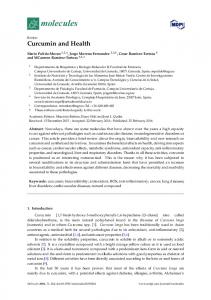
Transverse 1 μm-thick sections of the sciatic nerve were cut, mounted on slides, and stained with paraphenylenediamine. Representative image is shown in Fig. 1. The sections were examined under a light microscope and the cross-sectional areas were chosen using the three-window sampling method. Details of this sampling technique were described elsewhere [15]. Briefly, under 40x objective lens, three windows of 0.012 mm 2 were randomly placed, one in the middle and the other two in the periphery of fascicle. Images of these windows were imported into the microcomputer via a digital camera. Morphometric analysis was done to obtain the number of myelinated fibers, axon diameter, myelinated fiber density, myelin thickness and g ratio using the Image-Pro Plus software. The values derived from the three windows were extrapolated to the whole nerve. DRG morphometry
The DRG were serially cut into 2 μm-thick sections and stained with toluidine blue. Representative image is shown in Fig. 1. The estimation for total number of neurons in each ganglion was http://dx.doi.org/10.5607/en.2015.24.2.139
Curcumin and Cisplatin Neuropathy
Fig. 1. Representative images of dorsal root ganglion (A) and sciatic nerve (B) used for morphometric analyses. Since the pathological changes could not be seen with eyes and images from different groups looked similar, only representative images from the control group are shown. The sections of ganglion and nerve were stained with toluidine blue and paraphenylenediamine, respectively. In A, arrows and arrowheads indicate nuclei and nucleoli of neurons, respectively. In B, asterisks and arrowheads indicate axons and myelin sheath, respectively. Scale bars represent 50 µm.
Fig. 2. Changes in the average body weight of the control (C), cisplatin (P) and cisplatin+curcumin (M) groups. The graph shows means and SEM. *p